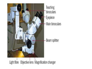
Operating Microscope AND Slit
Lamp
Operating microscope and binocular
slit lamp microscope:
The operating microscope and binocular slit lamp
microscope are compound microscope
• Microscope: Magnify an object at a near.
• Telescope: Magnify an object at a distance
• The compound microscope consists of two convex
lenses, the objective and eyepiece lenses.
• The compound microscope consists of two convex
lenses, the objective and eyepiece lenses. The
object O to be studied is placed just outside the
anterior focal point, Fo, of the objective lens.
• A real, inverted, magnified image, is formed
some distance behind the objective lens.
The eyepiece lens, is placed so that the image
formed by the objective falls at or close to its
principal focal plane, Fe. The eyepiece thus acts as
a loupe and further magnifies the image seen by
the observer. The final image I is vertically and
horizontally inverted.
• Porro prisms are incorporated. in clinical
microscopes to obtain an erect non-inverted
image and to shorten the physical length of the
instrument.
An operating or surgical microscope..
 An operating or surgical microscope is an optical
instrument that provides the surgeon with a…..
 Stereoscopic
 high quality magnified and
 illuminated image of the small structures in the
surgical area.
The optical components of a basic
stereo microscope consist of the..
a) binocular head,
b) a magnification changer,
c) the objective lens and
d) an illuminator which beams light through the
objective lens and onto the operating field.
The binocular head consists of:
 Two telescopes with adjustable eyepieces for
users with refractive error.
 The magnification can be changed by turning a
knob (which selects different magnification
lenses) or by using a motorized zoom controlled
by a foot pedal
The working distance:
• The working distance is the distance from the
microscope objective lens to the point of focus of
the optical system. This value is fixed and is
dependent on the chosen focal length of the
objective lens.
The working distance:
• The choice of working distance depends on the
type of surgery.
• For modern ophthalmic surgery that involves
delicate work in the posterior chamber, objective
focal lengths of 150 mm, 175 mm and 200 mm
are commonly used.
 The optical system often includes a beam splitter
and a second set of teaching binoculars so that
two people can view the operation
simultaneously.
Caring for the operating microscope
 Keep the microscope in a dry, cool and well-
ventilated place to prevent fungus growth on the
optics (lenses).
 Every week, clean the optics according to the
optical cleaning instructions.
 If fungus growth is detected, clean according to
the instructions.
Caring for the operating microscope
 To protect it from dust when not in use, drape a
cover over the microscope.
 Vinyl coverings are preferred because they do
not shed lint (like cloth coverings do).
 However, their use should be avoided in humid
environments since they can trap moisture,
which increases the risk of fungal growth.
Caring for the operating microscope
 Wipe down the external surfaces with a damp
cloth soaked in hot, soapy water.
 Cover the foot pedal with a clear plastic bag to
prevent surgical and cleaning fluids from
entering and damaging the electronics.
 Lift the foot pedal off the floor when washing the
floor.
Caring for the operating microscope
• Use a voltage stabilizer with the microscope.
This will prevent sudden increases in voltage
from destroying the bulbs and will ensure that
the illumination provided remains constant.
Caring for the operating microscope
• Before using, test the controls of the foot pedal
(the x,y movement, zoom, focus, light on and
off).
• Before using, check that the suspension arm can
be fixed into position to ensure that it does not
fall on the patient.
• Avoid kinking or bending the fiber optic cables.
Before using the operating microscope:
• When replacing the bulbs, avoid touching them
with your fingers. The oil left as fingerprints on
the bulb can shorten its life.
• Do not move the microscope while the bulb is
still hot because strong vibrations may damage
the filament.
• Every six months, clean and oil the wheels and
the brakes. Remove any surplus oil when done.

10 slit lamp and operating microscope

  • 1.
  • 3.
    Operating microscope andbinocular slit lamp microscope: The operating microscope and binocular slit lamp microscope are compound microscope • Microscope: Magnify an object at a near. • Telescope: Magnify an object at a distance • The compound microscope consists of two convex lenses, the objective and eyepiece lenses.
  • 5.
    • The compoundmicroscope consists of two convex lenses, the objective and eyepiece lenses. The object O to be studied is placed just outside the anterior focal point, Fo, of the objective lens. • A real, inverted, magnified image, is formed some distance behind the objective lens.
  • 6.
    The eyepiece lens,is placed so that the image formed by the objective falls at or close to its principal focal plane, Fe. The eyepiece thus acts as a loupe and further magnifies the image seen by the observer. The final image I is vertically and horizontally inverted.
  • 7.
    • Porro prismsare incorporated. in clinical microscopes to obtain an erect non-inverted image and to shorten the physical length of the instrument.
  • 12.
    An operating orsurgical microscope..  An operating or surgical microscope is an optical instrument that provides the surgeon with a…..  Stereoscopic  high quality magnified and  illuminated image of the small structures in the surgical area.
  • 13.
    The optical componentsof a basic stereo microscope consist of the.. a) binocular head, b) a magnification changer, c) the objective lens and d) an illuminator which beams light through the objective lens and onto the operating field.
  • 14.
    The binocular headconsists of:  Two telescopes with adjustable eyepieces for users with refractive error.  The magnification can be changed by turning a knob (which selects different magnification lenses) or by using a motorized zoom controlled by a foot pedal
  • 16.
    The working distance: •The working distance is the distance from the microscope objective lens to the point of focus of the optical system. This value is fixed and is dependent on the chosen focal length of the objective lens.
  • 17.
    The working distance: •The choice of working distance depends on the type of surgery. • For modern ophthalmic surgery that involves delicate work in the posterior chamber, objective focal lengths of 150 mm, 175 mm and 200 mm are commonly used.
  • 19.
     The opticalsystem often includes a beam splitter and a second set of teaching binoculars so that two people can view the operation simultaneously.
  • 20.
    Caring for theoperating microscope  Keep the microscope in a dry, cool and well- ventilated place to prevent fungus growth on the optics (lenses).  Every week, clean the optics according to the optical cleaning instructions.  If fungus growth is detected, clean according to the instructions.
  • 21.
    Caring for theoperating microscope  To protect it from dust when not in use, drape a cover over the microscope.  Vinyl coverings are preferred because they do not shed lint (like cloth coverings do).  However, their use should be avoided in humid environments since they can trap moisture, which increases the risk of fungal growth.
  • 22.
    Caring for theoperating microscope  Wipe down the external surfaces with a damp cloth soaked in hot, soapy water.  Cover the foot pedal with a clear plastic bag to prevent surgical and cleaning fluids from entering and damaging the electronics.  Lift the foot pedal off the floor when washing the floor.
  • 23.
    Caring for theoperating microscope • Use a voltage stabilizer with the microscope. This will prevent sudden increases in voltage from destroying the bulbs and will ensure that the illumination provided remains constant.
  • 24.
    Caring for theoperating microscope • Before using, test the controls of the foot pedal (the x,y movement, zoom, focus, light on and off). • Before using, check that the suspension arm can be fixed into position to ensure that it does not fall on the patient. • Avoid kinking or bending the fiber optic cables.
  • 25.
    Before using theoperating microscope: • When replacing the bulbs, avoid touching them with your fingers. The oil left as fingerprints on the bulb can shorten its life. • Do not move the microscope while the bulb is still hot because strong vibrations may damage the filament. • Every six months, clean and oil the wheels and the brakes. Remove any surplus oil when done.