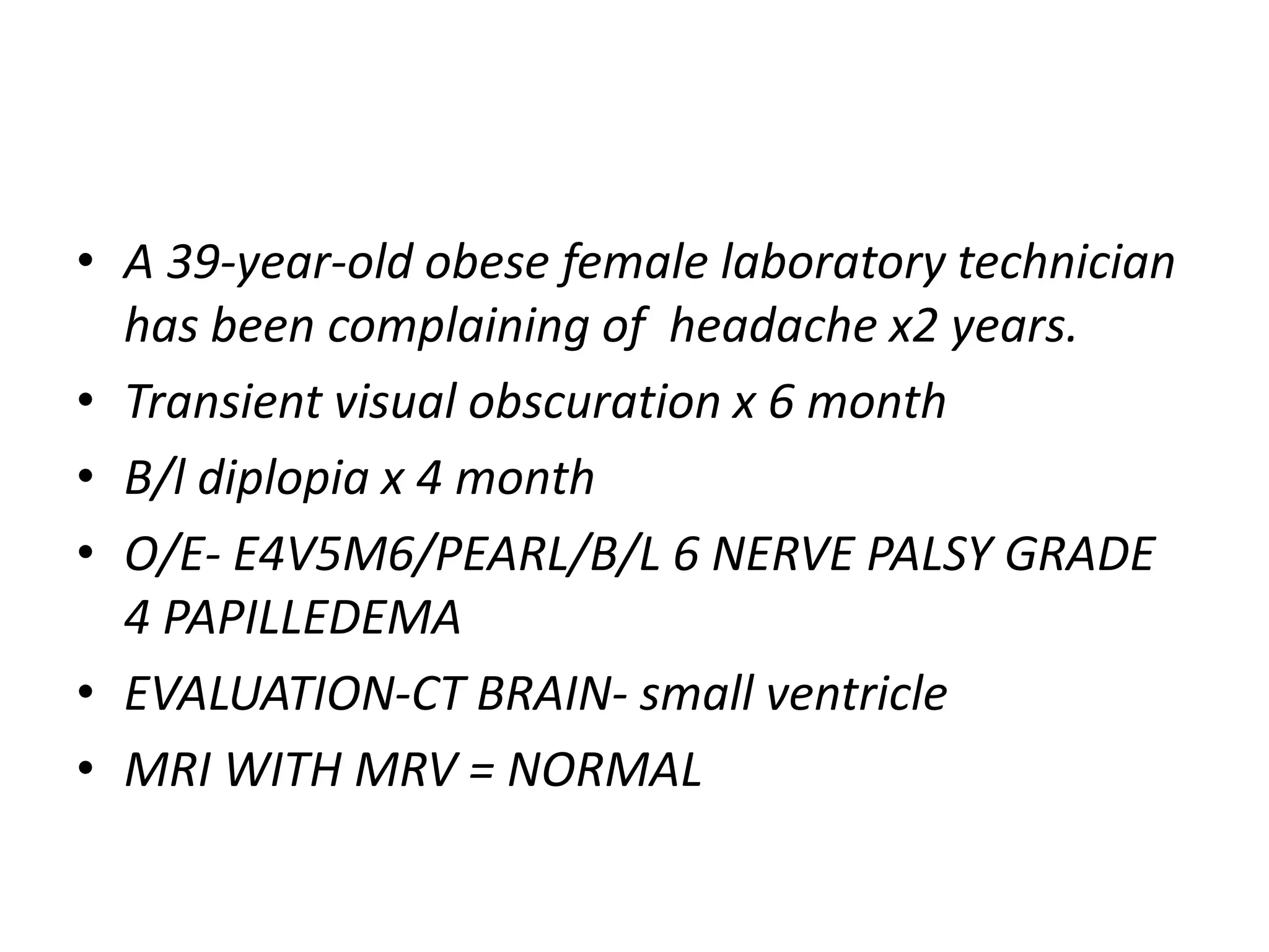
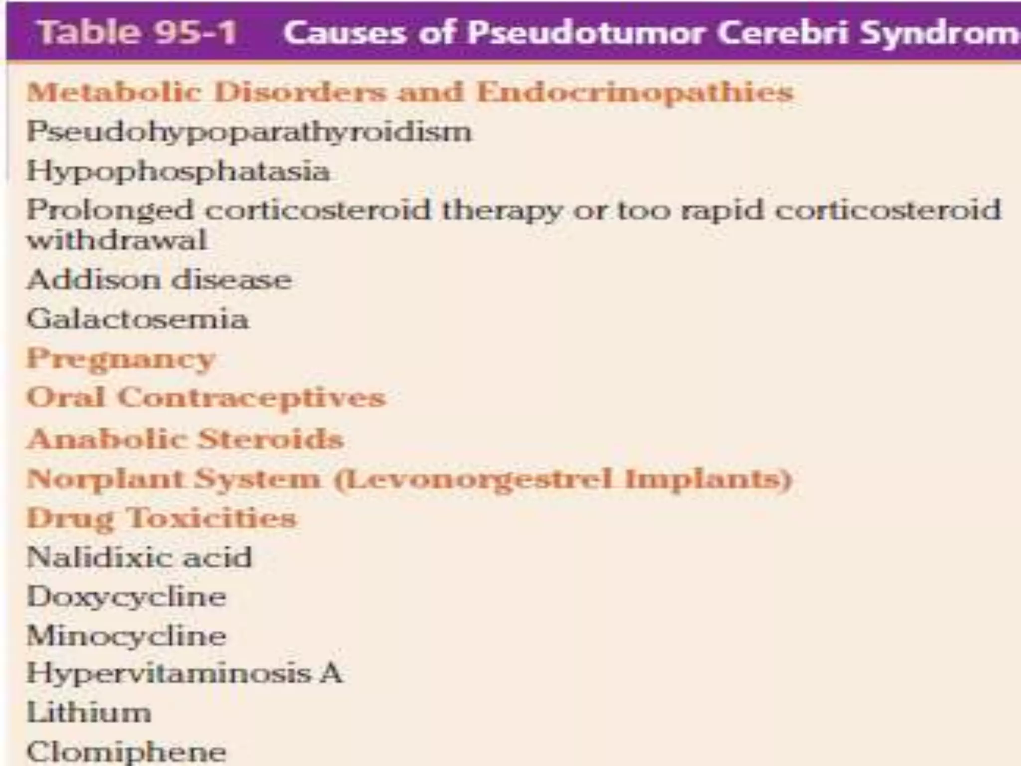
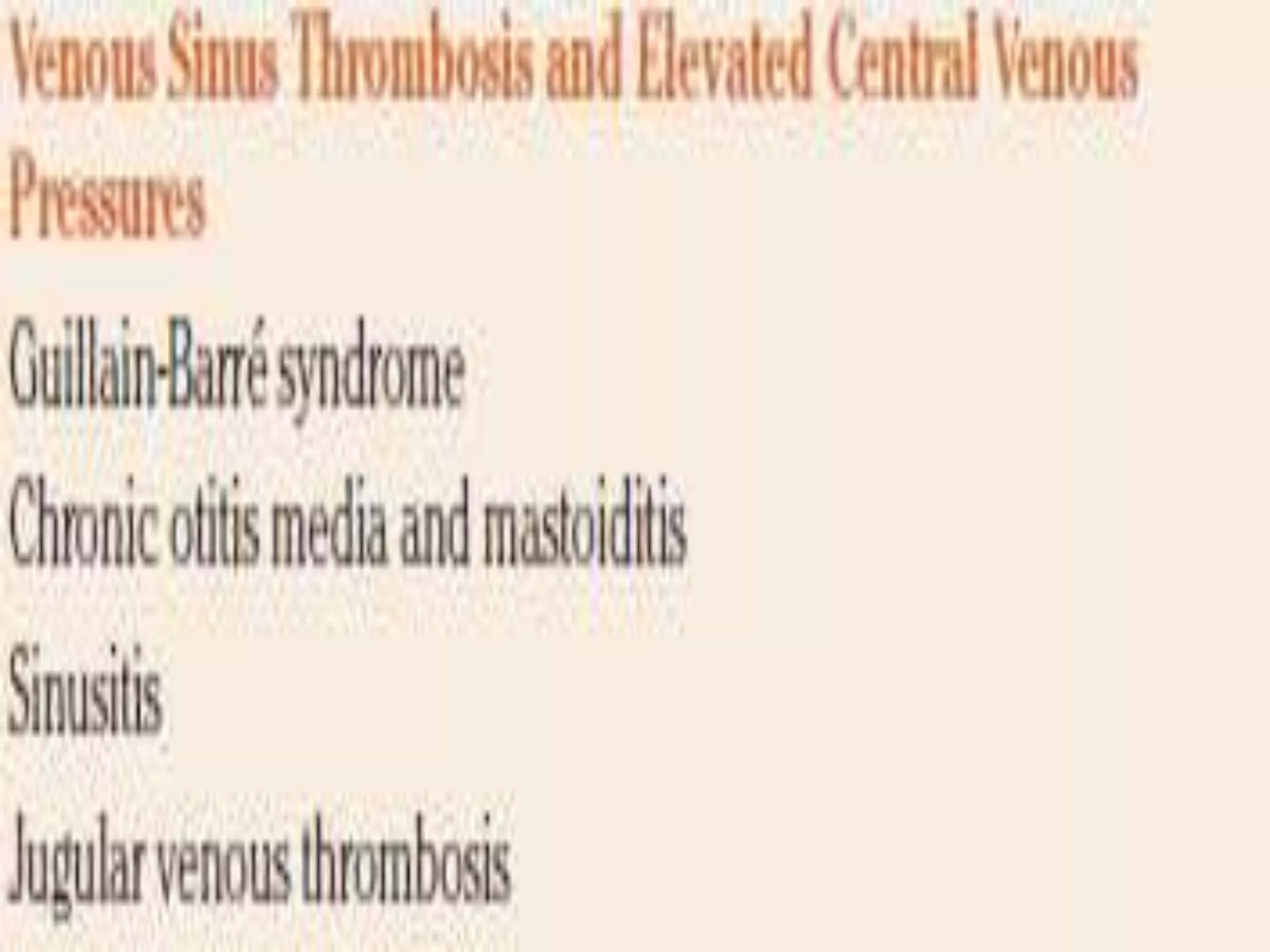
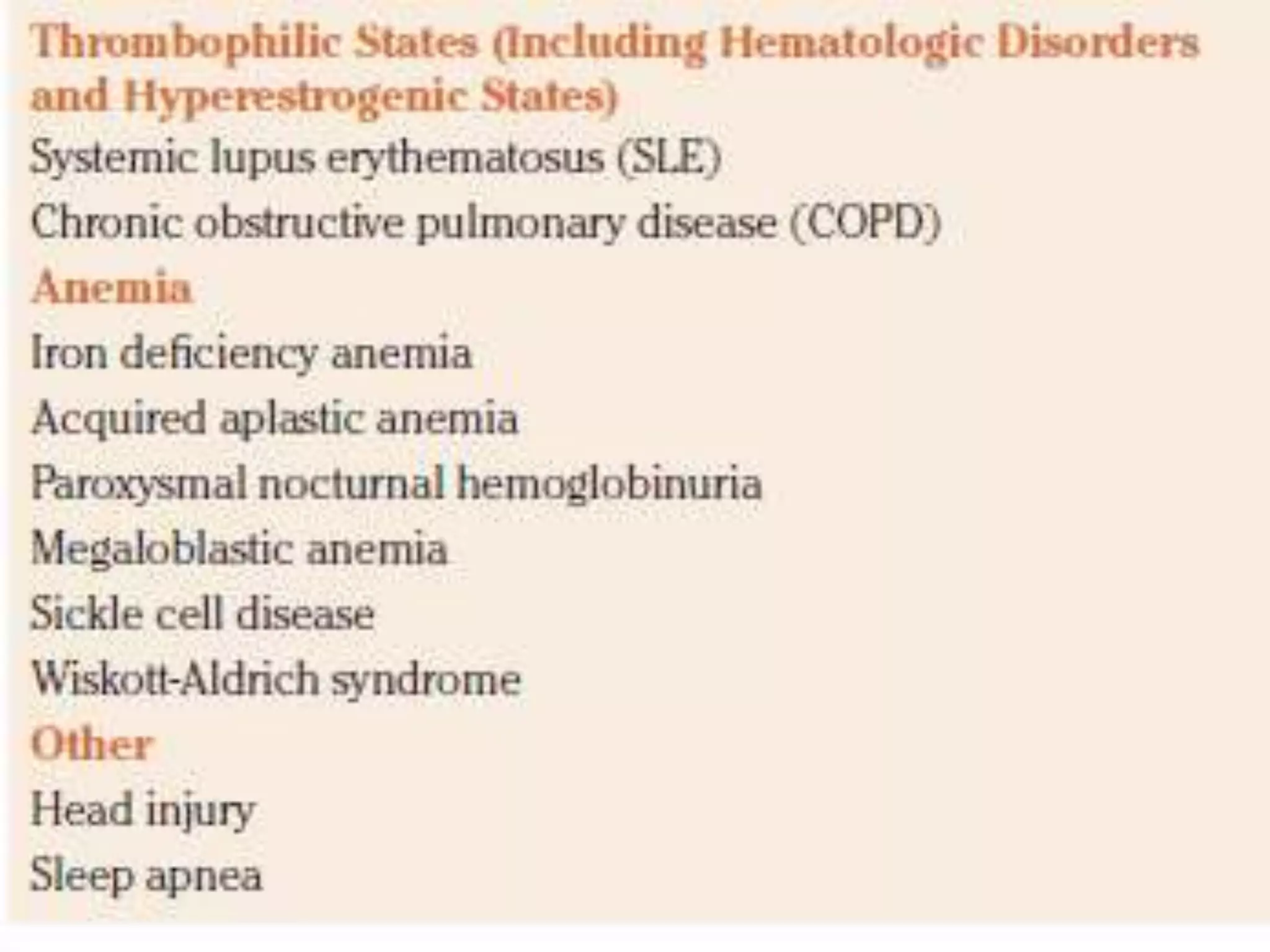
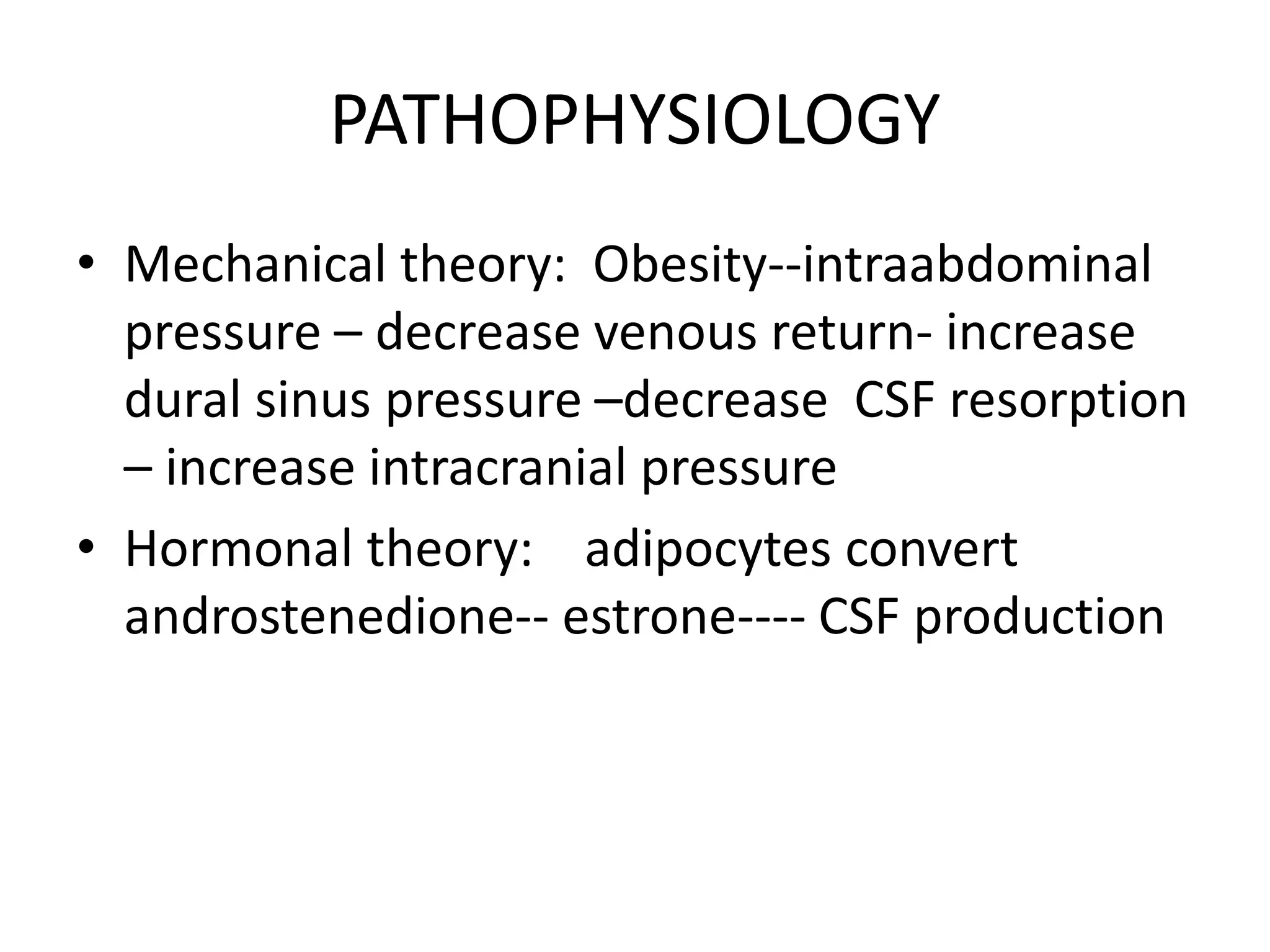
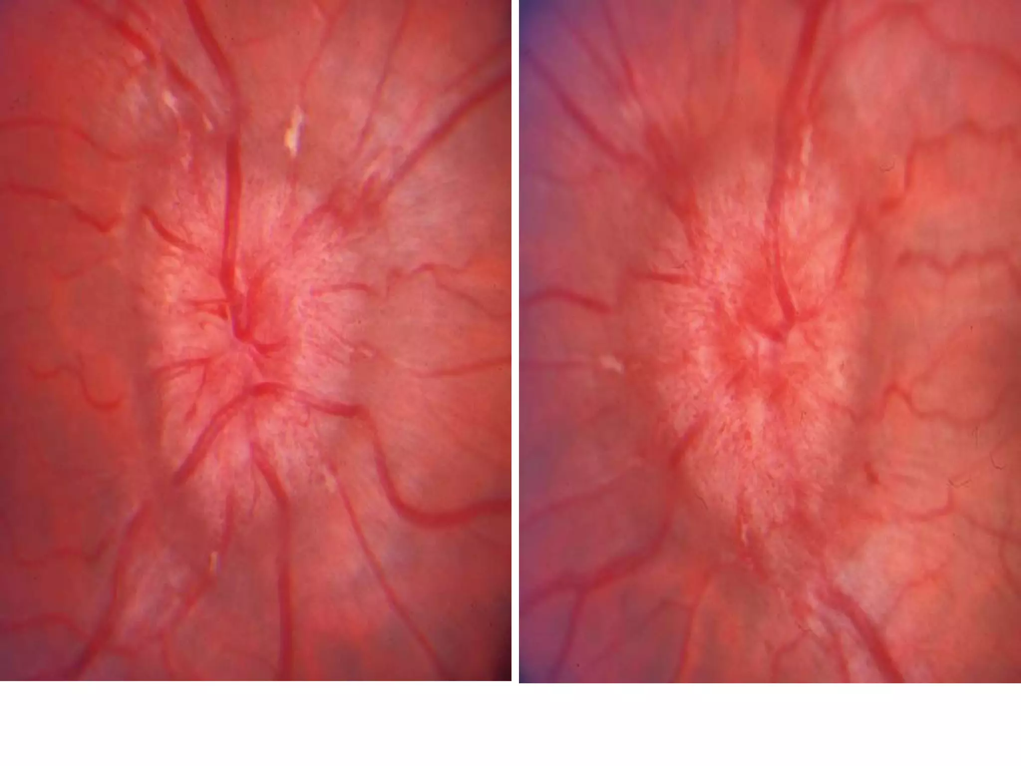
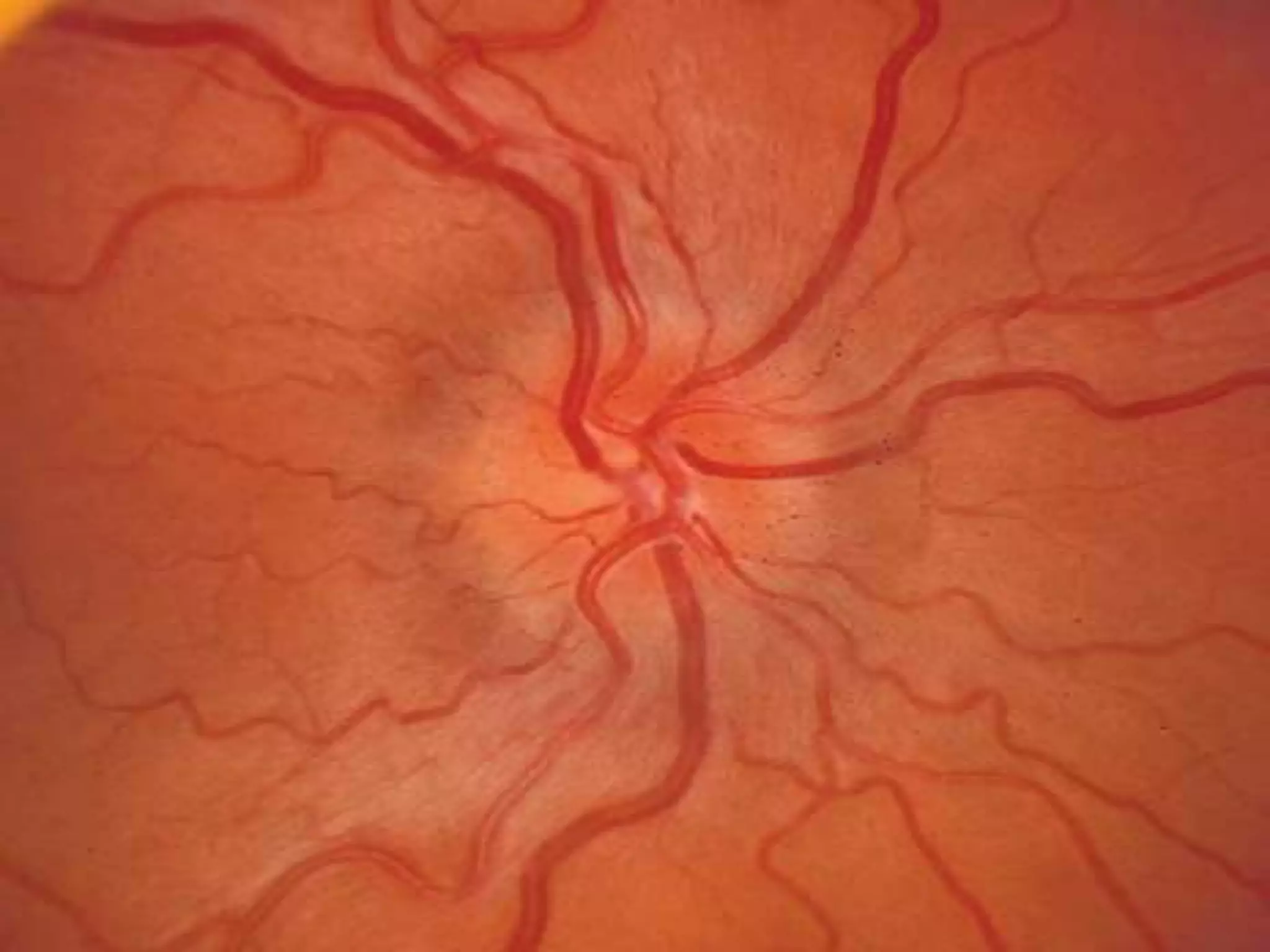
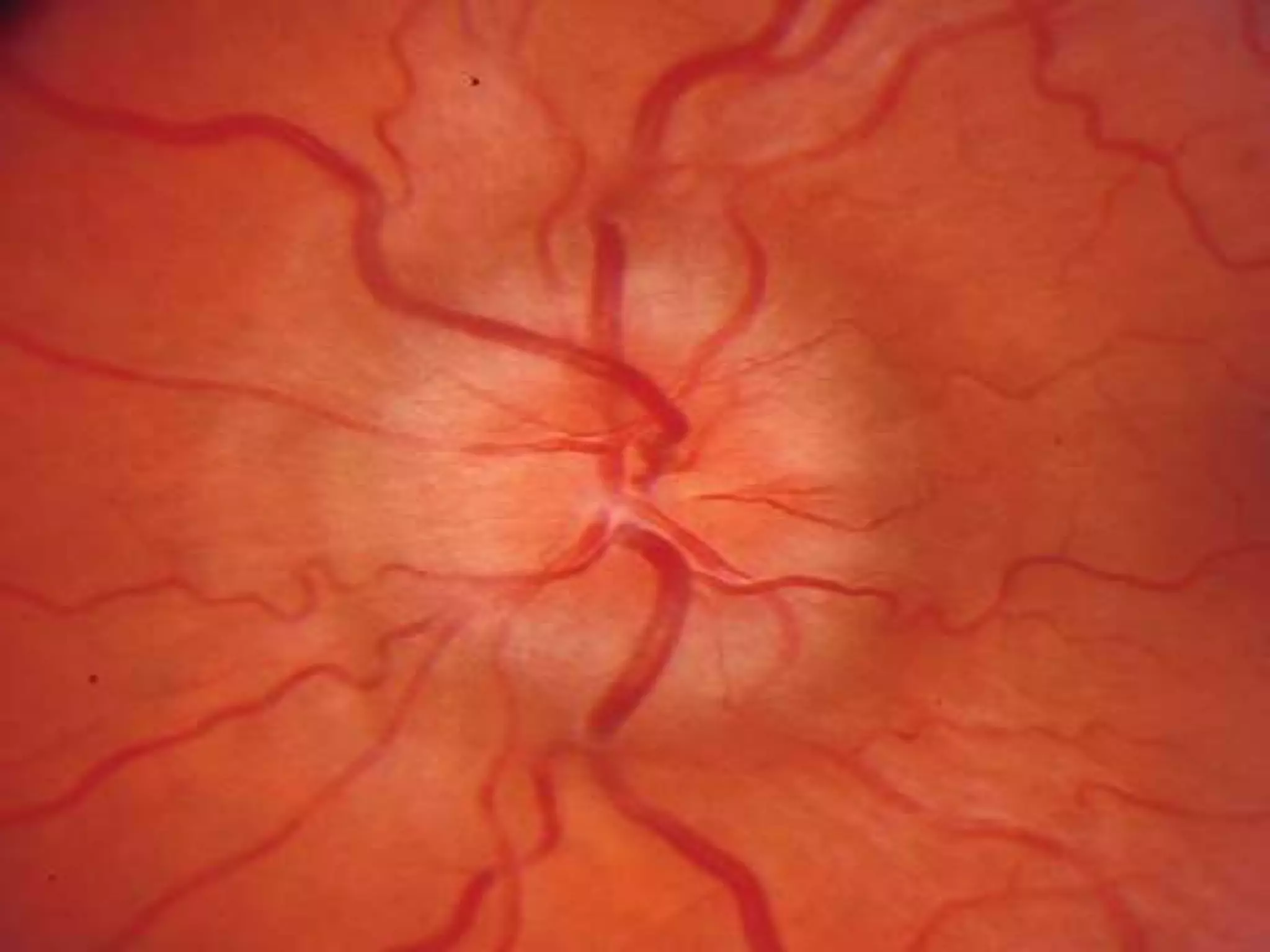
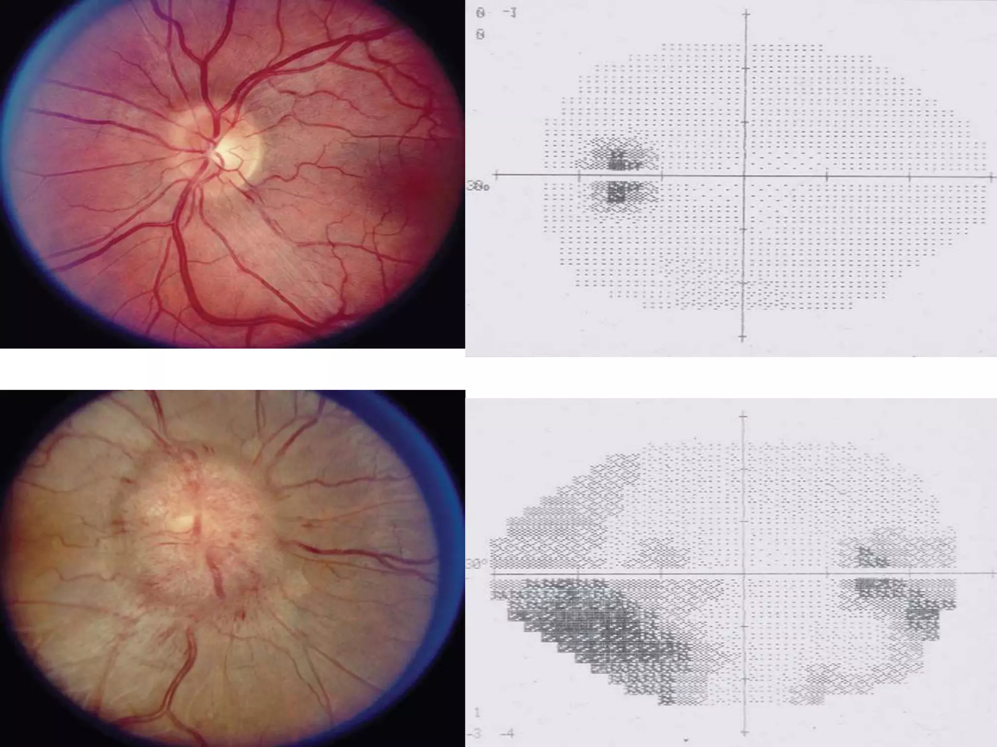
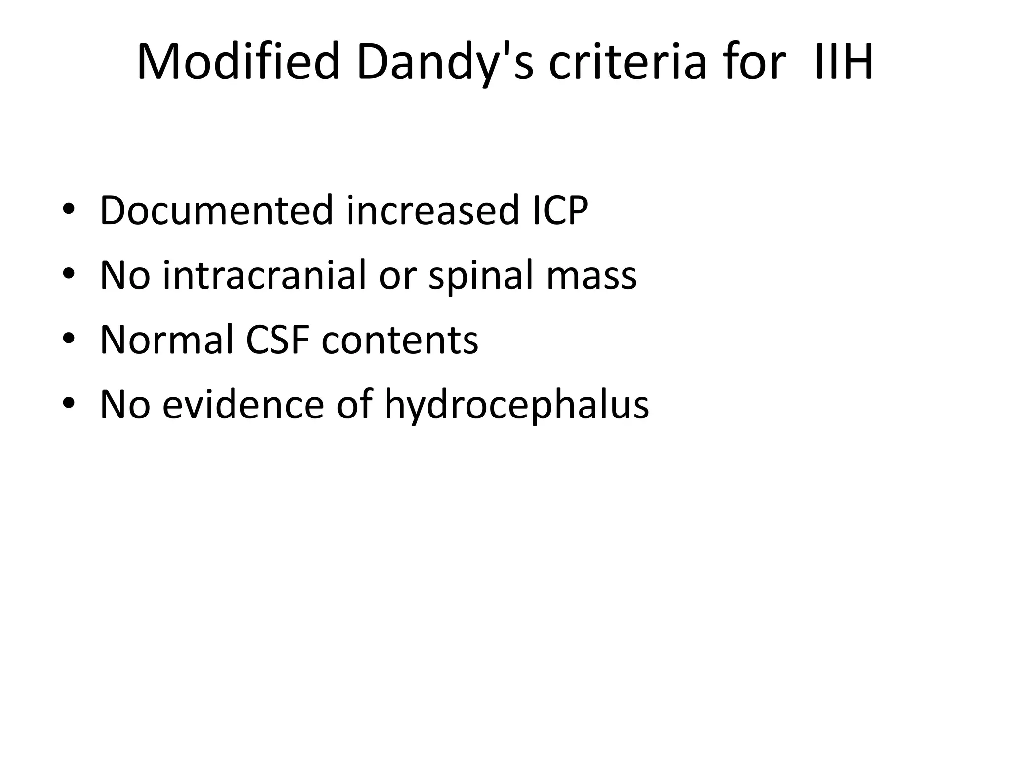
Pseudotumor cerebri
Dr. Amit
• A 39-year-old obese female laboratory technician
has been complaining of headache x2 years.
• Transient visual obscuration x 6 month
• B/l diplopia x 4 month
• O/E- E4V5M6/PEARL/B/L 6 NERVE PALSY GRADE
4 PAPILLEDEMA
• EVALUATION-CT BRAIN- small ventricle
• MRI WITH MRV = NORMAL
condition characterized by increased
intracranial pressure (ICP) without evidence
of dilated ventricles or a mass lesion by
imaging, normal cerebrospinal fluid (CSF)
content, and papilledema occurring in most
cases
• The condition was probably first described by
Quincke in 1897
• the term pseudotumor cerebri was given by
NONNE in 1914
• Foley suggested the condition “benign
intracranial hypertension
Pseudotumor Cerebri
• idiopathic intracranial hypertension (IIH)
• secondary pseudotumor cerebri
EPIDEMIOLOGY
• The idiopathic form ( IIH) represents about
90% of cases
• A disorder of obese females of childbearing
age
EPIDEMIOLOGY
• In the United States
• Annual incidence in Iowa and Louisiana (Durcan,
1988)
• the incidence to be approximately 0.9 per
100,000 in the general population
• female-to-male ratio of 8 : 1.
• The incidence increases to 3.5 per 100,000 in
women aged 20 to 44 years
• 13 per 100,000 in women who are 10% over ideal
weight
Secondary PTC
• About 10% of cases
• May develop at any age
• Equal frequency in both sexes
PATHOPHYSIOLOGY
• Mechanical theory: Obesity--intraabdominal
pressure – decrease venous return- increase
dural sinus pressure –decrease CSF resorption
– increase intracranial pressure
• Hormonal theory: adipocytes convert
androstenedione-- estrone---- CSF production
• Recent theory suggest defect at the level of
archanoid granulation
• Transverse sinus / Sup.sagital sinus
thrombosis, stenosis
SYMPTOMS AND SIGNS
• Headache-most common symptom
• Occurs in approximately 90% of cases
transient obscurations of vision (TOVs
• TOVs indicate the presence of optic disc
swelling
• 70% of patients
• unilateral or bilateral
• generally last only a few seconds
• A small percentage of patients with PTC
complain of visual loss
• Diplopia- present in approximately 40% of
patients
• Pulsatile tinnitus
• Focal neurological deficits in patients with PTC
are extremely uncommon
Papilledema
• Papilledema is the diagnostic hallmark of PTC
• Present in almost all patients.
• It is almost always bilateral and symmetrical
Papilledema Grading System (Frisén
Scale)
• STAGE 0—NORMAL OPTIC DISC
• Blurring of the nasal, superior, and inferior
poles in inverse proportion to disc diameter
• Radial nerve fiber layer without tortuosity
• Rare obscuration of a major blood vessel,
usually on the upper pole
STAGE 1—VERY EARLY PAPILLEDEMA
Obscuration of the nasal border of the disc
No elevation of the disc borders
Disruption of the normal arrangement of
radial nerve fiber layers with
a grayish opacity accentuating the nerve fiber
layer bundles
Normal temporal disc margin
STAGE 2—EARLY PAPILLEDEMA
• Obscuration of all borders
• Elevation of the nasal border
• Complete peripapillary halo
STAGE 3—MODERATE PAPILLEDEMA
• Obscurations of all borders
• Increased diameter of the optic nerve head
• Obscuration of one or more segments of
major blood vessels leaving the disc
• Peripapillary halo—irregular outer fringe with
finger-like extensions
STAGE 4—MARKED PAPILLEDEMA
• Elevation of the entire nerve head
• Obscuration of all borders
• Peripapillary halo
• Total obscuration on the disc of a segment of a
major blood vessel
STAGE 5—SEVERE PAPILLEDEMA
• Dome-shaped protrusions representing
anterior expansion of the optic nerve head
• Narrow and smoothly demarcated
peripapillary halo
• Obliteration of the optic cup
COMPLICATIONS
• The most feared complication of PTC is
permanent visual loss
• In one long-term study performed before the era
of modern neuroimaging,
• 57 patients with a diagnosis of PTC were
monitored for 5 to 41 years.
• Severe visual impairment occurred in one or both
• eyes in 14 patients (24.5%)
• 7 patients had the visual loss
• In a more recent prospective study of 50
patients treated for IIH
• visual acuity was worse than 20/20 in 13%.
Modified Dandy's criteria for IIH
• Documented increased ICP
• No intracranial or spinal mass
• Normal CSF contents
• No evidence of hydrocephalus
Neuroimaging
CT BRAIN
• Usuallu normal
• Occasionally slit ventricles may seen
MRI
• Intracranial abnormalities are usually absent
or minimal
• 1. Flattening of the posterior sclera: occurs in
80%
• 2. Enhancement of the prelaminar optic nerve: in
50%
• 3. Distention of the perioptic subarachnoid space:
in 45%
• 4. Vertical tortuosity of the orbital optic nerve: in
40%
• 5. Intraocular protrusion of the prelaminar optic
nerve: in 30%
Venography
• To rule-out dural sinus or venous thrombosis
Examination of Cerebrospinal Fluid
• CSF pressure: > 25 diagnostic
• Pressures 20-24.9 are nondiagnostic
• Pressure < 20 is normal
CSF composition:
• Normal glucose and cell count
TREATMENT
• Treatment of PTC is a teamwork requiring
concurrent management from more than one
physician.
• A neurologist, ophthalmologist, primary care
physician, and neurosurgeon
• The presence and severity of symptoms such
as headache
• The degree of visual loss at initial examination
• The rate of progression of visual loss
• The presence of an identifiable underlying
cause
• Detection of factors known to be associated
with a poorer visual prognosis
• Pt with mild papilledema with no headache
• Close observation
• Treat underlying cause
Treatment Related to Obesity
• Kupersmith et al. reported improvement in
headaches and papilledema in 58 obese
females with IIH who lost 7% to 10% of their
weight over a 3-month period
MEDICAL MANAGEMENT
• Mild- moderate headche
• Good visual function
• Acetazolamide (Diamox)
• Methazolamide
• Topiramate
• Diuretics such as furosemide
• Systemic corticosteroids
Surgical Procedures
• Moderate or severe papilledema
• Deterioration of visual acuity or worsening of
visual fields despite medical therapy
• CSF diversion to lower ICP
• ONSF
• stenting of venous sinuses
• Bariatric surgery for obese patients
Shunt Surgery
Moderate to sever headache
progressive visual disturbance
• Ventri peritoneal shunt
• Lumboperitoneal shunt
• Ventri atrial shunt
• Tulipan et al placed ventriculoperitoneal (VP)
shunts in seven patients
• Papilledema resolved in five patients
• two patients had mild residual papilledema
• Headaches resolved in all
Optic Nerve Sheath Fenestration
• Moderate to sever visual disturbance
• Mild headache
• Banta et al. described the outcomes of 158
ONSFs performed in 86 patients with IIH
• Improvement or stabilization of visual acuity
and visual fields was observed in 94% and 88%
patients
• Most common being diplopia (34.86%) and
anisocoria (7.5%)
• Sergott et al. reported the use of a modified
ONSF IN 32 pt. wherein multiple longitudinal
fenestrations were created and subarachnoid
adhesions lysed. These authors noted
improvement in 21 of 23 patients (91.3%) with
IIH
COMPLICATION
• Early/Late visual failure
Venous Sinus Stenting
• Moderate to sever headache
• Progressive visual disturbance
• MRV – Dural sinus thrombosis / stenosis
• Manometer showing raised pressure gradient
across stenosis
• Bussiere et al. reported transverse sinus
stenosis in 13 patients with IIH, 10 of whom
underwent stenting of the affected sin
• Almost complete resolution of papilledema
was seen in eight patients and significantly
improved in two others
Management of IIH in Pregnancy
• Rule out dural sinus thrmbosis
• Avoid use of acetazolamide in first trimester
• Shunt revision
Fulminant Pseudotumor Cerebri
significant visual field loss, central visual
acuity loss, and marked papilledema on initial
examination.
• Insertion of a lumbar drain until a definitive
shunt procedure or ONSF
CONCLUSION
• Early diagnosis
• Early treatment
• Follow up
• Thank you

Presentation1 pseudotumor

  • 1.
  • 2.
    • A 39-year-oldobese female laboratory technician has been complaining of headache x2 years. • Transient visual obscuration x 6 month • B/l diplopia x 4 month • O/E- E4V5M6/PEARL/B/L 6 NERVE PALSY GRADE 4 PAPILLEDEMA • EVALUATION-CT BRAIN- small ventricle • MRI WITH MRV = NORMAL
  • 3.
    condition characterized byincreased intracranial pressure (ICP) without evidence of dilated ventricles or a mass lesion by imaging, normal cerebrospinal fluid (CSF) content, and papilledema occurring in most cases
  • 4.
    • The conditionwas probably first described by Quincke in 1897 • the term pseudotumor cerebri was given by NONNE in 1914 • Foley suggested the condition “benign intracranial hypertension
  • 5.
    Pseudotumor Cerebri • idiopathicintracranial hypertension (IIH) • secondary pseudotumor cerebri
  • 6.
    EPIDEMIOLOGY • The idiopathicform ( IIH) represents about 90% of cases • A disorder of obese females of childbearing age
  • 7.
    EPIDEMIOLOGY • In theUnited States • Annual incidence in Iowa and Louisiana (Durcan, 1988) • the incidence to be approximately 0.9 per 100,000 in the general population • female-to-male ratio of 8 : 1. • The incidence increases to 3.5 per 100,000 in women aged 20 to 44 years • 13 per 100,000 in women who are 10% over ideal weight
  • 8.
    Secondary PTC • About10% of cases • May develop at any age • Equal frequency in both sexes
  • 12.
    PATHOPHYSIOLOGY • Mechanical theory:Obesity--intraabdominal pressure – decrease venous return- increase dural sinus pressure –decrease CSF resorption – increase intracranial pressure • Hormonal theory: adipocytes convert androstenedione-- estrone---- CSF production
  • 13.
    • Recent theorysuggest defect at the level of archanoid granulation • Transverse sinus / Sup.sagital sinus thrombosis, stenosis
  • 14.
    SYMPTOMS AND SIGNS •Headache-most common symptom • Occurs in approximately 90% of cases
  • 15.
    transient obscurations ofvision (TOVs • TOVs indicate the presence of optic disc swelling • 70% of patients • unilateral or bilateral • generally last only a few seconds
  • 16.
    • A smallpercentage of patients with PTC complain of visual loss
  • 17.
    • Diplopia- presentin approximately 40% of patients • Pulsatile tinnitus • Focal neurological deficits in patients with PTC are extremely uncommon
  • 18.
    Papilledema • Papilledema isthe diagnostic hallmark of PTC • Present in almost all patients. • It is almost always bilateral and symmetrical
  • 20.
    Papilledema Grading System(Frisén Scale) • STAGE 0—NORMAL OPTIC DISC • Blurring of the nasal, superior, and inferior poles in inverse proportion to disc diameter • Radial nerve fiber layer without tortuosity • Rare obscuration of a major blood vessel, usually on the upper pole
  • 21.
    STAGE 1—VERY EARLYPAPILLEDEMA Obscuration of the nasal border of the disc No elevation of the disc borders Disruption of the normal arrangement of radial nerve fiber layers with a grayish opacity accentuating the nerve fiber layer bundles Normal temporal disc margin
  • 23.
    STAGE 2—EARLY PAPILLEDEMA •Obscuration of all borders • Elevation of the nasal border • Complete peripapillary halo
  • 25.
    STAGE 3—MODERATE PAPILLEDEMA •Obscurations of all borders • Increased diameter of the optic nerve head • Obscuration of one or more segments of major blood vessels leaving the disc • Peripapillary halo—irregular outer fringe with finger-like extensions
  • 27.
    STAGE 4—MARKED PAPILLEDEMA •Elevation of the entire nerve head • Obscuration of all borders • Peripapillary halo • Total obscuration on the disc of a segment of a major blood vessel
  • 29.
    STAGE 5—SEVERE PAPILLEDEMA •Dome-shaped protrusions representing anterior expansion of the optic nerve head • Narrow and smoothly demarcated peripapillary halo • Obliteration of the optic cup
  • 31.
    COMPLICATIONS • The mostfeared complication of PTC is permanent visual loss • In one long-term study performed before the era of modern neuroimaging, • 57 patients with a diagnosis of PTC were monitored for 5 to 41 years. • Severe visual impairment occurred in one or both • eyes in 14 patients (24.5%) • 7 patients had the visual loss
  • 32.
    • In amore recent prospective study of 50 patients treated for IIH • visual acuity was worse than 20/20 in 13%.
  • 35.
    Modified Dandy's criteriafor IIH • Documented increased ICP • No intracranial or spinal mass • Normal CSF contents • No evidence of hydrocephalus
  • 36.
  • 37.
    CT BRAIN • Usuallunormal • Occasionally slit ventricles may seen
  • 38.
    MRI • Intracranial abnormalitiesare usually absent or minimal
  • 39.
    • 1. Flatteningof the posterior sclera: occurs in 80% • 2. Enhancement of the prelaminar optic nerve: in 50% • 3. Distention of the perioptic subarachnoid space: in 45% • 4. Vertical tortuosity of the orbital optic nerve: in 40% • 5. Intraocular protrusion of the prelaminar optic nerve: in 30%
  • 40.
    Venography • To rule-outdural sinus or venous thrombosis
  • 41.
    Examination of CerebrospinalFluid • CSF pressure: > 25 diagnostic • Pressures 20-24.9 are nondiagnostic • Pressure < 20 is normal
  • 42.
    CSF composition: • Normalglucose and cell count
  • 43.
    TREATMENT • Treatment ofPTC is a teamwork requiring concurrent management from more than one physician. • A neurologist, ophthalmologist, primary care physician, and neurosurgeon
  • 44.
    • The presenceand severity of symptoms such as headache • The degree of visual loss at initial examination • The rate of progression of visual loss • The presence of an identifiable underlying cause • Detection of factors known to be associated with a poorer visual prognosis
  • 45.
    • Pt withmild papilledema with no headache • Close observation • Treat underlying cause
  • 46.
    Treatment Related toObesity • Kupersmith et al. reported improvement in headaches and papilledema in 58 obese females with IIH who lost 7% to 10% of their weight over a 3-month period
  • 47.
    MEDICAL MANAGEMENT • Mild-moderate headche • Good visual function
  • 48.
    • Acetazolamide (Diamox) •Methazolamide • Topiramate • Diuretics such as furosemide • Systemic corticosteroids
  • 49.
    Surgical Procedures • Moderateor severe papilledema • Deterioration of visual acuity or worsening of visual fields despite medical therapy
  • 50.
    • CSF diversionto lower ICP • ONSF • stenting of venous sinuses • Bariatric surgery for obese patients
  • 51.
    Shunt Surgery Moderate tosever headache progressive visual disturbance
  • 52.
    • Ventri peritonealshunt • Lumboperitoneal shunt • Ventri atrial shunt
  • 53.
    • Tulipan etal placed ventriculoperitoneal (VP) shunts in seven patients • Papilledema resolved in five patients • two patients had mild residual papilledema • Headaches resolved in all
  • 54.
    Optic Nerve SheathFenestration • Moderate to sever visual disturbance • Mild headache
  • 55.
    • Banta etal. described the outcomes of 158 ONSFs performed in 86 patients with IIH • Improvement or stabilization of visual acuity and visual fields was observed in 94% and 88% patients • Most common being diplopia (34.86%) and anisocoria (7.5%)
  • 56.
    • Sergott etal. reported the use of a modified ONSF IN 32 pt. wherein multiple longitudinal fenestrations were created and subarachnoid adhesions lysed. These authors noted improvement in 21 of 23 patients (91.3%) with IIH
  • 57.
  • 58.
    Venous Sinus Stenting •Moderate to sever headache • Progressive visual disturbance • MRV – Dural sinus thrombosis / stenosis • Manometer showing raised pressure gradient across stenosis
  • 59.
    • Bussiere etal. reported transverse sinus stenosis in 13 patients with IIH, 10 of whom underwent stenting of the affected sin • Almost complete resolution of papilledema was seen in eight patients and significantly improved in two others
  • 60.
    Management of IIHin Pregnancy • Rule out dural sinus thrmbosis • Avoid use of acetazolamide in first trimester • Shunt revision
  • 61.
    Fulminant Pseudotumor Cerebri significantvisual field loss, central visual acuity loss, and marked papilledema on initial examination. • Insertion of a lumbar drain until a definitive shunt procedure or ONSF
  • 62.
    CONCLUSION • Early diagnosis •Early treatment • Follow up
  • 63.