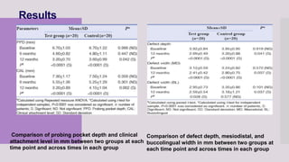
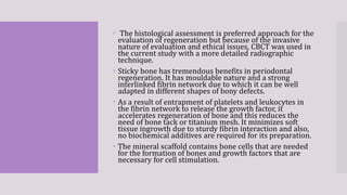
Randomized, controlled clinical
trial to evaluate efficacy of sticky
bone and concentrated growth
factor in the management of
intrabony defects: 12 months
follow-up study
Dental research journal 2022
Guided by:
DrTanvi Hirani
Presented by:
Rutu dabhi
Introduction
 One of the most complex processes of wound healing is potentially
the regeneration of damaged periodontal tissue.
 An innovative component of regenerative medicine is the
concentrated growth factor (CGF). It is known to be the third-
generation platelet concentrates prepared using continuous
differential centrifugation. CGF contains more growth factors and
has a harder fibrin structure and thicker elasticity than first-
generation Platelet-rich plasma (PRP) and second-generation
platelet-rich fibrin (PRF).
 This matrix of CGF aids in wound healing. It also contains
immunological cells that help regulate inflammation and decrease
the risk of infection.
 CGF is known to promote osteogenic differentiation and cell
proliferation, thereby enabling bone formation; tissues healing and
improving the quality of the newly formed bone.
 Sticky bone is a novel form of graft material that is also known
as an enriched bone graft matrix with growth factors, prepared
using autologous fibrin glue (AFG). AFG acts as a scaffold for
migrating fibroblast as well as a hemostatic barrier. It induces
and encourages angiogenesis and stimulates mesenchymal cells.
It gives stability to the bone graft material within the defect and
has the property of biocompatibility and biodegradability. This
facilitates tissue healing and decreases bone resorption during
the healing period.
 the present study aimed to evaluate the effect of AFG enriched
matrix (sticky bone) and CGFs in the treatment of intrabony
osseous defects by cone-beam computed tomography (CBCT).
The hypothesis of this study was that the AFG enriched bone
graft matrix (sticky bone) was more effective than the CGFs in
the treatment of intrabony osseous defects.
Inclusion
criteria:
 The age range 20–45 years with a mean age of 39 years.
 Forty intrabony defects were selected from 20 patients (9 males
and 11 females) with Stage III (Grade A) periodontitis,
 Patients with probing pocket depth (PPD) ≥6 mm and
 clinical attachment level (CAL) ≥5 mm with the
 minimum of two osseous defects (either two or three walled)
with a depth of ≥3 mm
 Patients showing radiographic evidence of intrabony defects as
initially revealed by radiovisiographs and later on confirmed
and standardized by CBCT were selected.
Exclusion
criteria:
 Patients with systemic diseases, allergies, or drug
usage,
 With a history of periodontal treatment in the
previous 6 months and
 Pregnant/lactating women were excluded from the
study.
Nonsurgical
periodontal
therapy

Nonsurgical periodontal therapy was given to the
patients followed by oral hygiene instructions.
 The patients were re-evaluated 6 weeks after initial
therapy to assess the status of complete oral hygiene.
 Then, the selected sites were randomly assigned as
test group (sticky bone) and control group (CGF).
Clinical
parameters
 The clinical parameters involving
 plaque index (PI), gingival index (GI),PPD, and CAL
were recorded using periodontal probe at baseline
after 6 and 12 months.
 Custom-made occlusal acrylic stents with grooves
were made to standardize the probe angulation and
position.
 Intrabony defects were assessed using CBCT at
baseline and after 12 months follow-up.
Schematic of reference points for measurement of Cone Beam Computed
Tomography parameters. (a) Defect depth and Mesiodistal Width. (b)
Buccolingual Width
Preparation
of
concentrated
growth factor
 CGF was prepared according to the protocol given by
Rodella et al.
 9 ml of blood was drawn in a sterile test tube without
anticoagulant.
 These tubes were then immediately centrifuged. The type
of Centrifuge used was Table Top single Phase Remi
Compact Laboratory Centrifuge R 4C, using specific
centrifugation protocol as follows: 30 s-acceleration, 2
min–2700 rpm, 4 min-2400 rpm, 4 min–2700 rpm, 3 min
3000 rpm, 36 s-deceleration, and stop.
 At the end of the process, three blood layers were
obtained: The upper layer containing platelet-poor
plasma, the middle layer with fibrin-rich gel with
aggregated platelets, and CGFs and the lower layer
comprised red blood cell (RBC)
Preparation
of sticky
bone
 Sticky bone was formulated according to the protocol
given by Sohn et al.
 10 ml of venous blood was drawn from the antecubital
fossa of the patient and collected in the test tubes.
 Noncoated test tubes having red-colored caps were used.
 It was then centrifuged at 2400–2700 rpm. The
centrifugation time for AFG varies from 2 to 12 min. The
centrifuge was stopped after 2 min. AFG obtained was
separated from the test tube.
 The noncoated tube showed 2 different layers.
 The upper layer comprised of AFG while the bottom layer
consisted of the RBC which was discarded.
 The upper layer of AFG was collected with the syringe and
mixed with particulate bone powder and allowed for 5–10
min for polymerization to produce yellow-colored sticky
bone
Surgical
periodontal
therapy
 After the administration of local anesthesia#,
 intra-sulcular incision extending at least one tooth mesial
and distal to the surgical site preserving the interdental
papilla wherever possible was given and a
 full-thickness mucoperiosteal flap was reflected.
 After the complete debridement of intrabony defects, the
CGF was placed in the defect site in the control group
while the sticky bone was placed in the test group.
 Immediately after placing the CGF and sticky bone, the
reflected flap was approximated and repositioned.
 The interrupted sutures and periodontal dressing were
placed at surgical sites.
Surgical procedure for Test Group and Control Group.
(a) Reflection of flap for Test Group. (b) Placement of Sticky Bone in defects for Test Group.
(c) Suture placement for Test Group. (d) Periodontal Pack Placement for Test Group.
(e) Reflection of flap for Control Group. (f) Placement of Concentrated Growth Factors in
defects for Control Group. (g) Suture placement for Control Group. (h) Periodontal Pack
Placement for Control Group.
 Postoperative instructions and antibiotics and
analgesic were prescribed twice daily for 7 days.
 The patients were advised Chlorhexidine oral rinse
(10 ml twice daily) and refrained from chewing hard
and sticky foods.
 In addition to this, they were instructed and
motivated to use toothbrush with soft bristles for the
next 12 months.
Postsurgical measurements
Cone Beam Computed Tomography for Test Group and Control Group.
(a) Sagittal view at baseline for Test Group. (b) Transverse view at baseline for Test Group.
(c) Sagittal view at 12 months for Test Group. (d) Transverse view at 12 months for Test Group.
(e) Sagittal view at baseline for Control Group. (f) Transverse view at baseline for Control Group.
(g) Sagittal view at 12 months for Control Group. (h) Transverse view at 12 months for Control
Group.
Results
Comparison of defect depth, mesiodistal, and
buccolingual width in mm between two groups at
each time point and across times in each group
Comparison of probing pocket depth and clinical
attachment level in mm between two groups at each
time point and across times in each group
Discussion
 Platelet concentrates provide rich source of growth
factors and therefore inhibit hemorrhage, tissue
adherence, promote healing, minimize pain and
accelerate the formation of new tissues.
 AFG is a biological product prepared using patient's
own blood and has benefits like decreased bleeding,
reduced scarring, and serous fluid collection.
 The histological assessment is preferred approach for the
evaluation of regeneration but because of the invasive
nature of evaluation and ethical issues, CBCT was used in
the current study with a more detailed radiographic
technique.
 Sticky bone has tremendous benefits in periodontal
regeneration. It has mouldable nature and a strong
interlinked fibrin network due to which it can be well
adapted in different shapes of bony defects.
 As a result of entrapment of platelets and leukocytes in
the fibrin network to release the growth factor, it
accelerates regeneration of bone and this reduces the
need of bone tack or titanium mesh. It minimizes soft
tissue ingrowth due to sturdy fibrin interaction and also,
no biochemical additives are required for its preparation.
 The mineral scaffold contains bone cells that are needed
for the formation of bones and growth factors that are
necessary for cell stimulation.
Conclusion
 Within the confines of the study, it can be
concluded that the sticky bone is more
effective in gaining the CAL, reduction in PPD
and radiographic outcomes like defect depth
as compared to CGF alone.
 Thus, can be preferred over CGF for the
treatment of intrabony osseous defects.
Furthermore, the use of CBCT proved to be a
better replacement over the invasive
histologic evaluation.

comparison between sticky bone and concentrated growth factor

  • 1.
    Randomized, controlled clinical trialto evaluate efficacy of sticky bone and concentrated growth factor in the management of intrabony defects: 12 months follow-up study Dental research journal 2022 Guided by: DrTanvi Hirani Presented by: Rutu dabhi
  • 2.
    Introduction  One ofthe most complex processes of wound healing is potentially the regeneration of damaged periodontal tissue.  An innovative component of regenerative medicine is the concentrated growth factor (CGF). It is known to be the third- generation platelet concentrates prepared using continuous differential centrifugation. CGF contains more growth factors and has a harder fibrin structure and thicker elasticity than first- generation Platelet-rich plasma (PRP) and second-generation platelet-rich fibrin (PRF).  This matrix of CGF aids in wound healing. It also contains immunological cells that help regulate inflammation and decrease the risk of infection.  CGF is known to promote osteogenic differentiation and cell proliferation, thereby enabling bone formation; tissues healing and improving the quality of the newly formed bone.
  • 3.
     Sticky boneis a novel form of graft material that is also known as an enriched bone graft matrix with growth factors, prepared using autologous fibrin glue (AFG). AFG acts as a scaffold for migrating fibroblast as well as a hemostatic barrier. It induces and encourages angiogenesis and stimulates mesenchymal cells. It gives stability to the bone graft material within the defect and has the property of biocompatibility and biodegradability. This facilitates tissue healing and decreases bone resorption during the healing period.  the present study aimed to evaluate the effect of AFG enriched matrix (sticky bone) and CGFs in the treatment of intrabony osseous defects by cone-beam computed tomography (CBCT). The hypothesis of this study was that the AFG enriched bone graft matrix (sticky bone) was more effective than the CGFs in the treatment of intrabony osseous defects.
  • 4.
    Inclusion criteria:  The agerange 20–45 years with a mean age of 39 years.  Forty intrabony defects were selected from 20 patients (9 males and 11 females) with Stage III (Grade A) periodontitis,  Patients with probing pocket depth (PPD) ≥6 mm and  clinical attachment level (CAL) ≥5 mm with the  minimum of two osseous defects (either two or three walled) with a depth of ≥3 mm  Patients showing radiographic evidence of intrabony defects as initially revealed by radiovisiographs and later on confirmed and standardized by CBCT were selected.
  • 5.
    Exclusion criteria:  Patients withsystemic diseases, allergies, or drug usage,  With a history of periodontal treatment in the previous 6 months and  Pregnant/lactating women were excluded from the study.
  • 6.
    Nonsurgical periodontal therapy  Nonsurgical periodontal therapywas given to the patients followed by oral hygiene instructions.  The patients were re-evaluated 6 weeks after initial therapy to assess the status of complete oral hygiene.  Then, the selected sites were randomly assigned as test group (sticky bone) and control group (CGF).
  • 7.
    Clinical parameters  The clinicalparameters involving  plaque index (PI), gingival index (GI),PPD, and CAL were recorded using periodontal probe at baseline after 6 and 12 months.  Custom-made occlusal acrylic stents with grooves were made to standardize the probe angulation and position.  Intrabony defects were assessed using CBCT at baseline and after 12 months follow-up.
  • 8.
    Schematic of referencepoints for measurement of Cone Beam Computed Tomography parameters. (a) Defect depth and Mesiodistal Width. (b) Buccolingual Width
  • 9.
    Preparation of concentrated growth factor  CGFwas prepared according to the protocol given by Rodella et al.  9 ml of blood was drawn in a sterile test tube without anticoagulant.  These tubes were then immediately centrifuged. The type of Centrifuge used was Table Top single Phase Remi Compact Laboratory Centrifuge R 4C, using specific centrifugation protocol as follows: 30 s-acceleration, 2 min–2700 rpm, 4 min-2400 rpm, 4 min–2700 rpm, 3 min 3000 rpm, 36 s-deceleration, and stop.  At the end of the process, three blood layers were obtained: The upper layer containing platelet-poor plasma, the middle layer with fibrin-rich gel with aggregated platelets, and CGFs and the lower layer comprised red blood cell (RBC)
  • 10.
    Preparation of sticky bone  Stickybone was formulated according to the protocol given by Sohn et al.  10 ml of venous blood was drawn from the antecubital fossa of the patient and collected in the test tubes.  Noncoated test tubes having red-colored caps were used.  It was then centrifuged at 2400–2700 rpm. The centrifugation time for AFG varies from 2 to 12 min. The centrifuge was stopped after 2 min. AFG obtained was separated from the test tube.  The noncoated tube showed 2 different layers.  The upper layer comprised of AFG while the bottom layer consisted of the RBC which was discarded.  The upper layer of AFG was collected with the syringe and mixed with particulate bone powder and allowed for 5–10 min for polymerization to produce yellow-colored sticky bone
  • 11.
    Surgical periodontal therapy  After theadministration of local anesthesia#,  intra-sulcular incision extending at least one tooth mesial and distal to the surgical site preserving the interdental papilla wherever possible was given and a  full-thickness mucoperiosteal flap was reflected.  After the complete debridement of intrabony defects, the CGF was placed in the defect site in the control group while the sticky bone was placed in the test group.  Immediately after placing the CGF and sticky bone, the reflected flap was approximated and repositioned.  The interrupted sutures and periodontal dressing were placed at surgical sites.
  • 12.
    Surgical procedure forTest Group and Control Group. (a) Reflection of flap for Test Group. (b) Placement of Sticky Bone in defects for Test Group. (c) Suture placement for Test Group. (d) Periodontal Pack Placement for Test Group. (e) Reflection of flap for Control Group. (f) Placement of Concentrated Growth Factors in defects for Control Group. (g) Suture placement for Control Group. (h) Periodontal Pack Placement for Control Group.
  • 13.
     Postoperative instructionsand antibiotics and analgesic were prescribed twice daily for 7 days.  The patients were advised Chlorhexidine oral rinse (10 ml twice daily) and refrained from chewing hard and sticky foods.  In addition to this, they were instructed and motivated to use toothbrush with soft bristles for the next 12 months.
  • 14.
    Postsurgical measurements Cone BeamComputed Tomography for Test Group and Control Group. (a) Sagittal view at baseline for Test Group. (b) Transverse view at baseline for Test Group. (c) Sagittal view at 12 months for Test Group. (d) Transverse view at 12 months for Test Group. (e) Sagittal view at baseline for Control Group. (f) Transverse view at baseline for Control Group. (g) Sagittal view at 12 months for Control Group. (h) Transverse view at 12 months for Control Group.
  • 15.
    Results Comparison of defectdepth, mesiodistal, and buccolingual width in mm between two groups at each time point and across times in each group Comparison of probing pocket depth and clinical attachment level in mm between two groups at each time point and across times in each group
  • 16.
    Discussion  Platelet concentratesprovide rich source of growth factors and therefore inhibit hemorrhage, tissue adherence, promote healing, minimize pain and accelerate the formation of new tissues.  AFG is a biological product prepared using patient's own blood and has benefits like decreased bleeding, reduced scarring, and serous fluid collection.
  • 17.
     The histologicalassessment is preferred approach for the evaluation of regeneration but because of the invasive nature of evaluation and ethical issues, CBCT was used in the current study with a more detailed radiographic technique.  Sticky bone has tremendous benefits in periodontal regeneration. It has mouldable nature and a strong interlinked fibrin network due to which it can be well adapted in different shapes of bony defects.  As a result of entrapment of platelets and leukocytes in the fibrin network to release the growth factor, it accelerates regeneration of bone and this reduces the need of bone tack or titanium mesh. It minimizes soft tissue ingrowth due to sturdy fibrin interaction and also, no biochemical additives are required for its preparation.  The mineral scaffold contains bone cells that are needed for the formation of bones and growth factors that are necessary for cell stimulation.
  • 18.
    Conclusion  Within theconfines of the study, it can be concluded that the sticky bone is more effective in gaining the CAL, reduction in PPD and radiographic outcomes like defect depth as compared to CGF alone.  Thus, can be preferred over CGF for the treatment of intrabony osseous defects. Furthermore, the use of CBCT proved to be a better replacement over the invasive histologic evaluation.

Editor's Notes

  • #9 The depth of the defect was measured as distance from the alveolar crest to base of the defect (AC), whereas, the Mesiodistal (MD) distance was recorded from AC-AC' as shown in [Figure 2]a. Buccolingual (BL) bone defect width was measured from B-L analyzed by CBCT as shown in [Figure 2]b. The lowest discontinuous point of the periodontal ligament was considered as the landmark of the base of the defect