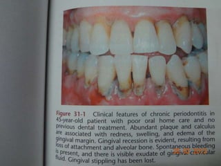
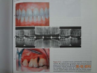
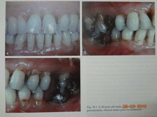
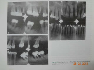
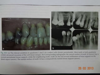
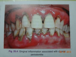
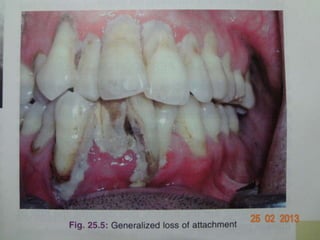
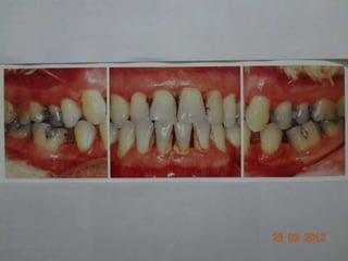
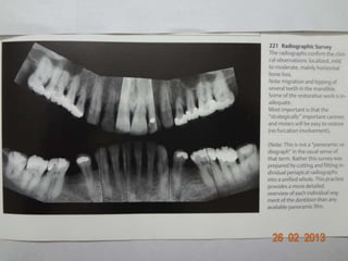
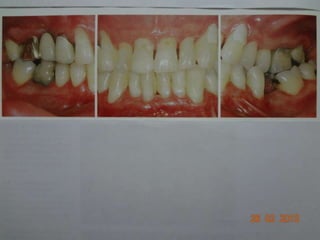
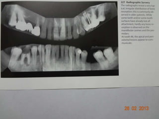
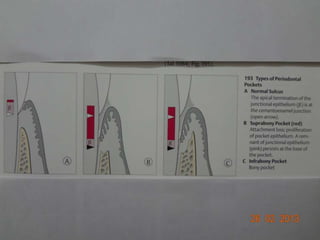
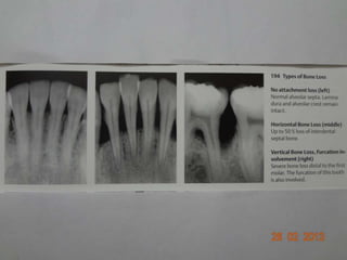
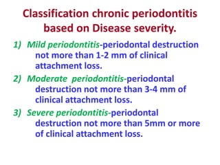
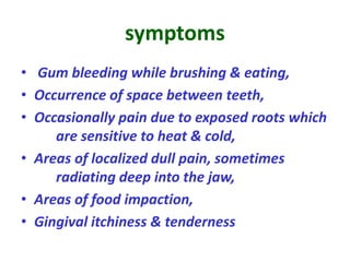
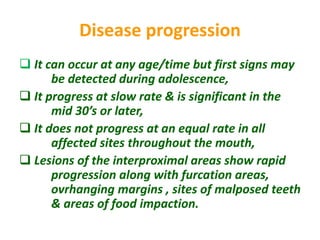
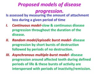
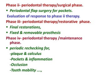
Chronic periodontitis
-Adult periodontitis.
-Chronic Adult periodontitis.
Chronic periodontitis
It is defined as an infectious disease
resulting in inflammation within the
supporting tissues of the teeth,
progressive attachment loss and bone
loss.
Clinical features.
Presence of supra & subgingival plaque and
calculus formation.
Gingival inflammation.
Pocket formation.
Loss of periodontal attachment.
Loss of alveolar bone.
Suppuration occasionally.
In pt’s with poor oral hygiene, the
gingiva shows following features
 Pale red to magenta color,
 Slight to moderate swelling,
 Loss of stippling,
 Blunted /rolled gingival margins and
flattened/ cratered papillae.
In pt’s with regular home care
measures,
 Changes associated with color, contour, of the
gingiva may not be visible by inspection,
Thickened & fibrotic gingiva,
 Presence of periodontal pockets,
 Bleeding on probing with periodontal probing
of pockets,
 GCF & suppuration is found in the pocket,
 Horizontal & vertical bone loss,
 Tooth mobility in advanced cases
Clinical diagnosis
 Detection of chronic inflammatory changes in
the marginal gingiva,
 Presence of periodontal pockets,
 Loss of clinical attachment,
 Radiographic evidence of bone loss.
Differential diagnosis from that of
aggressive periodontitis.
• Age of the pt,
• Rate of disease progression over time,
• Familial nature of aggressive disease,
• Relative absence of local factors in aggressive
disease.
Disease distribution
• Chronic periodontitis is considered as a
‘site specific disease’.
• Because clinical signs of the disease
occur on one surface of a tooth ,while
the other surfaces maintain normal
attachment levels
Classification of chronic periodontitis
based on disease distribution
A. Localized periodontitis - less than 30% of the
sites assessed in the mouth demonstrate
attachment loss & bone loss.
B. Generalized periodontitis - 30% or more of
the sites assessed in the mouth
demonstrate attachment loss & bone loss.
 vertical bone loss- attachment & bone loss
on one tooth surface is greater than that on
an adjacent surface & is associated with intra
bony pockets.
 horizontal bone loss- attachment & bone loss
proceeds at a uniform rate on the majority of
tooth surfaces & is associated with supra
bony pockets.
Classification chronic periodontitis
based on Disease severity.
1) Mild periodontitis-periodontal destruction
not more than 1-2 mm of clinical
attachment loss.
2) Moderate periodontitis-periodontal
destruction not more than 3-4 mm of
clinical attachment loss.
3) Severe periodontitis-periodontal
destruction not more than 5mm or more
of clinical attachment loss.
symptoms
• Gum bleeding while brushing & eating,
• Occurrence of space between teeth,
• Occasionally pain due to exposed roots which
are sensitive to heat & cold,
• Areas of localized dull pain, sometimes
radiating deep into the jaw,
• Areas of food impaction,
• Gingival itchiness & tenderness
Disease progression
 It can occur at any age/time but first signs may
be detected during adolescence,
 It progress at slow rate & is significant in the
mid 30’s or later,
 It does not progress at an equal rate in all
affected sites throughout the mouth,
 Lesions of the interproximal areas show rapid
progression along with furcation areas,
ovrhanging margins , sites of malposed teeth
& areas of food impaction.
Proposed models of disease
progression.
Is assessed by measuring the amount of attachment
loss during a given period of time
I. Continuous model-slow & continuous disease
progression throughout the duration of the
disease.
II. Random model/episodic burst model- disease
progression by short bursts of destruction
followed by periods of no destruction.
III. Asynchronous multiple-burst model- disease
progression around affected teeth during defined
periods of life & these bursts of activity are
interspersed with periods of inactivity/remission.
prevalence
• It increases in prevalence & severity with age.
• Both the genders are equally affected,
• Periodontitis is age associated, not an age
related disease-it is not the age of the
individual that causes the increase in disease
prevalence but rather the length of time that
the periodontal tissues are challenged by
chronic plaque accumulation.
Risk factors
 Prior history of periodontitis,
-if not successfully treated,
-in pt’s with poor oral hygiene maintenance
 Local factors,
-dental plaque with porphyromonus
gingivalis, treponema denticola &
tannerella forsythia.
-dental calculus & other predisposing factors
• Systemic factors.
- diabetes mellitus.
• Environmental factors-
- smoking,
- emotional stress.
• Genetic factors
- genetic variation or polymorphism in the
genes encoding IL-1 and IL-1β is associated
with more aggressive form of chronic
periodontitis.
Treatment of chronic periodontitis.
Treatment plan.
Preliminary phase/ emergency phase
 Not required
Phase i- periodontal therapy / nonsurgical / etiotrophic
phase.
Plaque control & pt education.
 Scaling & polishing,
 Root planing,
 Correction of T.F.O / occlusal therapy,
 Antimicrobial therapy-systemic & local,
 Minor orthodontic therapy,
 Provisional splinting,
 Excavation of caries & restoration
 correction of restorative & prosthetic irritational factors
Evaluation of response to phase I therapy.
Phase ii- periodontal therapy/surgical phase.
 Periodontal flap surgery for pockets.
Evaluation of response to phase ii therapy.
Phase iii- periodontal therapy/restorative phase.
 Final restorations,
 Fixed & removable prosthesis
Phase iv- periodontal therapy /maintenance
phase.
 periodic rechecking for,
-plaque & calculus
-Pockets & inflammation
-Occlusion
-Tooth mobility ….,

chronic periodontitis.pptx

  • 1.
  • 2.
    Chronic periodontitis It isdefined as an infectious disease resulting in inflammation within the supporting tissues of the teeth, progressive attachment loss and bone loss.
  • 3.
    Clinical features. Presence ofsupra & subgingival plaque and calculus formation. Gingival inflammation. Pocket formation. Loss of periodontal attachment. Loss of alveolar bone. Suppuration occasionally.
  • 20.
    In pt’s withpoor oral hygiene, the gingiva shows following features  Pale red to magenta color,  Slight to moderate swelling,  Loss of stippling,  Blunted /rolled gingival margins and flattened/ cratered papillae.
  • 21.
    In pt’s withregular home care measures,  Changes associated with color, contour, of the gingiva may not be visible by inspection, Thickened & fibrotic gingiva,  Presence of periodontal pockets,  Bleeding on probing with periodontal probing of pockets,  GCF & suppuration is found in the pocket,  Horizontal & vertical bone loss,  Tooth mobility in advanced cases
  • 22.
    Clinical diagnosis  Detectionof chronic inflammatory changes in the marginal gingiva,  Presence of periodontal pockets,  Loss of clinical attachment,  Radiographic evidence of bone loss.
  • 23.
    Differential diagnosis fromthat of aggressive periodontitis. • Age of the pt, • Rate of disease progression over time, • Familial nature of aggressive disease, • Relative absence of local factors in aggressive disease.
  • 24.
    Disease distribution • Chronicperiodontitis is considered as a ‘site specific disease’. • Because clinical signs of the disease occur on one surface of a tooth ,while the other surfaces maintain normal attachment levels
  • 25.
    Classification of chronicperiodontitis based on disease distribution A. Localized periodontitis - less than 30% of the sites assessed in the mouth demonstrate attachment loss & bone loss. B. Generalized periodontitis - 30% or more of the sites assessed in the mouth demonstrate attachment loss & bone loss.
  • 26.
     vertical boneloss- attachment & bone loss on one tooth surface is greater than that on an adjacent surface & is associated with intra bony pockets.  horizontal bone loss- attachment & bone loss proceeds at a uniform rate on the majority of tooth surfaces & is associated with supra bony pockets.
  • 27.
    Classification chronic periodontitis basedon Disease severity. 1) Mild periodontitis-periodontal destruction not more than 1-2 mm of clinical attachment loss. 2) Moderate periodontitis-periodontal destruction not more than 3-4 mm of clinical attachment loss. 3) Severe periodontitis-periodontal destruction not more than 5mm or more of clinical attachment loss.
  • 28.
    symptoms • Gum bleedingwhile brushing & eating, • Occurrence of space between teeth, • Occasionally pain due to exposed roots which are sensitive to heat & cold, • Areas of localized dull pain, sometimes radiating deep into the jaw, • Areas of food impaction, • Gingival itchiness & tenderness
  • 29.
    Disease progression  Itcan occur at any age/time but first signs may be detected during adolescence,  It progress at slow rate & is significant in the mid 30’s or later,  It does not progress at an equal rate in all affected sites throughout the mouth,  Lesions of the interproximal areas show rapid progression along with furcation areas, ovrhanging margins , sites of malposed teeth & areas of food impaction.
  • 30.
    Proposed models ofdisease progression. Is assessed by measuring the amount of attachment loss during a given period of time I. Continuous model-slow & continuous disease progression throughout the duration of the disease. II. Random model/episodic burst model- disease progression by short bursts of destruction followed by periods of no destruction. III. Asynchronous multiple-burst model- disease progression around affected teeth during defined periods of life & these bursts of activity are interspersed with periods of inactivity/remission.
  • 31.
    prevalence • It increasesin prevalence & severity with age. • Both the genders are equally affected, • Periodontitis is age associated, not an age related disease-it is not the age of the individual that causes the increase in disease prevalence but rather the length of time that the periodontal tissues are challenged by chronic plaque accumulation.
  • 32.
    Risk factors  Priorhistory of periodontitis, -if not successfully treated, -in pt’s with poor oral hygiene maintenance  Local factors, -dental plaque with porphyromonus gingivalis, treponema denticola & tannerella forsythia. -dental calculus & other predisposing factors
  • 33.
    • Systemic factors. -diabetes mellitus. • Environmental factors- - smoking, - emotional stress. • Genetic factors - genetic variation or polymorphism in the genes encoding IL-1 and IL-1β is associated with more aggressive form of chronic periodontitis.
  • 34.
    Treatment of chronicperiodontitis. Treatment plan. Preliminary phase/ emergency phase  Not required Phase i- periodontal therapy / nonsurgical / etiotrophic phase. Plaque control & pt education.  Scaling & polishing,  Root planing,  Correction of T.F.O / occlusal therapy,  Antimicrobial therapy-systemic & local,  Minor orthodontic therapy,  Provisional splinting,  Excavation of caries & restoration  correction of restorative & prosthetic irritational factors Evaluation of response to phase I therapy.
  • 35.
    Phase ii- periodontaltherapy/surgical phase.  Periodontal flap surgery for pockets. Evaluation of response to phase ii therapy. Phase iii- periodontal therapy/restorative phase.  Final restorations,  Fixed & removable prosthesis Phase iv- periodontal therapy /maintenance phase.  periodic rechecking for, -plaque & calculus -Pockets & inflammation -Occlusion -Tooth mobility ….,