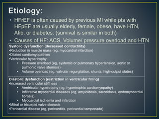
• Chronic HF: is a progressive clinical syndrome that can
result from any changes in cardiac structure or function
that impairs the ability of the ventricle to fill with or eject
blood. (affects ability of heart to contract (systolic) or
relax (diastolic)
• HFrEF: there is decrease in cardiac output (reduced
systolic function).
• HFpEF: Preserved LV systolic function (normal LVEF)
with presumed diastolic dysfunction.
• HF is the final common pathway for many cardiac d/o
including those affecting pericardium, heart valves, and
myocardium.
• Risk doubles with every decade in life
• Major cause for hospitalization among elderly
• 5-year mortality rates around 50% for all pt with HF.
• Around 40% of deaths from HF come as sudden cardiac
death
• About 50% of HF pts have HFpEF.
• HFrEF is often caused by previous MI while pts with
HFpEF are usually elderly, female, obese, have HTN,
Afib, or diabetes. (survival is similar in both)
• Causes of HF: ACS, Volume/ pressure overload and HTN
Systolic dysfunction (decreased contractility)
•Reduction in muscle mass (eg, myocardial infarction)
•Dilated cardiomyopathies
•Ventricular hypertrophy
• Pressure overload (eg, systemic or pulmonary hypertension, aortic or
pulmonic valve stenosis)
• Volume overload (eg, valvular regurgitation, shunts, high-output states)
Diastolic dysfunction (restriction in ventricular filling)
•Increased ventricular stiffness
• Ventricular hypertrophy (eg, hypertrophic cardiomyopathy)
• Infiltrative myocardial diseases (eg, amyloidosis, sarcoidosis, endomyocardial
fibrosis)
• Myocardial ischemia and infarction
•Mitral or tricuspid valve stenosis
•Pericardial disease (eg, pericarditis, pericardial tamponade)
• CO: volume of blood ejected per unit time (L/min)
CO= HR X SV
MAP= CO X systemic vascular resistance (SVR)
• HR is controlled by the ANS (autonomic nervous system)
• SV: Stroke volume or the volume of blood ejected during
systole, depends on preload, afterload, and contractility.
• 1) Tachycardia and increased contractility through SNS
activation
• 2) the Frank-starling mechanism (increase in Preload
causes increase in SV)
• 3) vasoconstriction
• 4) ventricular
hypertrophy and
remodeling
• Angiotensin II: It is a potent vasoconstrictor. Angiotensin
II facilitates release of NE. It promotes sodium retention
through direct effects on the renal tubules and by
stimulating aldosterone release.
• NE: causes tachycardia, vasoconstriction, and increased
contractility and plasma renin activity in HFrEF. Plasma
NE concentrations are elevated in correlation with the
degree of HF. Excess catecholamines increase the risk of
arrhythmias and can cause myocardial cell loss by
stimulating both necrosis and apoptosis.
• Aldosterone: aldosterone-mediated sodium retention
and its key role in volume overload and edema.
• Natriuretic peptides: has three members, atrial natriuretic
peptide (ANP), B-type natriuretic peptide (BNP), and C-type
natriuretic peptide (CNP). Of these, BNP is the most useful in
the diagnosis and management of HF. BNP and its related
biologically inactive peptide, NT-proBNP, are synthesized and
released from the ventricle in response to pressure or volume
overload. BNP or NT-proBNP plasma concentrations are
elevated in patients with HF functioning to increase natriuresis
and diuresis and attenuate activation of the RAAS and SNS.
BNP and NT-proBNP are biomarker for prognostic, diagnostic,
and therapeutic use.
• Arginine vasopressin: AVP is a pituitary peptide hormone
that regulates renal water excretion and plasma osmolality.
Plasma concentrations of AVP are elevated in patients with
HF. This causes (a) increased renal free water reabsorption in
the face of plasma hypoosmolality resulting in volume
overload and hyponatremia; (b) increased arterial
vasoconstriction that contributes to reduced CO; and (c)
stimulation of remodeling by cardiac hypertrophy and
• Cardiac events: Myocardial ischemia and infarction, Atrial
fibrillation, and HTN.
• Noncardiac events: Pulmonary infections (pneumococcal
and influenza), pulmonary embolism, diabetes,
worsening renal fcn, hypothyroidism, and
hyperthyroidism.
• Non-adherence with prescribed HF medications or
dietary recommendations.
• Some drugs may cause or exacerbate HF: 1) negative
inotropic effects 2) cardiotoxic 3) Na/Water retention 4)
other mechanism.
• Symptoms: Dyspnea, particularly on
exertion, Orthopnea, Paroxysmal nocturnal
dyspnea, Exercise intolerance, Tachypnea,
Cough, Fatigue, Nocturia, Hemoptysis,
Abdominal pain, Anorexia, Nausea,
Bloating, Poor appetite, early satiety,
Ascites, Mental status, changesWeight gain
or loss.
• Signs: Pulmonary rales, Pulmonary edema,
S3 gallop, mitral regurgitant murmur, Cool
extremities, Pleural effusion, Cheyne-Stokes
respiration, Tachycardia, Narrow pulse
pressure, Cardiomegaly, Peripheral edema,
Jugular venous distention, Hepatojugular
reflux, Hepatomegaly, Venous stasis
changes, Lateral displacement of apical
• Labs:
BNP > 100 pg/mL
NT-proBNP > 300 pg/mL
Electrocardiogram
Serum creatinine
Chest X-ray
Echocardiogram
Hyponatremia: serum sodium < 130 mEq/L
• New York Heart Association Functional Classification:
• I: Patients with cardiac disease but without limitations of
physical activity. Ordinary physical activity does not cause
undue fatigue, dyspnea, or palpitation.
• II: Patients with cardiac disease that results in slight limitations
of physical activity. Ordinary physical activity results in fatigue,
palpitation, dyspnea, or angina.
• III: Patients with cardiac disease that results in marked
limitation of physical activity. Although patients are comfortable
at rest, less than ordinary activity will lead to symptoms.
• IV: Patients with cardiac disease that results in an inability to
carry on physical activity without discomfort. Symptoms of
congestive heart failure are present even at rest. With any
physical activity, increased discomfort is experienced.
• Improve pts quality of life, relieve/ reduce symptoms,
prevent or minimize hospitalization, slow progression of
the disease and prolong survival.
• Non- Pharmacological therapy:
• smoking and alcohol cessastion.
• Exercise tailored to pt, restrict when decompensated.
• Na restriction: <2g/day
• General fluid restriction: <2L/d if Hyponatremia or
persistent fluid overload
• Daily wt measurements and logging: intervene if wt
increases >1 kg in 1 day or > 2kg over 5 days.
• Implantable devices.
• Symptom-targeted treatment
• Disease-targeted treatment
• Mechanism-targeted treatment
Recommendations
Diuretics
•A loop or a thiazide diuretic should be considered for patients with volume overload. However,
with more severe volume overload or inadequate response to a thiazide, a loop diuretic should be
implemented. Caution is warranted not to lower preload excessively, which may reduce stroke
volume and cardiac output.
ACE inhibitors
•ACE inhibitors should be considered in all patients who have symptomatic atherosclerotic
cardiovascular disease or diabetes and one additional risk factor.
Angiotensin receptor blockers
•In patients who are intolerant of ACE inhibitors, an angiotensin receptor blocker can be
considered an alternative.
Aldosterone antagonists
•reduce the risk of hospitalization in patients that do not have contraindications or are at risk for
hyperkalemia.
β-Blockers considered in pts w/:
•Myocardial infarction
•Hypertension
•Atrial fibrillation requiring ventricular rate control
Calcium channel blockers
•In patients with atrial fibrillation warranting ventricular rate control who either are intolerant to or
have not responded to a β-blocker, diltiazem or verapamil should be considered.
•for symptom-limiting angina and HTN
• As advanced, refractory, or end-stage HF.
• A poor quality of life, and are at high risk for morbidity
and mortality. Recurrent hospitalizations
• Referred to HF management programs including:
 Mechanical circulatory support, continuous IV positive
inotropic therapy, and cardiac transplantation
 Restriction of sodium and fluid( High doses of diuretics
or mechanical methods of fluid removal such as
ultrafiltration may be required).
• For patients with fluid o/l -> decrease edema by reduction
of preload
• Chronic use to maintain euvolemia.
• Relief symptoms but does not prolong survival. Improve
QOL and exercise tolerance.
• Use with caution, 2/2 risk of overdiuresis which lead to a
reduction in CO (HoTN), renal perfusion, and symptoms
of volume depletion.
• Start with low dose, adjust dose according to daily wt
measurements. Intervene if wt increases >1 kg in 1 day
or > 2kg over 5 days.
• Thiazide diuretics: hydrochlorothiazide used in pt with
mild fluid o/l and high BP.
• Loop diuretics restore and maintain euvolemia in HF.
• Loop diuretics: Furosemide, torsemide and bumetanide.
Only pharmacokinetic differences.
• “ceiling effect” overcome it with increasing frequency or
dose or adding thiazide (metolazone).
• S.E: Hypovolemia, hypotension, hyponatremia,
hypokalemia, hypomagnesemia, hyperuricemia, renal
dysfunction, thirst.
• decrease development of severe HF, and reduce
reinfarction and HF hospitalization rates, improve
symptoms, slow disease progression, and decrease
mortality in patients with HFrEF.
• S.E. of ACEi: 1) Hypotension 2) renal insuffeciency 3)
hyperkalemia 4) cough 5) angioedema
• ACEi C/I in pregnancy, angioedema and renal artery
stenosis.
• Renal function and serum potassium should be evaluated
at baseline and within 1 to 2 weeks after therapy is
started.
• candesartan, losartan, or valsartan are recommended by
the guidelines.
• ARBs reduced mortality and hospitalizations and
improved symptoms.
• Used in pts. who can’t tolerate ACE inhibitors.
• Blood pressure, renal function, and serum potassium
should be evaluated within 1 to 2 weeks after initiation of
therapy and after increases in dose.
• S.E: hyperkalemia, decrease in renal function and
hypotension.
• Candesartan and valsartan have no drug drug
interactions.
• Metoprolol succinate, carvedilol, and bisoprolol reduce
mortality.
• Carvedilol preferred in pts with uncontrolled BP.
• metoprolol or bisoprolol may be preferred in patients with
low BP or dizziness and in patients with significant airway
disease.
• S.E: bradycardia or heart block, hypotension, fatigue,
impaired glycemic control in diabetic patients,
bronchospasm in patients with asthma, and worsening
HF. May mask hypoglycemia s/s. except sweating
• monitor vital signs and carefully assess for signs and
symptoms of worsening HF during β-blocker initiation and
uptitration.
• The strategy of starting a β-blocker prior to an ACE
inhibitor is safe and effective
• Start low and go slow with up titration. Dose should not
be doubled more than every 2 weeks.
• β-blockers slow disease progression, decrease
hospitalizations, and improve survival, improve quality of
life in HFrEF.
• β-blocker are C/I uncontrolled bronchospastic disease,
symptomatic bradycardia, advanced heart block without a
pacemaker, and acute decompensated HF.
• Spironolactone and eplerenone block the
mineralocorticoid receptor, the target site for aldosterone.
• It improves symptoms, reduce the risk of HF
hospitalization, and increase survival in select patients
• 2 groups benefit from this drug: those with mild to
moderately severe HFrEF (NYHA class II-IV) who are
receiving standard therapy and those with left ventricular
dysfunction and either acute HF or diabetes early after
MI.
• It has high incidence of hyperkalemia and may cause
gynecomastia and menstrual irregularities.
• Monitor renal fcn. & K levels
• Nitrates result in venodilation and decreased preload.
• Hydralazine is a direct-acting arterial vasodilator causing
a decrease in SVR and resultant increases in SV and
CO. reduces oxidative stress.
• useful in patients unable to tolerate either an ACE
inhibitor or ARB because of renal insufficiency,
hyperkalemia, or possibly hypotension.
• Problems: 1) need for frequent dosing 2) high cost 3)
adverse effects e.g. dizziness, HA, and GI distress.
• have positive inotropic effect on heart.
• improves cardiac function, quality of life, exercise
tolerance, and HF symptoms in patients with HFrEF.
• the target SDC should be 0.5 to 0.9 ng/mL (0.6-1.2
nmol/L). Cam ne achieved with daily dose of 0.125mg.
• Help control ventricular response in pts with HFrEF.
• blood samples for measurement of SDCs should be
collected at least 6 hours and preferably 12 hours or
more after the last dose.
• provide symptom-targeted treatment in patients with
HFpEF by decreasing HR and increasing exercise
tolerance.
• nondihydropyridines (verapamil and diltiazem) are the
most effective because they lower heart rate in addition
to lowering BP. Used to treat the co-morbidities of
hypertension and atrial fibrillation in patients with HFpEF.
• Initial daily doses are verapamil 120 to 240 mg, diltiazem
90 to 120 mg, and amlodipine 2.5 mg.
• S.E.: bradycardia, heart block, peripheral edema and HA.
• NDHP CCB Contraindicated in heart block.
• HYPERTENSION:
Both HFrEF and HFpEF
 ACE inhibitors or ARBs, β-blockers, and diuretics,ARB,
aldosterone antagonist, ISDN/hydralazine, or a second-
generation calcium channel blocker such as amlodipine.
• ANGINA:
• Nitrates and β-blockers , amlodipine and felodipine
appear to be safe
• ATRIAL FIBRILLATION:Digoxin , β-Blockers OR
Combination therapy with digoxin and a β-blocker.
Calcium channel blockers with negative inotropic effects
such as verapamil or diltiazem.
• DIABETES: the treatment of diabetes in this population
remains uncertain.
• Evaluation :
o (a) functional capacity
o (b) volume status
o (c) laboratory monitoring

Chronic Heart Failure- Pharmacotherapy

  • 2.
    • Chronic HF:is a progressive clinical syndrome that can result from any changes in cardiac structure or function that impairs the ability of the ventricle to fill with or eject blood. (affects ability of heart to contract (systolic) or relax (diastolic) • HFrEF: there is decrease in cardiac output (reduced systolic function). • HFpEF: Preserved LV systolic function (normal LVEF) with presumed diastolic dysfunction. • HF is the final common pathway for many cardiac d/o including those affecting pericardium, heart valves, and myocardium.
  • 3.
    • Risk doubleswith every decade in life • Major cause for hospitalization among elderly • 5-year mortality rates around 50% for all pt with HF. • Around 40% of deaths from HF come as sudden cardiac death • About 50% of HF pts have HFpEF.
  • 4.
    • HFrEF isoften caused by previous MI while pts with HFpEF are usually elderly, female, obese, have HTN, Afib, or diabetes. (survival is similar in both) • Causes of HF: ACS, Volume/ pressure overload and HTN Systolic dysfunction (decreased contractility) •Reduction in muscle mass (eg, myocardial infarction) •Dilated cardiomyopathies •Ventricular hypertrophy • Pressure overload (eg, systemic or pulmonary hypertension, aortic or pulmonic valve stenosis) • Volume overload (eg, valvular regurgitation, shunts, high-output states) Diastolic dysfunction (restriction in ventricular filling) •Increased ventricular stiffness • Ventricular hypertrophy (eg, hypertrophic cardiomyopathy) • Infiltrative myocardial diseases (eg, amyloidosis, sarcoidosis, endomyocardial fibrosis) • Myocardial ischemia and infarction •Mitral or tricuspid valve stenosis •Pericardial disease (eg, pericarditis, pericardial tamponade)
  • 5.
    • CO: volumeof blood ejected per unit time (L/min) CO= HR X SV MAP= CO X systemic vascular resistance (SVR) • HR is controlled by the ANS (autonomic nervous system) • SV: Stroke volume or the volume of blood ejected during systole, depends on preload, afterload, and contractility.
  • 8.
    • 1) Tachycardiaand increased contractility through SNS activation • 2) the Frank-starling mechanism (increase in Preload causes increase in SV) • 3) vasoconstriction • 4) ventricular hypertrophy and remodeling
  • 10.
    • Angiotensin II:It is a potent vasoconstrictor. Angiotensin II facilitates release of NE. It promotes sodium retention through direct effects on the renal tubules and by stimulating aldosterone release. • NE: causes tachycardia, vasoconstriction, and increased contractility and plasma renin activity in HFrEF. Plasma NE concentrations are elevated in correlation with the degree of HF. Excess catecholamines increase the risk of arrhythmias and can cause myocardial cell loss by stimulating both necrosis and apoptosis. • Aldosterone: aldosterone-mediated sodium retention and its key role in volume overload and edema.
  • 11.
    • Natriuretic peptides:has three members, atrial natriuretic peptide (ANP), B-type natriuretic peptide (BNP), and C-type natriuretic peptide (CNP). Of these, BNP is the most useful in the diagnosis and management of HF. BNP and its related biologically inactive peptide, NT-proBNP, are synthesized and released from the ventricle in response to pressure or volume overload. BNP or NT-proBNP plasma concentrations are elevated in patients with HF functioning to increase natriuresis and diuresis and attenuate activation of the RAAS and SNS. BNP and NT-proBNP are biomarker for prognostic, diagnostic, and therapeutic use. • Arginine vasopressin: AVP is a pituitary peptide hormone that regulates renal water excretion and plasma osmolality. Plasma concentrations of AVP are elevated in patients with HF. This causes (a) increased renal free water reabsorption in the face of plasma hypoosmolality resulting in volume overload and hyponatremia; (b) increased arterial vasoconstriction that contributes to reduced CO; and (c) stimulation of remodeling by cardiac hypertrophy and
  • 12.
    • Cardiac events:Myocardial ischemia and infarction, Atrial fibrillation, and HTN. • Noncardiac events: Pulmonary infections (pneumococcal and influenza), pulmonary embolism, diabetes, worsening renal fcn, hypothyroidism, and hyperthyroidism. • Non-adherence with prescribed HF medications or dietary recommendations. • Some drugs may cause or exacerbate HF: 1) negative inotropic effects 2) cardiotoxic 3) Na/Water retention 4) other mechanism.
  • 13.
    • Symptoms: Dyspnea,particularly on exertion, Orthopnea, Paroxysmal nocturnal dyspnea, Exercise intolerance, Tachypnea, Cough, Fatigue, Nocturia, Hemoptysis, Abdominal pain, Anorexia, Nausea, Bloating, Poor appetite, early satiety, Ascites, Mental status, changesWeight gain or loss. • Signs: Pulmonary rales, Pulmonary edema, S3 gallop, mitral regurgitant murmur, Cool extremities, Pleural effusion, Cheyne-Stokes respiration, Tachycardia, Narrow pulse pressure, Cardiomegaly, Peripheral edema, Jugular venous distention, Hepatojugular reflux, Hepatomegaly, Venous stasis changes, Lateral displacement of apical
  • 14.
    • Labs: BNP >100 pg/mL NT-proBNP > 300 pg/mL Electrocardiogram Serum creatinine Chest X-ray Echocardiogram Hyponatremia: serum sodium < 130 mEq/L
  • 15.
    • New YorkHeart Association Functional Classification: • I: Patients with cardiac disease but without limitations of physical activity. Ordinary physical activity does not cause undue fatigue, dyspnea, or palpitation. • II: Patients with cardiac disease that results in slight limitations of physical activity. Ordinary physical activity results in fatigue, palpitation, dyspnea, or angina. • III: Patients with cardiac disease that results in marked limitation of physical activity. Although patients are comfortable at rest, less than ordinary activity will lead to symptoms. • IV: Patients with cardiac disease that results in an inability to carry on physical activity without discomfort. Symptoms of congestive heart failure are present even at rest. With any physical activity, increased discomfort is experienced.
  • 17.
    • Improve ptsquality of life, relieve/ reduce symptoms, prevent or minimize hospitalization, slow progression of the disease and prolong survival. • Non- Pharmacological therapy: • smoking and alcohol cessastion. • Exercise tailored to pt, restrict when decompensated. • Na restriction: <2g/day • General fluid restriction: <2L/d if Hyponatremia or persistent fluid overload • Daily wt measurements and logging: intervene if wt increases >1 kg in 1 day or > 2kg over 5 days. • Implantable devices.
  • 18.
    • Symptom-targeted treatment •Disease-targeted treatment • Mechanism-targeted treatment
  • 19.
    Recommendations Diuretics •A loop ora thiazide diuretic should be considered for patients with volume overload. However, with more severe volume overload or inadequate response to a thiazide, a loop diuretic should be implemented. Caution is warranted not to lower preload excessively, which may reduce stroke volume and cardiac output. ACE inhibitors •ACE inhibitors should be considered in all patients who have symptomatic atherosclerotic cardiovascular disease or diabetes and one additional risk factor. Angiotensin receptor blockers •In patients who are intolerant of ACE inhibitors, an angiotensin receptor blocker can be considered an alternative. Aldosterone antagonists •reduce the risk of hospitalization in patients that do not have contraindications or are at risk for hyperkalemia. β-Blockers considered in pts w/: •Myocardial infarction •Hypertension •Atrial fibrillation requiring ventricular rate control Calcium channel blockers •In patients with atrial fibrillation warranting ventricular rate control who either are intolerant to or have not responded to a β-blocker, diltiazem or verapamil should be considered. •for symptom-limiting angina and HTN
  • 22.
    • As advanced,refractory, or end-stage HF. • A poor quality of life, and are at high risk for morbidity and mortality. Recurrent hospitalizations • Referred to HF management programs including:  Mechanical circulatory support, continuous IV positive inotropic therapy, and cardiac transplantation  Restriction of sodium and fluid( High doses of diuretics or mechanical methods of fluid removal such as ultrafiltration may be required).
  • 23.
    • For patientswith fluid o/l -> decrease edema by reduction of preload • Chronic use to maintain euvolemia. • Relief symptoms but does not prolong survival. Improve QOL and exercise tolerance. • Use with caution, 2/2 risk of overdiuresis which lead to a reduction in CO (HoTN), renal perfusion, and symptoms of volume depletion. • Start with low dose, adjust dose according to daily wt measurements. Intervene if wt increases >1 kg in 1 day or > 2kg over 5 days.
  • 24.
    • Thiazide diuretics:hydrochlorothiazide used in pt with mild fluid o/l and high BP. • Loop diuretics restore and maintain euvolemia in HF. • Loop diuretics: Furosemide, torsemide and bumetanide. Only pharmacokinetic differences. • “ceiling effect” overcome it with increasing frequency or dose or adding thiazide (metolazone). • S.E: Hypovolemia, hypotension, hyponatremia, hypokalemia, hypomagnesemia, hyperuricemia, renal dysfunction, thirst.
  • 25.
    • decrease developmentof severe HF, and reduce reinfarction and HF hospitalization rates, improve symptoms, slow disease progression, and decrease mortality in patients with HFrEF. • S.E. of ACEi: 1) Hypotension 2) renal insuffeciency 3) hyperkalemia 4) cough 5) angioedema • ACEi C/I in pregnancy, angioedema and renal artery stenosis. • Renal function and serum potassium should be evaluated at baseline and within 1 to 2 weeks after therapy is started.
  • 26.
    • candesartan, losartan,or valsartan are recommended by the guidelines. • ARBs reduced mortality and hospitalizations and improved symptoms. • Used in pts. who can’t tolerate ACE inhibitors. • Blood pressure, renal function, and serum potassium should be evaluated within 1 to 2 weeks after initiation of therapy and after increases in dose. • S.E: hyperkalemia, decrease in renal function and hypotension. • Candesartan and valsartan have no drug drug interactions.
  • 27.
    • Metoprolol succinate,carvedilol, and bisoprolol reduce mortality. • Carvedilol preferred in pts with uncontrolled BP. • metoprolol or bisoprolol may be preferred in patients with low BP or dizziness and in patients with significant airway disease. • S.E: bradycardia or heart block, hypotension, fatigue, impaired glycemic control in diabetic patients, bronchospasm in patients with asthma, and worsening HF. May mask hypoglycemia s/s. except sweating • monitor vital signs and carefully assess for signs and symptoms of worsening HF during β-blocker initiation and uptitration. • The strategy of starting a β-blocker prior to an ACE inhibitor is safe and effective
  • 28.
    • Start lowand go slow with up titration. Dose should not be doubled more than every 2 weeks. • β-blockers slow disease progression, decrease hospitalizations, and improve survival, improve quality of life in HFrEF. • β-blocker are C/I uncontrolled bronchospastic disease, symptomatic bradycardia, advanced heart block without a pacemaker, and acute decompensated HF.
  • 29.
    • Spironolactone andeplerenone block the mineralocorticoid receptor, the target site for aldosterone. • It improves symptoms, reduce the risk of HF hospitalization, and increase survival in select patients • 2 groups benefit from this drug: those with mild to moderately severe HFrEF (NYHA class II-IV) who are receiving standard therapy and those with left ventricular dysfunction and either acute HF or diabetes early after MI. • It has high incidence of hyperkalemia and may cause gynecomastia and menstrual irregularities. • Monitor renal fcn. & K levels
  • 30.
    • Nitrates resultin venodilation and decreased preload. • Hydralazine is a direct-acting arterial vasodilator causing a decrease in SVR and resultant increases in SV and CO. reduces oxidative stress. • useful in patients unable to tolerate either an ACE inhibitor or ARB because of renal insufficiency, hyperkalemia, or possibly hypotension. • Problems: 1) need for frequent dosing 2) high cost 3) adverse effects e.g. dizziness, HA, and GI distress.
  • 31.
    • have positiveinotropic effect on heart. • improves cardiac function, quality of life, exercise tolerance, and HF symptoms in patients with HFrEF. • the target SDC should be 0.5 to 0.9 ng/mL (0.6-1.2 nmol/L). Cam ne achieved with daily dose of 0.125mg. • Help control ventricular response in pts with HFrEF. • blood samples for measurement of SDCs should be collected at least 6 hours and preferably 12 hours or more after the last dose.
  • 32.
    • provide symptom-targetedtreatment in patients with HFpEF by decreasing HR and increasing exercise tolerance. • nondihydropyridines (verapamil and diltiazem) are the most effective because they lower heart rate in addition to lowering BP. Used to treat the co-morbidities of hypertension and atrial fibrillation in patients with HFpEF. • Initial daily doses are verapamil 120 to 240 mg, diltiazem 90 to 120 mg, and amlodipine 2.5 mg. • S.E.: bradycardia, heart block, peripheral edema and HA. • NDHP CCB Contraindicated in heart block.
  • 33.
    • HYPERTENSION: Both HFrEFand HFpEF  ACE inhibitors or ARBs, β-blockers, and diuretics,ARB, aldosterone antagonist, ISDN/hydralazine, or a second- generation calcium channel blocker such as amlodipine. • ANGINA: • Nitrates and β-blockers , amlodipine and felodipine appear to be safe • ATRIAL FIBRILLATION:Digoxin , β-Blockers OR Combination therapy with digoxin and a β-blocker. Calcium channel blockers with negative inotropic effects such as verapamil or diltiazem. • DIABETES: the treatment of diabetes in this population remains uncertain.
  • 34.
    • Evaluation : o(a) functional capacity o (b) volume status o (c) laboratory monitoring

Editor's Notes

  • #6 Note preload: more blood stretches the walls of ventricles, higher contractiliy Afterload: anything that increases contractility primarly SVR Contractility : purely intrinsic to heart muscle, ability of myocyte to shorten and cause contraction and pumping to the heart.
  • #8 myocardial relaxation and filling are impaired and incomplete. The ventricle is unable to accept an adequate volume of blood from the venous system, does not fill at low pressure, and/or is unable to maintain normal SV. The ventricular chamber is not enlarged and EF may be normal or elevated.
  • #9 HR is increased due to release of NE. increasing NE increases myocardial oxygen demand. If ischemia is induced or worsened both diastolic and systolic function may become impaired and SV decreases. This mechanism usually helps maintain CO and can be seen as increased MVO2, shortened diastolic filling time, ventricular arrhythmias and increased myocardial death. Renal perfusion in HF is reduced due to decrease in CO and redistribution of blood away from nonvital organs this activates RAAS system to maintain blood pressure and increase renal sodium / water retention. This mechanism usually optimize stroke volume via frank-starling mechanism. Can be seen by pulmonary /systemic congestion and edema. And increased MVO2 vasoconstriction occurs in patients with HFrEF to help redistribute blood flow away from nonessential organs to coronary and cerebral circulations to support BP, neurohormones that contribute to vasoconstriction: NE, angiotensin II, endothelin-1 (ET-1), neuropeptide Y, urotensin II, and arginine vasopressin (AVP).This mechanism usually maintain BP, and can be seen as increased MVO2, increased afterload and decrease stroke volume. It describe increase in ventricular muscle mass. this mechanism helps maintain CO, reduces myocarial wall stress and decreases MVO2, causes diastolic and systolic dysfunction, increase myocardial cell death and ischemia and increase risk of arrhythmia and fibrosis.
  • #10 Key components of the pathophysiology of cardiac remodeling. Myocardial injury (eg, myocardial infarction) results in the activation of a number of hemodynamic and neurohormonal compensatory responses in an attempt to maintain circulatory homeostasis. Chronic activation of the neurohormonal systems results in a cascade of events that affect the myocardium at the molecular and cellular levels. These events lead to the changes in ventricular size, shape, structure, and function known as ventricular remodeling. The alterations in ventricular function result in further deterioration in cardiac systolic and diastolic functions that further promotes the remodeling process.
  • #13 Negative Inotropic Effect  Antiarrhythmics (eg, disopyramide, flecainide, propafenone)  Beta-blockers (eg, propranolol, metoprolol, carvedilol)  Calcium channel blockers (eg, verapamil, diltiazem)  Itraconazole Cardiotoxic  Doxorubicin, Epirubicin, Daunomycin, Cyclophosphamide, Trastuzumab, Bevacizumab, Mitoxantrone, Ifosfamide,Mitomycin, Lapatinib, Sunitinib, Imatinib, Ethanol, Amphetamines (eg, cocaine, methamphetamine) Sodium and Water Retention  NSAIDs, COX-2inhibitors, Rosiglitazone and pioglitazone, Glucocorticoids, Androgens and estrogens, Salicylates (high dose), Sodium-containing drugs (eg, carbenicillin disodium, ticarcillin disodium) Uncertain Mechanism  Adalimumab, Dronedarone, Etanercept, Infliximab
  • #15 Electrocardiogram may be normal or it could show numerous abnormalities including acute ST-T wave changes from myocardial ischemia, atrial fibrillation, bradycardia, left ventricular hypertrophy Serum creatinine: It may be increased due to hypoperfusion. Preexisting renal dysfunction can contribute to volume overload Complete blood count useful to determine if heart failure due to reduced oxygen carrying capacity Chest X-ray: Useful for detection of cardiac enlargement, pulmonary edema, and pleural effusions Echocardiogram: Used to assess LV size, valve function, pericardial effusion, wall motion abnormalities, and ejection fraction Hyponatremia: serum sodium < 130 mEq/L is associated with reduced survival and may indicate worsening volume overload and/or disease progression
  • #16 The NYHA system is primarily intended to classify symptoms according to the clinician’s subjective evaluation and does not recognize preventive measures or the progression of the disorder. A patient’s symptoms can change frequently over a short period of time due to changes in medications, diet, intercurrent illnesses, etc (PATIENT can go forward and backward in classes)
  • #19 Decrease pulmonary venous pressure -> Reduce left ventricular volume -> Diuretics, nitrates, salt restriction Decrease myocardial oxygen consumption-> Reduce heart rate, Control blood pressure-> β-blockers, diltiazem, verapamil, ACE inhibitors, ARBs, calcium channel blockers Maintain atrial contraction-> Restore and/or maintain sinus rhythm-> Cardioversion of atrial fibrillation Improve exercise tolerance-> As above -> Use positive inotropic agents with caution Prevent/treat myocardial ischemia->  β-blockers, diltiazem, verapamil, nitrates Prevent/regress ventricular hypertrophy -> Antihypertensive therapy Modify myocardial and extramyocardial mechanisms->  Possibly ACE inhibitors or ARBs, diuretics, spironolactone Modify intracellular and extracellular mechanisms -> Possibly ACE inhibitors or ARBs, spironolactone
  • #21 Treatment of Stage A Heart Failure Patients in Stage A do not have structural heart disease or HF symptoms but are at high risk for developing HF because of the presence of risk factors such as HTN, dyslipidemia, diabetes, obesity, metabolic syndrome, smoking, and coronary artery disease. ACE inhibitors or ARBs and statins are recommended for HF prevention in patients with multiple vascular risk factors Treatment of Stage B Heart Failure Patients in Stage B have structural heart disease, but do not have HF symptoms. This group includes patients with left ventricular hypertrophy, recent or remote MI, valvular disease, or LVEF less than 40%. In addition to the treatment measures outlined in Stage A, ACE inhibitors or ARBs and β-blockers are important components of therapy. Patients with a previous MI and reduced LVEF should also receive an ACE inhibitor or ARB, evidence-based β-blockers, and a statin.
  • #22 Treatment of Stage C HF  Patients with structural heart disease and previous or current symptoms are classified in Stage C and include both HFrEF and HFpEF. In addition to treatments in Stages A and B, patients with HFrEF in Stage C should be routinely treated with GDMT that includes an ACE inhibitor or ARB and an evidence-based β-blocker. Loop diuretics, aldosterone antagonists, and hydralazine–isosorbide dinitrate (ISDN) are also routinely used in these patients. Digoxin can also be considered in selected patients, as can two newly approved medications, ivabradine and sacubitril/valsartan. Nonpharmacologic therapy with devices such as an implantable cardioverter-defibrillator (ICD) or cardiac resynchronization therapy (CRT) with a biventricular pacemaker.
  • #23 These patients often undergo recurrent hospitalizations or cannot be discharged from the hospital without special interventions, have a poor quality of life, and are at high risk for morbidity and mortality Table 14-8 and 14-9 you can see each drug dosing and monitoring parameters.
  • #25 the peak effect with all the agents occurs in 30 to 90 minutes, with duration of 4 to 8 hours (longer for torsemide). Following IV administration, the diuretic effect begins within minutes. All three drugs are highly (greater than 95%) bound to serum albumin and enter the nephron by active secretion in the proximal tubule. The magnitude of effect is determined by the peak concentration achieved in the nephron, and there is a threshold concentration that must be achieved before any diuresis is seen. The biggest difference between the agents is bioavailability. Bioavailability of bumetanide and torsemide is essentially complete (80%-100%), whereas furosemide bioavailability exhibits marked intrapatient and interpatient variability. Coadministration of furosemide and bumetanide with food can decrease bioavailability significantly, whereas food has no effect on bioavailability of torsemide. 
  • #26 (symptoms of HoTN: dizziness, light-headedness, blurred vision, presyncope or syncope) risk factors for HoTN: hyponatremia <130mEq/L, hypovolemia and overdiuresis.