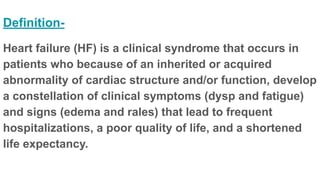
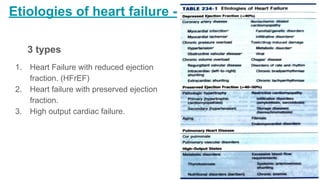
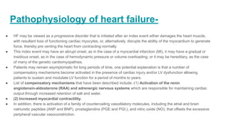
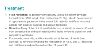
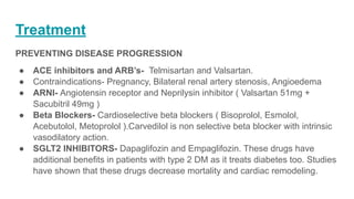
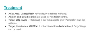
Heart Failure
Definition-
Heart failure (HF) is a clinical syndrome that occurs in
patients who because of an inherited or acquired
abnormality of cardiac structure and/or function, develop
a constellation of clinical symptoms (dysp and fatigue)
and signs (edema and rales) that lead to frequent
hospitalizations, a poor quality of life, and a shortened
life expectancy.
Etiologies of heart failure -
3 types
1. Heart Failure with reduced ejection
fraction. (HFrEF)
2. Heart failure with preserved ejection
fraction.
3. High output cardiac failure.
Classification of heart failure
Pathophysiology of heart failure-
● HF may be viewed as a progressive disorder that is initiated after an index event either damages the heart muscle,
with resultant loss of functioning cardiac myocytes, or, alternatively, disrupts the ability of the myocardium to generate
force, thereby pre venting the heart from contracting normally.
● This index event may have an abrupt onset, as in the case of a myocardial infarction (MI), it may have a gradual or
insidious onset, as in the case of hemodynamic pressure or volume overloading; or it may be hereditary, as the case
of many of the genetic cardiomyopathies.
● Patients may remain asymptomatic for long periods of time, one potential explanation is that a number of
compensatory mechanisms become activated in the presence of cardiac injury and/or LV dysfunction allowing
patients to sustain and modulate LV function for a period of months to years.
● List of compensatory mechanisms that have been described include -(1) Activation of the renin
angiotensin-aldosterone (RAA) and adrenergic nervous systems which are responsible for maintaining cardiac
output through increased retention of salt and water.
● (2) Increased myocardial contractility.
● In addition, there is activation of a family of countervailing vasodilatory molecules, including the atrial and brain
natriuretic peptides (ANP and BNP), prostaglandins (PGE and PGI,), and nitric oxide (NO), that offsets the excessive
peripheral vascular vasoconstriction.
Pathophysiology of heart failure-
● At some point patients become overtly symptomatic, with a resultant striking increase
in morbidity and mortality rates. Although the exact mechanisms that are responsible
for this transition are not known, as will be discussed below, the transition to
symptomatic HF is accompanied by increasing activation of neurohormonal,
adrenergic, and cytokine systems that lead to a series of adaptive changes within the
myocardium collectively referred to as LV remodeling.
Mechanisms of heart failure
● Systolic dysfunction
● LV remodeling develops in response to a series of complex events that occur
at the cellular and molecular levels.
● Changes include (1) myocyte hypertrophy, (2) alterations in the contractile
properties of the myocyte, (3) progressive loss of myocytes through necrosis,
apoptosis and autophagic cell death, (4) Beta-adrenergic desensitization, (5)
abnormal myocardial energetics and metabolism, and (6) Reorganization of
the extracellular matrix with dissolution of the organized structural collagen.
Mechanisms of heart failure-
● Diastolic dysfunction
● Myocardial relaxation is an adenosine triphosphate (ATP) dependent process that is
regulated by uptake of cytoplasmic calcium into the SR by SERCA2A and extrusion of
calcium by sarcolemmal pumps.
● Accordingly, reductions in ATP concentration, as occurs in ischemia, may interfere with
these processes and lead to slowed myocardial relaxation.
● Alternatively, if LV filling is delayed because LV compliance is reduced (e.g, from
hypertrophy or fibrosis), LV filling pressures will similarly remain elevated at end
diastole.Elevated LV end-diastolic filling pressures result in increases in pulmonary
capillary pressures, which can contribute to the dyspnea experienced by patients with
diastolic dysfunction.
● In addition to impaired myocardial relaxation, increased myo- cardial stiffness
secondary to cardiac hypertrophy and increased nyocardial collagen content may
contribute to diastolic failure
Risk factors associated with HF
● Type 2 Diabetes Mellitus
● Hypertension
● Obesity
● Smoking
● Alcoholism/ Drug use
● Lack of physical activity
Clinical manifestations of CHF-
Symptoms-
● The cardinal symptoms of HF are fatigue and shortness of breath.
● Orthopnea, which is defined as dyspnea in the recumbent position, is usually a later manifestation.
● Paroxysmal nocturnal dyspnea (PND)- This term refers to acute episodes of severe shortness of
breath and coughing that generally occur at night and awaken the patient from sleep.
● Cheyne-Stokes respiration- Also referred to as periodic respiration or cyclic respiration. It is
present of patients with advanced HF and usually is associated with low cardiac output.
● Patients with HF also may present with gastrointestinal symptoms. Anorexia, nausea, and early
satiety associated with abdominal pain and fullness are common complaints and may be related to
edema. of the bowel wall and/or a congested liver.
● Nocturia is common in HF and may contribute to insomnia.
● Cerebral symptoms such as confusion and sleep disturbances may also occur.
Signs of heart failure
● Tachycardia
● Tachypnea
● Increased JVP
● Pedal Edema
● Bibasilar crepitations
● S3 gallop
● Mitral regurgitation
Diagnosis
● Echocardiogram- A routine12-lead ECG is recommended. The major
importance of the ECG is to assess cardiac rhythm and determine the
presence of LV hypertrophy or a prior MI (presence or absence of Q waves)
as well as to determine QRS width to ascertain whether the patient may
benefit from resynchronization therapy. A normal ECG virtually excludes LV
systolic dysfunction.
● Chest X-Ray- A chest x-ray provides useful information about cardiac size
and shape, as well as the state of the pulmonary vasculature, and may
identify noncardiac causes of the patient's symptoms. It also shows
pulmonary effusion if present.
Diagnosis
● LV function assessment - Noninvasive cardiac imaging is essential for the
diagnosis, evaluation, and management of HF. The most useful test is the
two-dimensional (2-D) echocardiogram/Doppler, which can provide a
semiquantitative assessment of LV size and function as well as the presence
or absence of valvular and/or regional wall motion abnormalities (indicative of
a prior MI).
● The most useful index of LV function is the EF (stroke vol- ume divided by
end-diastolic volume). Because the EF is easy to measure by noninvasive
testing and easy to conceptualize, it has gained wide acceptance among
clinicians.
Diagnosis
● Biomarkers- Circulating levels of natriuretic peptides are useful adjunctive
tools in the diagnosis of patients with HF. Both B-type natriuretic peptide
(BNP) and N-terminal pro-BNP, which are released from the failing heart, are
relatively sensitive markers for the presence of HF with depressed EF; they
also are elevated in HF patients with a preserved EF.
Treatment
DEFINING AN APPROPRIATE THERAPEUTIC STRATEGY FOR CHRONIC HF-
● For patients who have developed LV systolic dysfunction but remain
asymptomatic (class I), the goal should be to slow disease progression by
blocking neuro- hormonal systems that lead to cardiac remodeling.
● For patients who have developed symptoms (class II-IV), the primary goal
should be to alleviate fluid retention, lessen disabil- ity, and reduce the risk of
further disease progression and death. These goals generally require a
strategy that combines diuretics (to control salt and water retention) with
neurohormonal inter- ventions (to minimize cardiac remodeling).
Treatment
MANAGEMENT OF HF WITH DEPRESSED EJECTION FRACTION (< 40%)
● General measures- Clinicians should aim to screen for and treat
comorbidities such as hypertension, CAD, diabetes mellitus, anemia, and
sleep-disordered breathing, as these conditions tend to exacerbate HF
● Activity- Although heavy physical labor is not recommended in HF, routine
modest exercise has been shown to be beneficial in patients with NYHA class
I-III HF. For euvolemic patients, regular isotonic exercise such as walking or
riding a stationary- bicycle ergometer, as tolerated, should be encouraged.
Exercise training results in reduced HF symptoms, increased exercise
capacity, and improved quality of life.
● Diet- Dietary restriction of sodium (2-3 g daily)
Treatment
● Fluid restriction- is generally unnecessary unless the patient develops
hyponatremia (<130 meq/L).Fluid restriction (<2 L/day) should be considered
in hyponatremic patients or those whose fluid retention is difficult to control
despite high doses of diuretics and sodium restriction.
● Diuretics- Many of the clinical manifestations of moderate to severe HF result
from excessive salt and water retention that leads to volume expansion and
congestive symptoms.
● Furosemide, torsemide, and bumetanide act at the loop of Henle (loop
diuretics) by reversibly inhibiting the reabsorption of Na, K, and Cl-.Thiazides
and metolazone reduce the reabsorption of Na and Cl.
Treatment
PREVENTING DISEASE PROGRESSION
● ACE inhibitors and ARB’s- Telmisartan and Valsartan.
● Contraindications- Pregnancy, Bilateral renal artery stenosis, Angioedema
● ARNI- Angiotensin receptor and Neprilysin inhibitor ( Valsartan 51mg +
Sacubitril 49mg )
● Beta Blockers- Cardioselective beta blockers ( Bisoprolol, Esmolol,
Acebutolol, Metoprolol ).Carvedilol is non selective beta blocker with intrinsic
vasodilatory action.
● SGLT2 INHIBITORS- Dapaglifozin and Empaglifozin. These drugs have
additional benefits in patients with type 2 DM as it treats diabetes too. Studies
have shown that these drugs decrease mortality and cardiac remodeling.
Treatment
● ACE/ ARB/ Dapaglifozin have shown to reduce mortality.
● Aspirin and Beta blockers are used for risk factor control.
● Target LDL levels - <100mg/dl in low risk patients and <70mg/dl in high risk
patients.
● Target Heart rate - <70BPM. If not achieved then Ivabradine( 2.5mg-10mg)
can be used.

Heart failure ppt.pdfjsnjaja52772$&-#+@1

  • 1.
  • 2.
    Definition- Heart failure (HF)is a clinical syndrome that occurs in patients who because of an inherited or acquired abnormality of cardiac structure and/or function, develop a constellation of clinical symptoms (dysp and fatigue) and signs (edema and rales) that lead to frequent hospitalizations, a poor quality of life, and a shortened life expectancy.
  • 3.
    Etiologies of heartfailure - 3 types 1. Heart Failure with reduced ejection fraction. (HFrEF) 2. Heart failure with preserved ejection fraction. 3. High output cardiac failure.
  • 4.
  • 5.
    Pathophysiology of heartfailure- ● HF may be viewed as a progressive disorder that is initiated after an index event either damages the heart muscle, with resultant loss of functioning cardiac myocytes, or, alternatively, disrupts the ability of the myocardium to generate force, thereby pre venting the heart from contracting normally. ● This index event may have an abrupt onset, as in the case of a myocardial infarction (MI), it may have a gradual or insidious onset, as in the case of hemodynamic pressure or volume overloading; or it may be hereditary, as the case of many of the genetic cardiomyopathies. ● Patients may remain asymptomatic for long periods of time, one potential explanation is that a number of compensatory mechanisms become activated in the presence of cardiac injury and/or LV dysfunction allowing patients to sustain and modulate LV function for a period of months to years. ● List of compensatory mechanisms that have been described include -(1) Activation of the renin angiotensin-aldosterone (RAA) and adrenergic nervous systems which are responsible for maintaining cardiac output through increased retention of salt and water. ● (2) Increased myocardial contractility. ● In addition, there is activation of a family of countervailing vasodilatory molecules, including the atrial and brain natriuretic peptides (ANP and BNP), prostaglandins (PGE and PGI,), and nitric oxide (NO), that offsets the excessive peripheral vascular vasoconstriction.
  • 6.
    Pathophysiology of heartfailure- ● At some point patients become overtly symptomatic, with a resultant striking increase in morbidity and mortality rates. Although the exact mechanisms that are responsible for this transition are not known, as will be discussed below, the transition to symptomatic HF is accompanied by increasing activation of neurohormonal, adrenergic, and cytokine systems that lead to a series of adaptive changes within the myocardium collectively referred to as LV remodeling.
  • 8.
    Mechanisms of heartfailure ● Systolic dysfunction ● LV remodeling develops in response to a series of complex events that occur at the cellular and molecular levels. ● Changes include (1) myocyte hypertrophy, (2) alterations in the contractile properties of the myocyte, (3) progressive loss of myocytes through necrosis, apoptosis and autophagic cell death, (4) Beta-adrenergic desensitization, (5) abnormal myocardial energetics and metabolism, and (6) Reorganization of the extracellular matrix with dissolution of the organized structural collagen.
  • 9.
  • 10.
    Mechanisms of heartfailure- ● Diastolic dysfunction ● Myocardial relaxation is an adenosine triphosphate (ATP) dependent process that is regulated by uptake of cytoplasmic calcium into the SR by SERCA2A and extrusion of calcium by sarcolemmal pumps. ● Accordingly, reductions in ATP concentration, as occurs in ischemia, may interfere with these processes and lead to slowed myocardial relaxation. ● Alternatively, if LV filling is delayed because LV compliance is reduced (e.g, from hypertrophy or fibrosis), LV filling pressures will similarly remain elevated at end diastole.Elevated LV end-diastolic filling pressures result in increases in pulmonary capillary pressures, which can contribute to the dyspnea experienced by patients with diastolic dysfunction. ● In addition to impaired myocardial relaxation, increased myo- cardial stiffness secondary to cardiac hypertrophy and increased nyocardial collagen content may contribute to diastolic failure
  • 11.
    Risk factors associatedwith HF ● Type 2 Diabetes Mellitus ● Hypertension ● Obesity ● Smoking ● Alcoholism/ Drug use ● Lack of physical activity
  • 12.
    Clinical manifestations ofCHF- Symptoms- ● The cardinal symptoms of HF are fatigue and shortness of breath. ● Orthopnea, which is defined as dyspnea in the recumbent position, is usually a later manifestation. ● Paroxysmal nocturnal dyspnea (PND)- This term refers to acute episodes of severe shortness of breath and coughing that generally occur at night and awaken the patient from sleep. ● Cheyne-Stokes respiration- Also referred to as periodic respiration or cyclic respiration. It is present of patients with advanced HF and usually is associated with low cardiac output. ● Patients with HF also may present with gastrointestinal symptoms. Anorexia, nausea, and early satiety associated with abdominal pain and fullness are common complaints and may be related to edema. of the bowel wall and/or a congested liver. ● Nocturia is common in HF and may contribute to insomnia. ● Cerebral symptoms such as confusion and sleep disturbances may also occur.
  • 13.
    Signs of heartfailure ● Tachycardia ● Tachypnea ● Increased JVP ● Pedal Edema ● Bibasilar crepitations ● S3 gallop ● Mitral regurgitation
  • 14.
    Diagnosis ● Echocardiogram- Aroutine12-lead ECG is recommended. The major importance of the ECG is to assess cardiac rhythm and determine the presence of LV hypertrophy or a prior MI (presence or absence of Q waves) as well as to determine QRS width to ascertain whether the patient may benefit from resynchronization therapy. A normal ECG virtually excludes LV systolic dysfunction. ● Chest X-Ray- A chest x-ray provides useful information about cardiac size and shape, as well as the state of the pulmonary vasculature, and may identify noncardiac causes of the patient's symptoms. It also shows pulmonary effusion if present.
  • 15.
    Diagnosis ● LV functionassessment - Noninvasive cardiac imaging is essential for the diagnosis, evaluation, and management of HF. The most useful test is the two-dimensional (2-D) echocardiogram/Doppler, which can provide a semiquantitative assessment of LV size and function as well as the presence or absence of valvular and/or regional wall motion abnormalities (indicative of a prior MI). ● The most useful index of LV function is the EF (stroke vol- ume divided by end-diastolic volume). Because the EF is easy to measure by noninvasive testing and easy to conceptualize, it has gained wide acceptance among clinicians.
  • 16.
    Diagnosis ● Biomarkers- Circulatinglevels of natriuretic peptides are useful adjunctive tools in the diagnosis of patients with HF. Both B-type natriuretic peptide (BNP) and N-terminal pro-BNP, which are released from the failing heart, are relatively sensitive markers for the presence of HF with depressed EF; they also are elevated in HF patients with a preserved EF.
  • 17.
    Treatment DEFINING AN APPROPRIATETHERAPEUTIC STRATEGY FOR CHRONIC HF- ● For patients who have developed LV systolic dysfunction but remain asymptomatic (class I), the goal should be to slow disease progression by blocking neuro- hormonal systems that lead to cardiac remodeling. ● For patients who have developed symptoms (class II-IV), the primary goal should be to alleviate fluid retention, lessen disabil- ity, and reduce the risk of further disease progression and death. These goals generally require a strategy that combines diuretics (to control salt and water retention) with neurohormonal inter- ventions (to minimize cardiac remodeling).
  • 18.
    Treatment MANAGEMENT OF HFWITH DEPRESSED EJECTION FRACTION (< 40%) ● General measures- Clinicians should aim to screen for and treat comorbidities such as hypertension, CAD, diabetes mellitus, anemia, and sleep-disordered breathing, as these conditions tend to exacerbate HF ● Activity- Although heavy physical labor is not recommended in HF, routine modest exercise has been shown to be beneficial in patients with NYHA class I-III HF. For euvolemic patients, regular isotonic exercise such as walking or riding a stationary- bicycle ergometer, as tolerated, should be encouraged. Exercise training results in reduced HF symptoms, increased exercise capacity, and improved quality of life. ● Diet- Dietary restriction of sodium (2-3 g daily)
  • 19.
    Treatment ● Fluid restriction-is generally unnecessary unless the patient develops hyponatremia (<130 meq/L).Fluid restriction (<2 L/day) should be considered in hyponatremic patients or those whose fluid retention is difficult to control despite high doses of diuretics and sodium restriction. ● Diuretics- Many of the clinical manifestations of moderate to severe HF result from excessive salt and water retention that leads to volume expansion and congestive symptoms. ● Furosemide, torsemide, and bumetanide act at the loop of Henle (loop diuretics) by reversibly inhibiting the reabsorption of Na, K, and Cl-.Thiazides and metolazone reduce the reabsorption of Na and Cl.
  • 20.
    Treatment PREVENTING DISEASE PROGRESSION ●ACE inhibitors and ARB’s- Telmisartan and Valsartan. ● Contraindications- Pregnancy, Bilateral renal artery stenosis, Angioedema ● ARNI- Angiotensin receptor and Neprilysin inhibitor ( Valsartan 51mg + Sacubitril 49mg ) ● Beta Blockers- Cardioselective beta blockers ( Bisoprolol, Esmolol, Acebutolol, Metoprolol ).Carvedilol is non selective beta blocker with intrinsic vasodilatory action. ● SGLT2 INHIBITORS- Dapaglifozin and Empaglifozin. These drugs have additional benefits in patients with type 2 DM as it treats diabetes too. Studies have shown that these drugs decrease mortality and cardiac remodeling.
  • 21.
    Treatment ● ACE/ ARB/Dapaglifozin have shown to reduce mortality. ● Aspirin and Beta blockers are used for risk factor control. ● Target LDL levels - <100mg/dl in low risk patients and <70mg/dl in high risk patients. ● Target Heart rate - <70BPM. If not achieved then Ivabradine( 2.5mg-10mg) can be used.