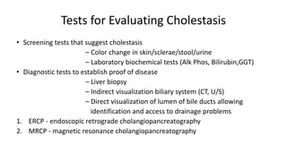
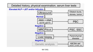
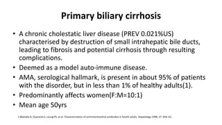
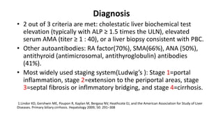
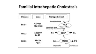
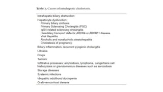
MANAGEMENT OF CHOLESTATIC
SYNDROMES
Gautam nath
Cholestasis
• Cholestatic liver disorders are caused by genetic defects,
mechanical aberrations, toxins, or dysregulations in the
immune system that damage the bile ducts and cause
accumulation of bile and liver tissue damage.
• Injuries to bile ducts or hepatocytes can lead to a range of
clinical presentations, from isolated abnormalities in liver
biochemistry, to liver failure or hepatobiliary malignancy;
congenital, immunologic, structural (obstructive/vascular), and
toxic factors can all contribute to disease.
Cell Biology
• Approximately 5% of cells in the liver are cholangiocytes; these
ciliated epithelial cells line the biliary tree, an intricate network
of interconnecting bile ducts that increase in diameter from the
ducts of Hering to the extrahepatic bile ducts.
• Cholangiocytes that line the large interlobular and major ducts
are predominantly involved in secretory functions, whereas that
line smaller bile duct branches, cholangioles, and ducts of
Hering have roles in inflammatory and proliferative
responses(1).
1.Alpini G, Glaser S, Robertson W, et al. Large but not small intrahepatic bile ducts are involved in secretin-regulated ductal
bile secretion. Am J Physiol 1997;272:G1064–G1074.
Cholestasis
• classically subdivided into 2 types
• Intrahepatic cholestasis: functional defect in bile formation at the level of
the hepatocyte,
• Extrahepatic cholestasis: obstruction to bile flow within the biliary tract.
Classification
INTRAHEPATIC:
• Primary biliary cirrhosis
• Primary sclerosing cholangitis
• IgG4-associated cholangitis
• Intrahepatic cholestasis of pregnancy
• Benign infiltrating disorders: e.g., amyloidosis, sarcoidosis and other
storage diseases.
• Drug-induced cholangiopathy
• Genetic disorders; PFIC, BRIC
• Paucity of bile ducts ; Alagille syndrome
• Non syndromic Paucity of bile ducts ; congenital infections(CMV, Rubella),
α1-antitrypsin deficiency, inborn errors of bile acid metabolism.
Classification
EXTRAHEPATIC:
• Lithiasis
• Tumours: Biliary, Pancreatic
• Biliary Atresia
• Ductal plate malform: Choledocal Cyst
• Spontaneous perforation of bile duct
• Bile plug syndrome
• Infection and inflammation
Tests for Evaluating Cholestasis
• Screening tests that suggest cholestasis
– Color change in skin/sclerae/stool/urine
– Laboratory biochemical tests (Alk Phos, Bilirubin,GGT)
• Diagnostic tests to establish proof of disease
– Liver biopsy
– Indirect visualization biliary system (CT, U/S)
– Direct visualization of lumen of bile ducts allowing
identification and access to drainage problems
1. ERCP - endoscopic retrograde cholangiopancreatography
2. MRCP - magnetic resonance cholangiopancreatography
Ref: EASL
Individual disorders
Primary biliary cirrhosis
• A chronic cholestatic liver disease (PREV 0.021%US)
characterised by destruction of small intrahepatic bile ducts,
leading to fibrosis and potential cirrhosis through resulting
complications.
• Deemed as a model auto-immune disease.
• AMA, serological hallmark, is present in about 95% of patients
with the disorder, but in less than 1% of healthy adults(1).
• Predominantly affects women(F:M=10:1)
• Mean age 50yrs
1.Mattalia A, Quaranta S, Leung PS, et al. Characterization of antimitochondrial antibodies in health adults. Hepatology 1998; 27: 656–61.
Diagnosis
• 2 out of 3 criteria are met: cholestatic liver biochemical test
elevation (typically with ALP ≥ 1.5 times the ULN), elevated
serum AMA (titer ≥ 1 : 40), or a liver biopsy consistent with PBC.
• Other autoantibodies: RA factor(70%), SMA(66%), ANA (50%),
antithyroid (antimicrosomal, antithyroglobulin) antibodies
(41%).
• Most widely used staging system(Ludwig’s ): Stage 1=portal
inflammation, stage 2=extension to the periportal areas, stage
3=septal fibrosis or inflmmatory bridging, and stage 4=cirrhosis.
1.Lindor KD, Gershwin ME, Poupon R, Kaplan M, Bergasa NV, Heathcote EJ, and the American Association for Study of Liver
Diseases. Primary biliary cirrhosis. Hepatology 2009; 50: 291–308
Treatment
UDCA
• Remains the only approved drug
• Has been shown to improve serum hepatic biochemistries, delay
histological progression, and delay the development of oesophageal
varices(1).
• Being a 7-β-epimer of the primary bile acid chenodeoxycholic acid.
UDCA inhibits intestinal absorption of bile acids, increases biliary
secretion of bile acids
• Besides having anti-inflammatory action, it stimulates secretion of a
bicarbonate-rich fluid from cholangiocytes, decreasing cholestasis.
• Augments micelle formation, decreasing the toxic effect of bile acids
to cell membranes(2).
1. Corpechot C, Carrat F, Bahr A, Chrétien Y, Poupon RE, Poupon R. The eff ect of ursodeoxycholic acid therapy on the natural course of primary biliary cirrhosis.
Gastroenterology 2005; 128: 297–303.
2. Poupon R. Ursodeoxycholic acid and bile-acid mimetics as therapeutic agents for cholestatic liver diseases: an overview of their mechanisms of action. Clin Res Hepatol
Gastroenterol 2012; 36 (suppl 1): S3–12.
Newer modalities
• Corticosteroids improved serum biochemistries and histology
but with substantial deterioration of bone mineral density,
precluding use as a long-term agent in patients already at risk
for osteopenia.
• Budesonide, in combination with UDCA, has shown biochemical
and histological improvement in small trials without the large
side-effect profile usually seen with corticosteroids(1).
• Drugs acting upon Nuclear receptor target, in the likes of
Obeticholic acid (Farnesoid X receptor), Bezafibrate/fenofibrate
(Peroxisome proliferator-activated receptor α), Vitamin D.
1. Zhu GQ, Shi KQ, Huang S, et al. Network meta-analysis of randomized controlled trials: effi cacy and safety of UDCA-based
therapies in primary biliary cirrhosis. Medicine (Baltimore) 2015; 94: e609.
Management Of Symptoms
• Bone Disease: supplemental calcium , vitamin D,
bisphosphonates, estrogen modulators (HRT, raloxifene)
• Fat-Soluble Vitamin Deficiency: Supplimentation of VITAMIN-
D,A,K,E
• Pruritus: Cholestyramine, Rifampin, UDCA ,opiate receptor
antagonist, Antihistamines
Liver Transplantation
• The best therapeutic alternative for patients with end-stage PBC.
• Indications for referral: Development of complications associated
with chronic cholestasis(disabling fatigue, intractable pruritus, and
severe muscle wasting, persistent increases in the serum bilirubin
level).
• One-year survival rates after liver transplantation are higher than
90%, with 5-year survival rates of 80% or higher in most transplant
centers (1).
• Universal use of UDCA has decreased the rate of Liver
transplantation(1).
1. Lee J, Belanger A, Doucette J, et al. Transplantation trends in primary biliary cirrhosis. Clin Gastroenterol Hepatol 2007; 5:1313-15.
Prognosis
• Response to UDCA is an important predictor of prognosis.
• Paris criteria (decrease in the serum ALP <3 X ULN and serum AST level
<2 X ULN plus a normal BIL) to use criteria after 6 months of UDCA(1).
• Modified Paris criteria (normal serum total bilirubin level and both an
ALP and AST <1.5 times the ULN at 12mo of UDCA)
• About 40% of patients will not have an adequate biochemical response
to UDCA, and these patients have a more rapid progression of disease
than those with normalisation of ALP(2).
• Patients without biochemical response had relative risk of 5·51 of death
or liver transplantation compared with those with a response(2).
1. Carbone M, Mells G, Pells G, et al. Sex and age are determinants of the clinical phenotype of primary biliary cirrhosis and response to ursodeoxycholic acid. Gastroenterology 2013;
144:560-9.
2. Parés A, Caballería L, Rodés J. Excellent long-term survival in patients with primary biliary cirrhosis and biochemical response to ursodeoxycholic Acid. Gastroenterology 2006; 130:
715–20
Sclerosing Cholangitis
• characterized by progressive, inflammatory, sclerosing, and
obliterative process affecting the extrahepatic and/or the
intrahepatic bile ducts.
Sclerosing Cholangitis
Primary (of unknown origin)
• Primary sclerosing cholangitis
(PSC)
• IgG4-related sclerosing cholangitis
(IgG4-SC)
Secondary Sclerosing Cholangitis
• Biliary lesion in PLHA
• CholangioCa
• CBD stone
• Post-op/bile duct injury
• Congenital biliary disorders
• Chemical agents/drug-induced
cholangitis
• Ischemic biliary stenosis
• Others
Primary Sclerosing Cholangitis
• Prevalence of 0.006% (US)
• Diagnosis of PSC is based on typical cholangiographic findings in the setting
of consistent clinical, biochemical, serologic, and histologic findings, as well
as exclusion of secondary causes of sclerosing cholangitis.
• Both the intrahepatic and extrahepatic bile ducts are involved in the
majority (up to 87%), although small percentage may have an isolated
lesion.
• ERCP is considered the standard for establishing a diagnosis but equivalent
sensitivity and specificity has been noted with MRCP(1)
1.Dave M, Elmunzer BJ, Dwamena BA, et al. Primary sclerosing cholangitis: Meta-analysis of
diagnostic performance of MR cholangiopancreatography. Radiology 2010; 256:387-96.
Association with IBD
• Approximately 90% of all patients with PSC have concomitant IBD
• Of patients with both PSC and IBD, approximately 85% to 90% have UC and
the remainder have Crohn’s colitis
• Association with IBD is stronger with more extensive colonic involvement;
the prevalence of PSC is approximately 5.5% in those with pancolitis, in
contrast to 0.5% in those with only distal colitis(1)
• Patients with concurrent IBD have a much earlier age of onset and much
higher rates of cancer, liver transplantation, and death than those who do
not(2)
1. Olsson R, Danielsson A, Jarnerot G, et al. Prevalence of primary sclerosing cholangitis in patients with ulcerative colitis. Gastroenterology 1991; 100:1319-23
2. Clin Gastroenterol Hepatol 2011; 9:1092-7.
Treatment
• No benefit to UDCA with regard to fatigue, pruritus, or development of
cholangiocarcinoma(1)
• Studies on use of high-dose (30 mg/kg/day) UDCA in the treatment of PSC,
has shown detrimental effects (2)
• Paucity of data against immune modulators, steroids, antifibrotic drugs.
• Symptomatic Mx of complications(pruritus, nutritional deficiencies, and
bacterial cholangitis)
1. Hepatology 2009; 50:808-14.
2. Aliment Pharmacol Ther 2011; 34:1185-92.
Endoscopic Management
• Patients most likely to benefit from endoscopic intervention are those with
one (or more) dominant stricture.
• Studies of endoscopic intervention in patients with PSC, however, have
typically been small, retrospective series and uncontrolled trials, and firm
conclusions are not available.
• Treatment mostly limited by complications of endotherapy along with
unsuitable patient profile.
Liver Transplantation
• Only therapy that has been shown conclusively to improve the natural
history.
• Indications: development of cirrhosis, portal HTN, recurrent cholangitis,
refractory pruritus(medical and endoscopic).
• Roux-en-Y choledochojejunostomy anastomosis, instead of a standard
choledochocholedochostomy, to prevent recurrent strictures and
cholangiocarcinoma.
• Preoperative chemoradiation in highly selected patients with
cholangiocarcinoma has shown promise in reducing the rate of tumor
recurrence after liver transplantation(1)
1.Am J Transplant 2002; 2:774-9.
IgG4-SC
• A type of Sclerosing Cholangitis
• Frequently associated with Autoimmune Pancreatitis (AIP)(1)
• Affects mostly elderly men.
• Mean age: 69.3 yrs(2)
• Sex: M/F=3.3(2)
2. J Hepatobiliary Pancreat Sci 2014;21:43–50
1. Yoshida et al. Chronic pancreatitis caused by an autoimmune abnormality. Proposal of the concept of autoimmune pancreatitis. Dig Dis Sci 1995;40:1561–1568
Diagnosis
• Difficult to discriminate IgG4-SC from these progressive or
malignant diseases on the basis of cholangiographic findings
alone
• Accurate diagnosis of IgG4-SC not associated with autoimmune
pancreatitis is particularly difficult
• Mostly relies upon histology, imaging, serology, other organ
involvments.
Treatment
• NO RCTs
• Early introduction of steroid therapy (Prednisolone: 0.5 to 0.6
mg/kg)(1)
• Rituximab: Few case reports have shown benefits in Steroid
non responsive IgG4-SC
1.Ghazale A et al. Immunoglobulin G4-associated cholangitis: clinical profile and response to therapy.
Gastroenterology. 2008;134:706–715.
PSC Vs IgG4-SC
PSC IgG4-SC
Age 40s or younger 50s or older
Association IBD Autoimmune pancreatitis
CC risk High Low
ERCP Multifocal stricturing Lower CBD intra-pancreatic, or
hilar stricture (tumour like).
Multifocal stricturing may occur
but less than PSC.
LAB pANCA Raised IgG4 level
Biopsy Not essential. Ductopenia. Diagnostic. Plasma cell
infiltration.
Therapy No specific therapy Steroids
Intrahepatic Cholestasis Of Pregnancy
• Most commonly seen in the third trimester, but can begin at any time
• Pruritus being the most common feature while other signs of
cholestasis are occaisional.
• Etiologies are still not fully understood, hormones and genetic factors
(ABCB4) are likely to be important in the pathogenesis of the
disease(1).
• Resolution following delivery(<6wks), chance of recurence.
1.Lammert, Frank et al. Journal of Hepatology , Volume 33 , Issue 6 , 1012 - 1021
Treatment
• Symptomatic treatment for pruritus.
• Vitamin k may help avoid the risk of hemorrhage at delivery, in case of
coagulopathy.
• A combination therapy of Sadenosyl-L-methionine (SAMe) and UDCA may
have a synergistic effect(1)
• Delivery by 35–37 completed weeks may be important to fetal outcome as
recent studies has demonstrated that in severe ICP (defined as bile acids
greater than 2.5mg%) the risk of stillbirth was 1.5% compared to 0.5% of
uncomplicated pregnancies(1).
1. Geenes V et al Association of severe intrahepatic cholestasis of pregnancy with adverse pregnancy outcomes: a prospective population-based case-control
study. Hepatology. 2014;59(4):1482-91.
Infiltrative/Granulomatous Diseases
• Often present with cholestasis:
elevated alkaline phosphatase
with or without jaundice
• Due to compression of small intrahepatic bile ducts by expanding
granulomas/infiltration.
• Examples: SARCOIDOSIS, TUBERCULOSIS, GRANULOMATOSIS WITH
POLYANGITIS
MANAGMENT
• Treatment of the primary etiology.
• Symptomatic treatment for the complications.
Drug-Induced Cholestasis
• The spectrum can range from acute reversible cholestasis to
chronic cholestasis with loss of bile ducts.
• Can be categorized into acute and chronic forms.
Drug-Induced Cholestasis (Acute)
Drug-Induced Cholestasis (Chronic)
• Vanishing Bile Duct Syndrome: a long list of drugs, most known
ones are: Prochlorperazine, Flucloxacillin, Amoxicillin-clavulanic
Acid, Azathioprine, Ibuprofen, Sulfonamides, Clindamycin,
Cotrimoxazole, Amitriptyline.
• Sclerosing Cholangitis-like Syndrome: Intralesional and
scolicidal agents such as Formaldehyde, Hypertonic saline and
Absolute alcohol.
Drug-Induced Cholestasis (Chronic)
• Symptoms can occur weeks to months after beginning
treatment.
• Often are accompanied by additional clinical features, such as
fever, chills n rigors, jaundice.
• Drugs that result in a vanishing bile duct syndrome can lead to
progressive cholestasis, with prolonged jaundice, pruritus, and,
occasionally, cirrhosis and liver failure
Management
• Careful medical history
• Mainstay of treatment is withdrawal of the drug. Most cholestatic hepatic
injury resolves with withdrawal of the offending medication.
CHOLESTASIS IN PAEDIATRIC POPULATION
33%
36%
3%
6%
4%
18%
INDIAN SCENARIO
UNKNOWN
B.ATRESIA
DUCTAL PAUCITY
UNDIFFERENTIATED
CHOLEDOCHAL CYST
INFECTIONS
REF: Bhatia et al. Indian Pediatr 2014
Biliary Atresia
• Complete obstruction of bile flow as a result of the destruction or absence of all or a
portion of the extrahepatic bile ducts.
• Incidence: 1 in 10,000 to 15,000 live births (1)
• Accounts for one third of cases of neonatal cholestasis (both worldwide (1) & India
(2))
• Most frequent cause of death from liver disease in newborns(3)
• Major reason (≈50% of all cases) for liver transplantation in children(3)
1. Balisteri et al. Semin Liver Dis 1987
2. Bhatia et al. Indian Pediatr 2014
3. Sokol et al. J Pediatr Gastroenterol Nutr 2003
• Most infants have a normal birth weight with at term delivery
• Female infants are affected more commonly
• Normal perinatal development with normal postnatal weight gain
• Prolonged jaundice with dark urine
• Acholic stools
•Sensitivity: 89.7%
•Specificity: 99.9%
Ref: Hsiao et al. Hepatology 2008
Treatment
• Exploratory laparotomy and operative cholangiography are necessary to
document the site of obstruction and direct attempts at surgical treatment
• Patent proximal portions of the bile ducts or cystic structures in the porta
hepatis allow conventional anastomosis with a segment of bowel in
approximately 10% of patients
Hepatoportoenterostomy (PE):
Kasai Procedure
• Removal of the atretic extrahepatic tissue and
• Roux-en-Y jejunal loop anastomosis to the hepatic hilum
• half the patients normalize their bilirubin after Kasai’s PE if performed within six
months
• About 20% of all patients undergoing Kasai’s PE during infancy survive into adulthood
with their native liver(1,2)
1. Sokol et al. Hepatology. 2007
2. Bassett et al. Clin Gastroenterol. 2008
Liver transplantation
• Essential in the management of children in whom
– portoenterostomy does not successfully restore bile flow
– in whom referral is late (≥ 120 days)
• Biliary atresia accounts for 40-50% of all liver transplants performed in
children
• 1-year survival rates > 90%
Choledochal Cyst
• Congenital anomalies of the biliary tract that are manifested by cystic dilatation
of the extrahepatic and intrahepatic bile ducts.
• Accounts for 4% of total neonatal cholestasis in India
• Female children are affected more commonly
• Often appears during the first months of life
• 80% present with
– cholestatic jaundice
– acholic stools
• Vomiting
• Irritability may occur
• Failure to thrive
Classification (Todani)
• Type I cysts accounting for 80% to 90% of cases, exhibit segmental or
diffuse fusiform dilatation of the bile duct. Ia-diffuse fusiform(entire
length CBD), Ib-focal fusiform, Ic-cylindrical CBD dilataion.
• Type II cysts consist of a true choledochal diverticulum.
• Type III cysts consist of dilatation of the intraduodenal portion of the bile
duct, or choledochocele.
• Type IV cysts may be subdivided into
– type IVa, or multiple intrahepatic and extrahepatic cysts,
– Type IVb, or multiple extrahepatic cysts.
• Type V, or Caroli’s disease: single or multiple dilatations of the intrahepatic
ductal system, should be viewed as a form of choledochal cyst
Treatment
• Surgical excision of the cyst with reconstruction of the extrahepatic biliary
tree.
• Biliary drainage is usually accomplished by a choledochojejunostomy with a
Roux-en-Y anastomosis
• Excision of the cyst reduces bile stasis and the risk of cholangitis and
cholangiocarcinoma
Alagille Syndrome or
Arteriohepatic Dysplasia
• Most common form of familial intrahepatic cholestasis
• Paucity of interlobular bile ducts is the hallmark of this condition
• Decreased ratio of the numbers of interlobular portal bile ducts to portal
tracts (<0.4)(1)
• Mutations in the jagged1 (JAG1) gene have been identified in 94% of
cases(2)
• Mutations in the gene encoding the NOTCH2 receptor who were negative
for JAG1 mutations (3)
1. Oda et al. Nat Genet 1997
2. McDaniell et al. Am J Hum Genet 2006
Dysmorphic facies
–Broad forehead
–Widely spaced & deeply set eyes
–Small and pointed mandible
–Flattened malar eminence
–Prominent ears
Treatment
• Adequate caloric intake
• Preventing or correcting fat-soluble vitamin deficiencies
• Symptomatic measures to relieve pruritus
• Partial external biliary diversion may be effective for treating severe pruritus
and hypercholesterolemia
• Liver Transplatation:
– 1- and 5-year survival rates were significantly lower in patients than biliary atresia
(82.9% and 78.4% vs. 89.9% and 84%).
Spontaneous Perforation of the Bile Duct
• Rare but distinct cause of Cholestasis in neonatal group of patients
• Perforation usually occurs at the junction of the cystic and bile ducts
Suggested Causes:
• Congenital weakness at the site of the perforation
• injury produced by infection.
• jaundice, acholic stools, dark urine, and FTT being the most common
manifestations.
Management
• Operative cholangiography is required to demonstrate the site of the
perforation
• Surgical treatment may involve simple drainage of the bilious ascites and
repair of the site of the perforation
• If the perforation is associated with obstruction of the bile duct, drainage via
a cholecystojejunostomy may be required
Familial Intrahepatic Cholestasis
PFIC-1 & BRIC-1
Defective gene: FIC1 (ATP8B1)
–Recurrent Cholestasis
–Extrahepatic Manifestation:
• diarrhea, pancreatitis,
• Respiratory symptoms & hearing loss
– Normal GGT
Mx
–Supplementation with fat-soluble vitamins,
–External biliary diversion
–PFIC1Liver Transplantation
PFIC-2 & BRIC-2
• Chromosome 2q24 defective gene: BSEP
(ABCB11)
– ↑ bile acid levels
– Intractable pruritus
– Intestinal malabsorption
– Failure to thrive & cholestasis
– Fibrosis  End-stage Liver Disease
• Normal GGT
• Mx Liver Transplantation
PFIC-3
• Defective gene: MDR3 (ABCB4)
– jaundice, pale stools
– Pruritus & hepatomegaly
– Fibrosis  End-stage Liver Disease
• ↑↑ GGT
• Mx Liver Transplantation
Thank you
Cholestatic syndromes

Cholestatic syndromes

  • 1.
  • 2.
    Cholestasis • Cholestatic liverdisorders are caused by genetic defects, mechanical aberrations, toxins, or dysregulations in the immune system that damage the bile ducts and cause accumulation of bile and liver tissue damage. • Injuries to bile ducts or hepatocytes can lead to a range of clinical presentations, from isolated abnormalities in liver biochemistry, to liver failure or hepatobiliary malignancy; congenital, immunologic, structural (obstructive/vascular), and toxic factors can all contribute to disease.
  • 3.
    Cell Biology • Approximately5% of cells in the liver are cholangiocytes; these ciliated epithelial cells line the biliary tree, an intricate network of interconnecting bile ducts that increase in diameter from the ducts of Hering to the extrahepatic bile ducts. • Cholangiocytes that line the large interlobular and major ducts are predominantly involved in secretory functions, whereas that line smaller bile duct branches, cholangioles, and ducts of Hering have roles in inflammatory and proliferative responses(1). 1.Alpini G, Glaser S, Robertson W, et al. Large but not small intrahepatic bile ducts are involved in secretin-regulated ductal bile secretion. Am J Physiol 1997;272:G1064–G1074.
  • 4.
    Cholestasis • classically subdividedinto 2 types • Intrahepatic cholestasis: functional defect in bile formation at the level of the hepatocyte, • Extrahepatic cholestasis: obstruction to bile flow within the biliary tract.
  • 5.
    Classification INTRAHEPATIC: • Primary biliarycirrhosis • Primary sclerosing cholangitis • IgG4-associated cholangitis • Intrahepatic cholestasis of pregnancy • Benign infiltrating disorders: e.g., amyloidosis, sarcoidosis and other storage diseases. • Drug-induced cholangiopathy • Genetic disorders; PFIC, BRIC • Paucity of bile ducts ; Alagille syndrome • Non syndromic Paucity of bile ducts ; congenital infections(CMV, Rubella), α1-antitrypsin deficiency, inborn errors of bile acid metabolism.
  • 6.
    Classification EXTRAHEPATIC: • Lithiasis • Tumours:Biliary, Pancreatic • Biliary Atresia • Ductal plate malform: Choledocal Cyst • Spontaneous perforation of bile duct • Bile plug syndrome • Infection and inflammation
  • 7.
    Tests for EvaluatingCholestasis • Screening tests that suggest cholestasis – Color change in skin/sclerae/stool/urine – Laboratory biochemical tests (Alk Phos, Bilirubin,GGT) • Diagnostic tests to establish proof of disease – Liver biopsy – Indirect visualization biliary system (CT, U/S) – Direct visualization of lumen of bile ducts allowing identification and access to drainage problems 1. ERCP - endoscopic retrograde cholangiopancreatography 2. MRCP - magnetic resonance cholangiopancreatography
  • 8.
  • 9.
  • 10.
    Primary biliary cirrhosis •A chronic cholestatic liver disease (PREV 0.021%US) characterised by destruction of small intrahepatic bile ducts, leading to fibrosis and potential cirrhosis through resulting complications. • Deemed as a model auto-immune disease. • AMA, serological hallmark, is present in about 95% of patients with the disorder, but in less than 1% of healthy adults(1). • Predominantly affects women(F:M=10:1) • Mean age 50yrs 1.Mattalia A, Quaranta S, Leung PS, et al. Characterization of antimitochondrial antibodies in health adults. Hepatology 1998; 27: 656–61.
  • 11.
    Diagnosis • 2 outof 3 criteria are met: cholestatic liver biochemical test elevation (typically with ALP ≥ 1.5 times the ULN), elevated serum AMA (titer ≥ 1 : 40), or a liver biopsy consistent with PBC. • Other autoantibodies: RA factor(70%), SMA(66%), ANA (50%), antithyroid (antimicrosomal, antithyroglobulin) antibodies (41%). • Most widely used staging system(Ludwig’s ): Stage 1=portal inflammation, stage 2=extension to the periportal areas, stage 3=septal fibrosis or inflmmatory bridging, and stage 4=cirrhosis. 1.Lindor KD, Gershwin ME, Poupon R, Kaplan M, Bergasa NV, Heathcote EJ, and the American Association for Study of Liver Diseases. Primary biliary cirrhosis. Hepatology 2009; 50: 291–308
  • 12.
  • 13.
    UDCA • Remains theonly approved drug • Has been shown to improve serum hepatic biochemistries, delay histological progression, and delay the development of oesophageal varices(1). • Being a 7-β-epimer of the primary bile acid chenodeoxycholic acid. UDCA inhibits intestinal absorption of bile acids, increases biliary secretion of bile acids • Besides having anti-inflammatory action, it stimulates secretion of a bicarbonate-rich fluid from cholangiocytes, decreasing cholestasis. • Augments micelle formation, decreasing the toxic effect of bile acids to cell membranes(2). 1. Corpechot C, Carrat F, Bahr A, Chrétien Y, Poupon RE, Poupon R. The eff ect of ursodeoxycholic acid therapy on the natural course of primary biliary cirrhosis. Gastroenterology 2005; 128: 297–303. 2. Poupon R. Ursodeoxycholic acid and bile-acid mimetics as therapeutic agents for cholestatic liver diseases: an overview of their mechanisms of action. Clin Res Hepatol Gastroenterol 2012; 36 (suppl 1): S3–12.
  • 14.
    Newer modalities • Corticosteroidsimproved serum biochemistries and histology but with substantial deterioration of bone mineral density, precluding use as a long-term agent in patients already at risk for osteopenia. • Budesonide, in combination with UDCA, has shown biochemical and histological improvement in small trials without the large side-effect profile usually seen with corticosteroids(1). • Drugs acting upon Nuclear receptor target, in the likes of Obeticholic acid (Farnesoid X receptor), Bezafibrate/fenofibrate (Peroxisome proliferator-activated receptor α), Vitamin D. 1. Zhu GQ, Shi KQ, Huang S, et al. Network meta-analysis of randomized controlled trials: effi cacy and safety of UDCA-based therapies in primary biliary cirrhosis. Medicine (Baltimore) 2015; 94: e609.
  • 15.
    Management Of Symptoms •Bone Disease: supplemental calcium , vitamin D, bisphosphonates, estrogen modulators (HRT, raloxifene) • Fat-Soluble Vitamin Deficiency: Supplimentation of VITAMIN- D,A,K,E • Pruritus: Cholestyramine, Rifampin, UDCA ,opiate receptor antagonist, Antihistamines
  • 16.
    Liver Transplantation • Thebest therapeutic alternative for patients with end-stage PBC. • Indications for referral: Development of complications associated with chronic cholestasis(disabling fatigue, intractable pruritus, and severe muscle wasting, persistent increases in the serum bilirubin level). • One-year survival rates after liver transplantation are higher than 90%, with 5-year survival rates of 80% or higher in most transplant centers (1). • Universal use of UDCA has decreased the rate of Liver transplantation(1). 1. Lee J, Belanger A, Doucette J, et al. Transplantation trends in primary biliary cirrhosis. Clin Gastroenterol Hepatol 2007; 5:1313-15.
  • 17.
    Prognosis • Response toUDCA is an important predictor of prognosis. • Paris criteria (decrease in the serum ALP <3 X ULN and serum AST level <2 X ULN plus a normal BIL) to use criteria after 6 months of UDCA(1). • Modified Paris criteria (normal serum total bilirubin level and both an ALP and AST <1.5 times the ULN at 12mo of UDCA) • About 40% of patients will not have an adequate biochemical response to UDCA, and these patients have a more rapid progression of disease than those with normalisation of ALP(2). • Patients without biochemical response had relative risk of 5·51 of death or liver transplantation compared with those with a response(2). 1. Carbone M, Mells G, Pells G, et al. Sex and age are determinants of the clinical phenotype of primary biliary cirrhosis and response to ursodeoxycholic acid. Gastroenterology 2013; 144:560-9. 2. Parés A, Caballería L, Rodés J. Excellent long-term survival in patients with primary biliary cirrhosis and biochemical response to ursodeoxycholic Acid. Gastroenterology 2006; 130: 715–20
  • 18.
    Sclerosing Cholangitis • characterizedby progressive, inflammatory, sclerosing, and obliterative process affecting the extrahepatic and/or the intrahepatic bile ducts.
  • 19.
    Sclerosing Cholangitis Primary (ofunknown origin) • Primary sclerosing cholangitis (PSC) • IgG4-related sclerosing cholangitis (IgG4-SC) Secondary Sclerosing Cholangitis • Biliary lesion in PLHA • CholangioCa • CBD stone • Post-op/bile duct injury • Congenital biliary disorders • Chemical agents/drug-induced cholangitis • Ischemic biliary stenosis • Others
  • 20.
    Primary Sclerosing Cholangitis •Prevalence of 0.006% (US) • Diagnosis of PSC is based on typical cholangiographic findings in the setting of consistent clinical, biochemical, serologic, and histologic findings, as well as exclusion of secondary causes of sclerosing cholangitis. • Both the intrahepatic and extrahepatic bile ducts are involved in the majority (up to 87%), although small percentage may have an isolated lesion. • ERCP is considered the standard for establishing a diagnosis but equivalent sensitivity and specificity has been noted with MRCP(1) 1.Dave M, Elmunzer BJ, Dwamena BA, et al. Primary sclerosing cholangitis: Meta-analysis of diagnostic performance of MR cholangiopancreatography. Radiology 2010; 256:387-96.
  • 21.
    Association with IBD •Approximately 90% of all patients with PSC have concomitant IBD • Of patients with both PSC and IBD, approximately 85% to 90% have UC and the remainder have Crohn’s colitis • Association with IBD is stronger with more extensive colonic involvement; the prevalence of PSC is approximately 5.5% in those with pancolitis, in contrast to 0.5% in those with only distal colitis(1) • Patients with concurrent IBD have a much earlier age of onset and much higher rates of cancer, liver transplantation, and death than those who do not(2) 1. Olsson R, Danielsson A, Jarnerot G, et al. Prevalence of primary sclerosing cholangitis in patients with ulcerative colitis. Gastroenterology 1991; 100:1319-23 2. Clin Gastroenterol Hepatol 2011; 9:1092-7.
  • 22.
    Treatment • No benefitto UDCA with regard to fatigue, pruritus, or development of cholangiocarcinoma(1) • Studies on use of high-dose (30 mg/kg/day) UDCA in the treatment of PSC, has shown detrimental effects (2) • Paucity of data against immune modulators, steroids, antifibrotic drugs. • Symptomatic Mx of complications(pruritus, nutritional deficiencies, and bacterial cholangitis) 1. Hepatology 2009; 50:808-14. 2. Aliment Pharmacol Ther 2011; 34:1185-92.
  • 23.
    Endoscopic Management • Patientsmost likely to benefit from endoscopic intervention are those with one (or more) dominant stricture. • Studies of endoscopic intervention in patients with PSC, however, have typically been small, retrospective series and uncontrolled trials, and firm conclusions are not available. • Treatment mostly limited by complications of endotherapy along with unsuitable patient profile.
  • 24.
    Liver Transplantation • Onlytherapy that has been shown conclusively to improve the natural history. • Indications: development of cirrhosis, portal HTN, recurrent cholangitis, refractory pruritus(medical and endoscopic). • Roux-en-Y choledochojejunostomy anastomosis, instead of a standard choledochocholedochostomy, to prevent recurrent strictures and cholangiocarcinoma. • Preoperative chemoradiation in highly selected patients with cholangiocarcinoma has shown promise in reducing the rate of tumor recurrence after liver transplantation(1) 1.Am J Transplant 2002; 2:774-9.
  • 25.
    IgG4-SC • A typeof Sclerosing Cholangitis • Frequently associated with Autoimmune Pancreatitis (AIP)(1) • Affects mostly elderly men. • Mean age: 69.3 yrs(2) • Sex: M/F=3.3(2) 2. J Hepatobiliary Pancreat Sci 2014;21:43–50 1. Yoshida et al. Chronic pancreatitis caused by an autoimmune abnormality. Proposal of the concept of autoimmune pancreatitis. Dig Dis Sci 1995;40:1561–1568
  • 26.
    Diagnosis • Difficult todiscriminate IgG4-SC from these progressive or malignant diseases on the basis of cholangiographic findings alone • Accurate diagnosis of IgG4-SC not associated with autoimmune pancreatitis is particularly difficult • Mostly relies upon histology, imaging, serology, other organ involvments.
  • 27.
    Treatment • NO RCTs •Early introduction of steroid therapy (Prednisolone: 0.5 to 0.6 mg/kg)(1) • Rituximab: Few case reports have shown benefits in Steroid non responsive IgG4-SC 1.Ghazale A et al. Immunoglobulin G4-associated cholangitis: clinical profile and response to therapy. Gastroenterology. 2008;134:706–715.
  • 28.
    PSC Vs IgG4-SC PSCIgG4-SC Age 40s or younger 50s or older Association IBD Autoimmune pancreatitis CC risk High Low ERCP Multifocal stricturing Lower CBD intra-pancreatic, or hilar stricture (tumour like). Multifocal stricturing may occur but less than PSC. LAB pANCA Raised IgG4 level Biopsy Not essential. Ductopenia. Diagnostic. Plasma cell infiltration. Therapy No specific therapy Steroids
  • 29.
    Intrahepatic Cholestasis OfPregnancy • Most commonly seen in the third trimester, but can begin at any time • Pruritus being the most common feature while other signs of cholestasis are occaisional. • Etiologies are still not fully understood, hormones and genetic factors (ABCB4) are likely to be important in the pathogenesis of the disease(1). • Resolution following delivery(<6wks), chance of recurence. 1.Lammert, Frank et al. Journal of Hepatology , Volume 33 , Issue 6 , 1012 - 1021
  • 30.
    Treatment • Symptomatic treatmentfor pruritus. • Vitamin k may help avoid the risk of hemorrhage at delivery, in case of coagulopathy. • A combination therapy of Sadenosyl-L-methionine (SAMe) and UDCA may have a synergistic effect(1) • Delivery by 35–37 completed weeks may be important to fetal outcome as recent studies has demonstrated that in severe ICP (defined as bile acids greater than 2.5mg%) the risk of stillbirth was 1.5% compared to 0.5% of uncomplicated pregnancies(1). 1. Geenes V et al Association of severe intrahepatic cholestasis of pregnancy with adverse pregnancy outcomes: a prospective population-based case-control study. Hepatology. 2014;59(4):1482-91.
  • 31.
    Infiltrative/Granulomatous Diseases • Oftenpresent with cholestasis: elevated alkaline phosphatase with or without jaundice • Due to compression of small intrahepatic bile ducts by expanding granulomas/infiltration. • Examples: SARCOIDOSIS, TUBERCULOSIS, GRANULOMATOSIS WITH POLYANGITIS
  • 32.
    MANAGMENT • Treatment ofthe primary etiology. • Symptomatic treatment for the complications.
  • 33.
    Drug-Induced Cholestasis • Thespectrum can range from acute reversible cholestasis to chronic cholestasis with loss of bile ducts. • Can be categorized into acute and chronic forms.
  • 34.
  • 35.
    Drug-Induced Cholestasis (Chronic) •Vanishing Bile Duct Syndrome: a long list of drugs, most known ones are: Prochlorperazine, Flucloxacillin, Amoxicillin-clavulanic Acid, Azathioprine, Ibuprofen, Sulfonamides, Clindamycin, Cotrimoxazole, Amitriptyline. • Sclerosing Cholangitis-like Syndrome: Intralesional and scolicidal agents such as Formaldehyde, Hypertonic saline and Absolute alcohol.
  • 36.
    Drug-Induced Cholestasis (Chronic) •Symptoms can occur weeks to months after beginning treatment. • Often are accompanied by additional clinical features, such as fever, chills n rigors, jaundice. • Drugs that result in a vanishing bile duct syndrome can lead to progressive cholestasis, with prolonged jaundice, pruritus, and, occasionally, cirrhosis and liver failure
  • 37.
    Management • Careful medicalhistory • Mainstay of treatment is withdrawal of the drug. Most cholestatic hepatic injury resolves with withdrawal of the offending medication.
  • 38.
  • 39.
  • 40.
    Biliary Atresia • Completeobstruction of bile flow as a result of the destruction or absence of all or a portion of the extrahepatic bile ducts. • Incidence: 1 in 10,000 to 15,000 live births (1) • Accounts for one third of cases of neonatal cholestasis (both worldwide (1) & India (2)) • Most frequent cause of death from liver disease in newborns(3) • Major reason (≈50% of all cases) for liver transplantation in children(3) 1. Balisteri et al. Semin Liver Dis 1987 2. Bhatia et al. Indian Pediatr 2014 3. Sokol et al. J Pediatr Gastroenterol Nutr 2003
  • 41.
    • Most infantshave a normal birth weight with at term delivery • Female infants are affected more commonly • Normal perinatal development with normal postnatal weight gain • Prolonged jaundice with dark urine • Acholic stools
  • 42.
  • 43.
    Treatment • Exploratory laparotomyand operative cholangiography are necessary to document the site of obstruction and direct attempts at surgical treatment • Patent proximal portions of the bile ducts or cystic structures in the porta hepatis allow conventional anastomosis with a segment of bowel in approximately 10% of patients
  • 44.
    Hepatoportoenterostomy (PE): Kasai Procedure •Removal of the atretic extrahepatic tissue and • Roux-en-Y jejunal loop anastomosis to the hepatic hilum • half the patients normalize their bilirubin after Kasai’s PE if performed within six months • About 20% of all patients undergoing Kasai’s PE during infancy survive into adulthood with their native liver(1,2) 1. Sokol et al. Hepatology. 2007 2. Bassett et al. Clin Gastroenterol. 2008
  • 45.
    Liver transplantation • Essentialin the management of children in whom – portoenterostomy does not successfully restore bile flow – in whom referral is late (≥ 120 days) • Biliary atresia accounts for 40-50% of all liver transplants performed in children • 1-year survival rates > 90%
  • 46.
    Choledochal Cyst • Congenitalanomalies of the biliary tract that are manifested by cystic dilatation of the extrahepatic and intrahepatic bile ducts. • Accounts for 4% of total neonatal cholestasis in India • Female children are affected more commonly • Often appears during the first months of life • 80% present with – cholestatic jaundice – acholic stools • Vomiting • Irritability may occur • Failure to thrive
  • 47.
    Classification (Todani) • TypeI cysts accounting for 80% to 90% of cases, exhibit segmental or diffuse fusiform dilatation of the bile duct. Ia-diffuse fusiform(entire length CBD), Ib-focal fusiform, Ic-cylindrical CBD dilataion. • Type II cysts consist of a true choledochal diverticulum. • Type III cysts consist of dilatation of the intraduodenal portion of the bile duct, or choledochocele. • Type IV cysts may be subdivided into – type IVa, or multiple intrahepatic and extrahepatic cysts, – Type IVb, or multiple extrahepatic cysts. • Type V, or Caroli’s disease: single or multiple dilatations of the intrahepatic ductal system, should be viewed as a form of choledochal cyst
  • 48.
    Treatment • Surgical excisionof the cyst with reconstruction of the extrahepatic biliary tree. • Biliary drainage is usually accomplished by a choledochojejunostomy with a Roux-en-Y anastomosis • Excision of the cyst reduces bile stasis and the risk of cholangitis and cholangiocarcinoma
  • 49.
    Alagille Syndrome or ArteriohepaticDysplasia • Most common form of familial intrahepatic cholestasis • Paucity of interlobular bile ducts is the hallmark of this condition • Decreased ratio of the numbers of interlobular portal bile ducts to portal tracts (<0.4)(1) • Mutations in the jagged1 (JAG1) gene have been identified in 94% of cases(2) • Mutations in the gene encoding the NOTCH2 receptor who were negative for JAG1 mutations (3) 1. Oda et al. Nat Genet 1997 2. McDaniell et al. Am J Hum Genet 2006
  • 50.
    Dysmorphic facies –Broad forehead –Widelyspaced & deeply set eyes –Small and pointed mandible –Flattened malar eminence –Prominent ears
  • 51.
    Treatment • Adequate caloricintake • Preventing or correcting fat-soluble vitamin deficiencies • Symptomatic measures to relieve pruritus • Partial external biliary diversion may be effective for treating severe pruritus and hypercholesterolemia • Liver Transplatation: – 1- and 5-year survival rates were significantly lower in patients than biliary atresia (82.9% and 78.4% vs. 89.9% and 84%).
  • 52.
    Spontaneous Perforation ofthe Bile Duct • Rare but distinct cause of Cholestasis in neonatal group of patients • Perforation usually occurs at the junction of the cystic and bile ducts Suggested Causes: • Congenital weakness at the site of the perforation • injury produced by infection. • jaundice, acholic stools, dark urine, and FTT being the most common manifestations.
  • 53.
    Management • Operative cholangiographyis required to demonstrate the site of the perforation • Surgical treatment may involve simple drainage of the bilious ascites and repair of the site of the perforation • If the perforation is associated with obstruction of the bile duct, drainage via a cholecystojejunostomy may be required
  • 54.
  • 55.
    PFIC-1 & BRIC-1 Defectivegene: FIC1 (ATP8B1) –Recurrent Cholestasis –Extrahepatic Manifestation: • diarrhea, pancreatitis, • Respiratory symptoms & hearing loss – Normal GGT Mx –Supplementation with fat-soluble vitamins, –External biliary diversion –PFIC1Liver Transplantation
  • 56.
    PFIC-2 & BRIC-2 •Chromosome 2q24 defective gene: BSEP (ABCB11) – ↑ bile acid levels – Intractable pruritus – Intestinal malabsorption – Failure to thrive & cholestasis – Fibrosis  End-stage Liver Disease • Normal GGT • Mx Liver Transplantation
  • 57.
    PFIC-3 • Defective gene:MDR3 (ABCB4) – jaundice, pale stools – Pruritus & hepatomegaly – Fibrosis  End-stage Liver Disease • ↑↑ GGT • Mx Liver Transplantation
  • 62.