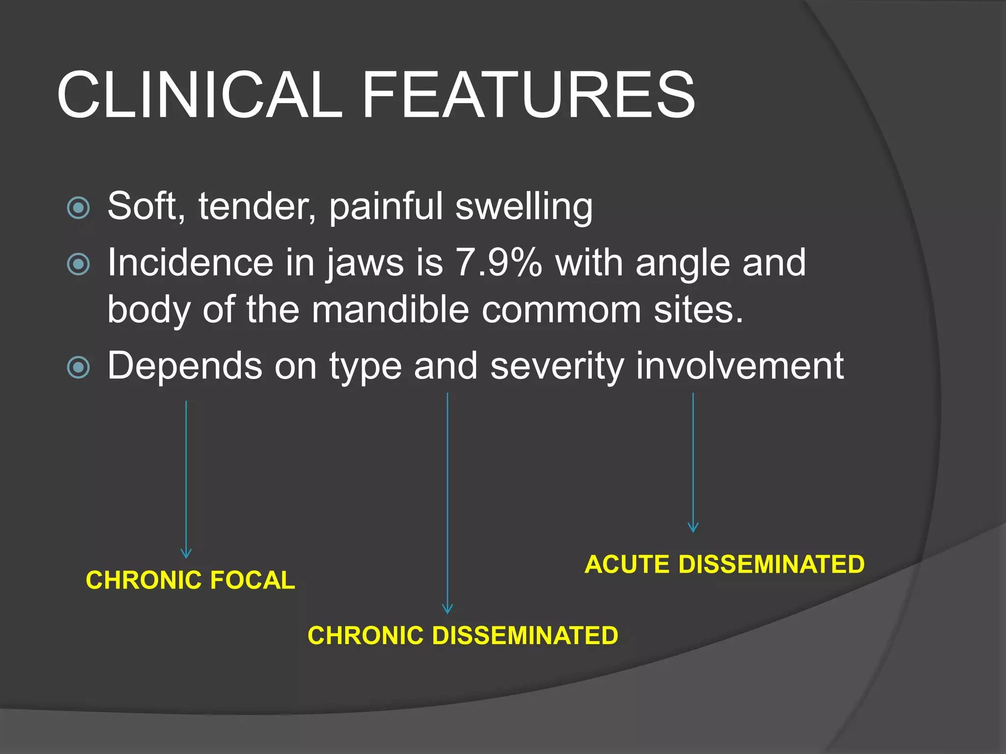
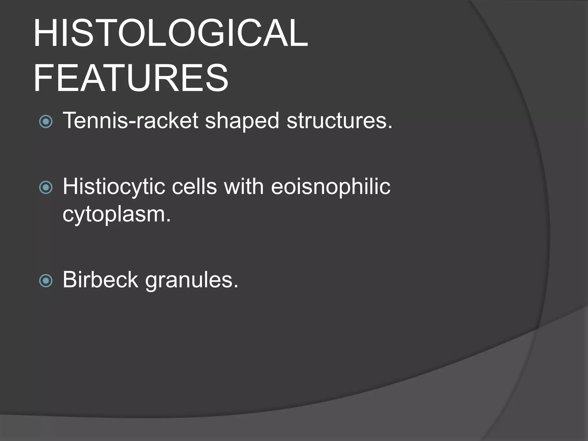
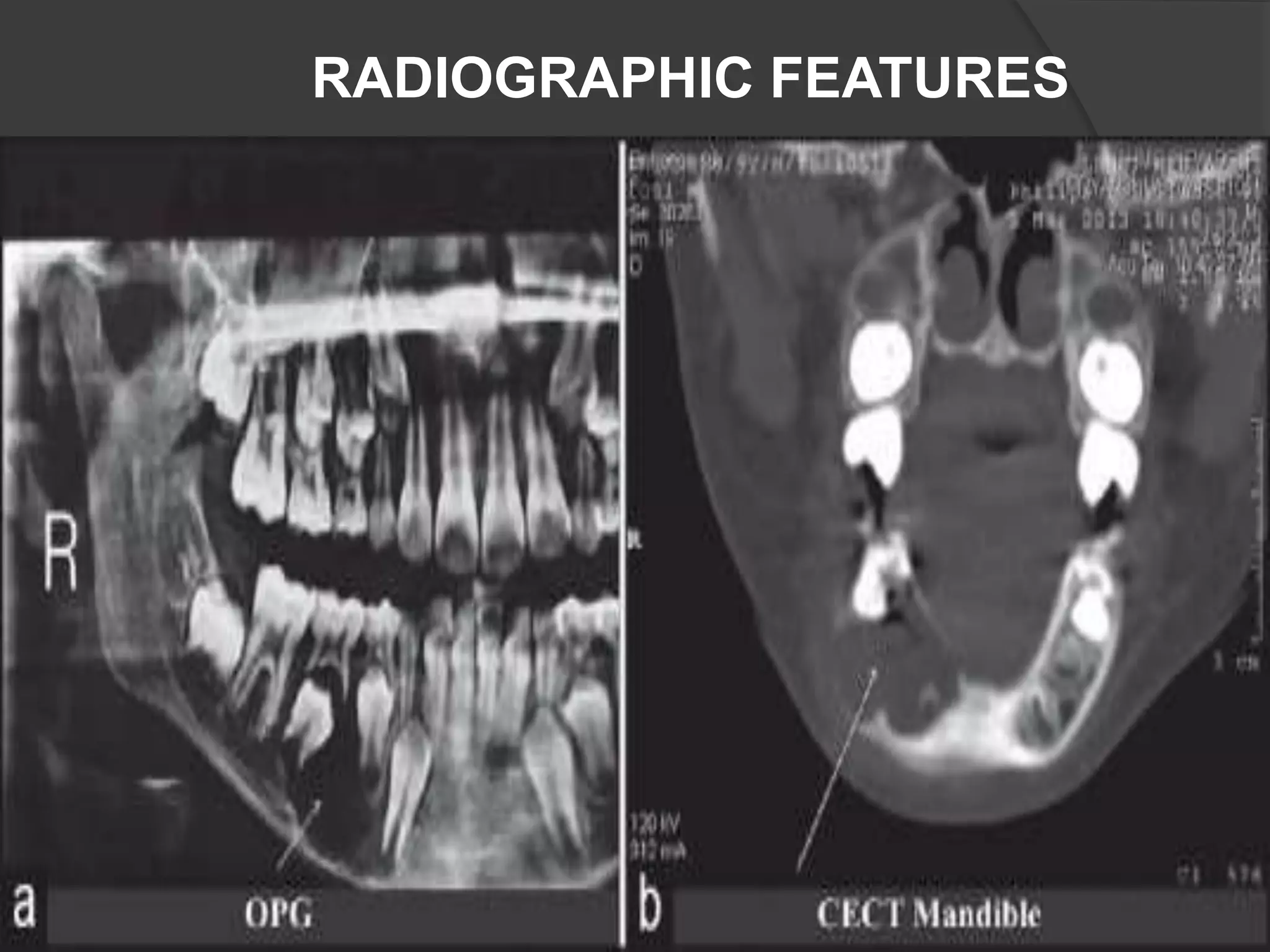
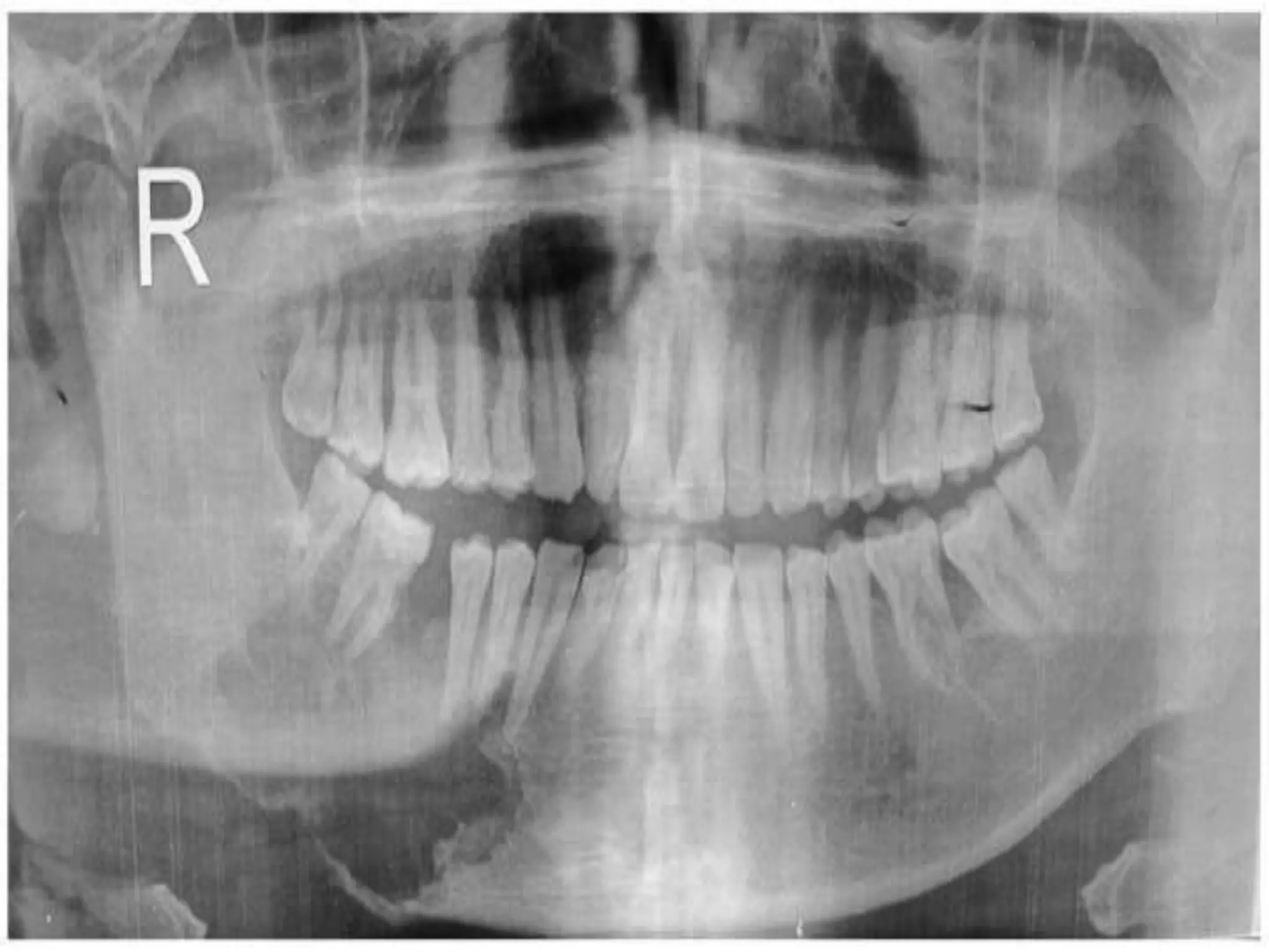
DR.RABIA SANNAM KHAN
MSC(UK)
OSTEOMA
DEFINITION
 Rare, Benign, Osteogenic neoplasm
characterized by proliferation of either
compact or cancellous bone usually in an
endosteal or periosteal location.
CLASSIFICATION
 Central Osteoma Arises from the
endosteum.
 Peripheral Osteoma Arises from the
periosteum.
 Extra Skeletal Soft Tissue Osteoma Arises
within the muscle.
ETIOLOGY
 Traumatic,
 Inflammatory
 Embryologic
 Associated with Gardner’s Syndrome.
CLINICALFEATURES
HISTOLOGICAL
FEATURES
 Consists of mature, lamellar bone or
cancellous bone.
 Classified as Compact/ivory,
Cancellous/spongy, or Mixed on histo-
pathologically.
RADIOGRAPHIC FEATURES
COMPLICATIONS /
TREATMENT
DIFFERENTIAL
DIAGNOSIS
 Exostoses.
 Ostoblastoma.
 Fibrous Dysplasia.
 Osteoma.
CEMENTOOSSIFYING
FIBROMA
DEFINITION
 (COFs) are rare, benign, non-
odontogenic tumors of the jaw,
characterized by massive deposition of
cementum,calcified material admixed
with fibrous tissue.
 JOF occurs mostly in children and
adolescents.
TYPES
 Conventional
 Juvenile
TRABECULAR PSAMMOMATOID
CLINICAL FEATURES
HISTOLOGICAL
FEATURES
 COFs are well-circumscribed, thin
isolated trabeculae of bones.
 Spherical deposits of calcified material,
resembling cementum.
 PJOF’s presents as eosinophilic
spherical structures.
RADIOGRAPHIC FEATURES
DIFFERENTIAL
DIAGNOSIS
 Fibrous dysplasia,
 Burkitt lymphoma,
 Osteosarcoma and
 Cystic lesions.
LANGERHAN CELL
HISTIOCYTOSIS
Definition
 (LCH) is characterized by an abnormal
proliferation of immature dendritic cells.
syndrome. The term “histiocytosis” denotes
proliferation of histiocytes and other
inflammatory cells, whereas the letter “X”
denotes unknown etiology of the disease.
TYPES
 The nomenclature — histiocytosis X was
coined by Lichenstein in 1953 to account
for 3 clinical varieties
 1) Eosinophilic Granuloma,
 2) Letterer-Siwe Disease and
 3) Hand–Schuller-Christian syndrome
ETIOLOGY
 Different hypothesis which include
 1) Disorder of immune regulation with
deficiency of T suppressor lymphocytes,
 2) A neoplastic proliferation of Langerhans
cells.
 or other etiological factors like viruses,
bacteria and genetic components.
CLINICAL FEATURES
 Soft, tender, painful swelling
 Incidence in jaws is 7.9% with angle and
body of the mandible commom sites.
 Depends on type and severity involvement
CHRONIC FOCAL
CHRONIC DISSEMINATED
ACUTE DISSEMINATED
HISTOLOGICAL
FEATURES
 Tennis-racket shaped structures.
 Histiocytic cells with eoisnophilic
cytoplasm.
 Birbeck granules.
RADIOGRAPHIC FEATURES
DIFFERENTIAL
DIAGNOSIS
 Tuberculous osteomyelitis,
 Ameloblastoma,
 Odontogenic keratocyst,
 Dentigerous cyst.
Osteoma lecture
Osteoma lecture

Osteoma lecture

Editor's Notes

  • #5 Medullary bone (endosteal), or on the bone surface as a polypoid or sessile growth (periosteal)
  • #6 Osteomas are exclusively found in membranous bones such as skull, facial, and jaw bones. 
  • #7 Traumatic theory of Gerber Injuries suffered during puberty may cause the growth of osteomas from bone sequestra. Inflammatory theory Sinusitis may stimulate osteoblastic proliferation or it can be a secondary symptom arising from obstruction of sinus drainage. Embryologic theory Osteoma arises from the remains of persistent embryologic cells located at the junction of the ethmoid and frontal sinuses. Their frequent relationship to Gardner’s syndromeis of crucial clinical significance.2 This autosomal dominant syndrome is characterised by the development of multiple osteomas and these may present as the initial clinical signs of the syndrome.
  • #8 Extraoral photograph shows a swelling on the left posterior body of the mandible. Intraoral view showing a well-defined, round swelling covered by normal oral mucosa on the buccal plate of the left posterior mandible.
  • #10 Compact/ IvoryMicroscopic features of peripheral osteoma consisting of mature lameller compact bone.
  • #11 Photomicrograph showing hard tissue composed of benign dense bone with minimal marrow spaces, and Haversian canals (haematoxylin & eosin stain x 10
  • #12 Panoramic radiograph showing a solitary, round, 3×3 cm well-defined radio-opaque mass without a radiolucent rim on the left side of the body of the mandible. The lesion extended distally of the second premolar till the mesial aspect of the second molar distal root.
  • #19 Intra oral view of buccal aspect with arrows showing anterior and inferior margins of the swelling
  • #20 Golg named these spherical structures as ‘psammoma- like bodies’. These particles vary in appearance such a concentric lamellated to irregular both having central basophilic area and a peripheral eosinophilic fringe. Ultra structure of psammoma- like bodies manifest with a dark rim of crystals. From these crystals spicules and needle-like crystalloids project toward the periphery resembling brush border
  • #21 Photomicrograph (a) Showing highly cellular fields with spheroidal cementum-like material (H&E stain, ×40). (b) Photomicrograph showing bony trabeculae rimmed by osteoblast with surrounding loose fibrous tissue (H&E stain, ×100). (c) Photomicrograph showing proliferating fibroblasts arranged in whorls and short fascicles with cementum-like material (H&E stain, ×100). (d) Photomicrograph Showing concentric cementum-like material (H&E stain, ×400)
  • #22 Magnified photomicrograph (40X) of psammoma- like bodies, indicated with an arrow showing peripheral brush border appearance
  • #23 Orthopantomogram showing expansile lytic lesion in the body of mandible involving the roots of premolars, first and second molars. The lateral aspect of the lesion shows faint radiopacity
  • #24 JUVENILE Occlusal radiograph marked with arrows showing buccal palatal extension of lesion with ossifications Intraoral periapical radiograph showing mixed radiodensity marked with an arrow, 14, 15 teeth show root divergence
  • #31 A pit in the right mandible. No pus is being discharged from the pit. The first molar is missing and the other molars are sloped anteriorly.
  • #33 (a) Histiocytes with nuclear groove, eosinophils (arrows), lymphocytes, multinucleated giant cell (inset) (Giemsa, ×400). (b) The arrow shows histiocytes with nuclear groove (Pap, ×1000)
  • #34 (a) The arrow shows the lytic lesion in the body of right mandible with floating teeth (orthopantomograph). (b) The arrow shows the lytic lesion in right mandible (contrast enhanced computed tomography mandible)
  • #35 Initial panoramic radiograph showing a radiolucent lesion with a moth-eaten margin in the right mandible. The lesion resulted in the fracture of the mandible. The involved teeth appear to be floating on the lesion.