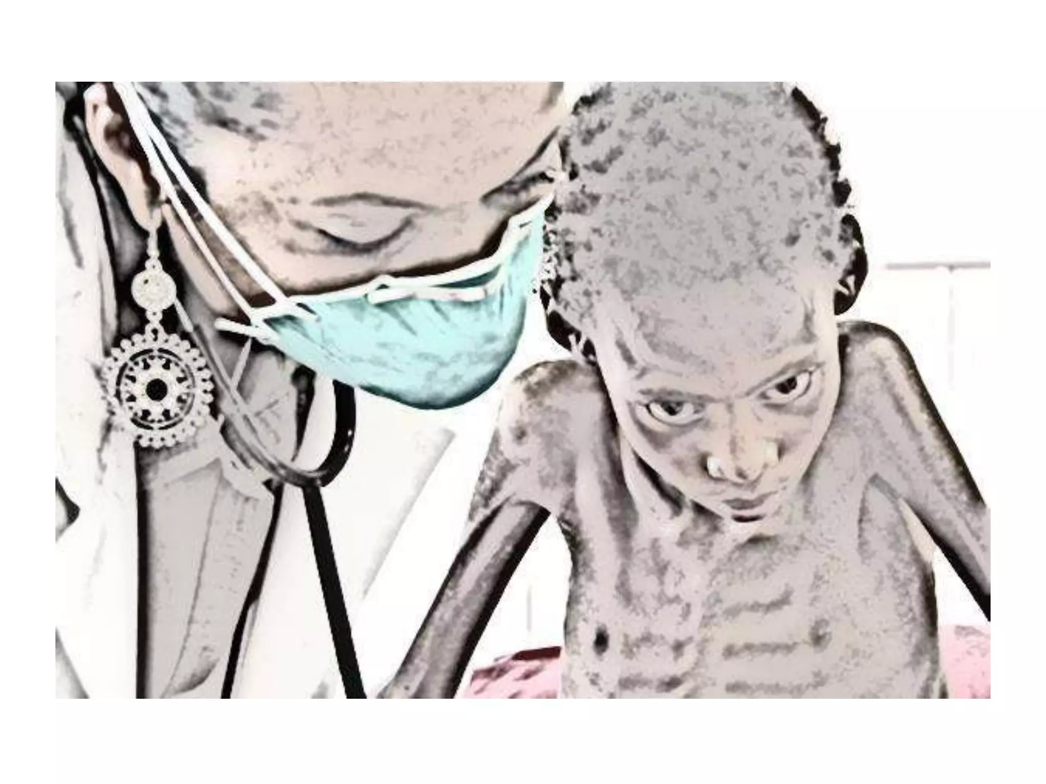
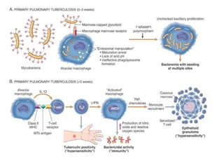
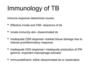
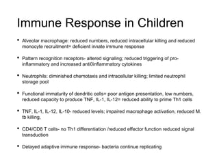
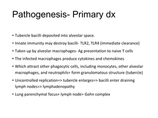
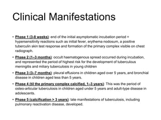
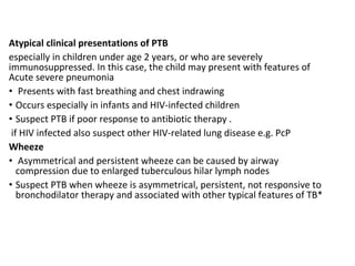
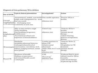
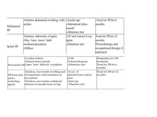
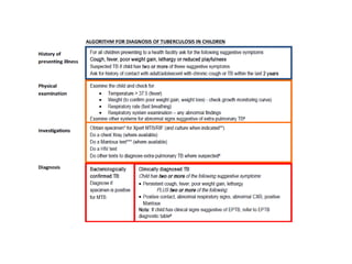
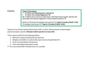
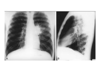
CHILDHOOD
TUBERCULOSIS
DR MOHAMED ADAN AHMED, DR JHUTHI
Natural History of Tb
Inhalation of aerosol droplets to one of four possible outcomes:
• Immediate clearance of the organism
• Primary disease: immediate onset of active disease
• Latent infection
• Reactivation disease: onset of active disease many years
following a period of latent infection
In children- have paucibacillary dx ie require smaller inoculum
to cause pathology and often have sputum negative dx due to
less cavitatory dx and have less forceful cough
Immunology of TB
Immune response determines course
• Effective innate and CMI- clearance of dx
• Innate immunity abn- disseminated dx
• Inadequate CD8 response- marked tissue damage due to
intense proinflammatory response
• Inadequate CD4 response= inadequate production of IFN
gamma- important macrophage activator
• Immunodeficient- either disseminated dx or reactivation
Immune Response in Children
• Alveolar macrophage: reduced numbers, reduced intracellular killing and reduced
monocyte recruitment= deficient innate immune response
• Pattern recognition receptors- altered signaling; reduced triggering of pro-
inflammatory and increased anti0inflammatory cytokines
• Neutrophils: diminished chemotaxis and intracellular killing; limited neutrophil
storage pool
• Functional immaturity of dendritic cells= poor antigen presentation, low numbers,
reduced capacity to produce TNF, IL-1, IL-12= reduced ability to prime Th1 cells
• TNF, IL-1, IL-12, IL-10- reduced levels; impaired macrophage activation, reduced M.
tb killing,
• CD4/CD8 T cells- no Th1 differentiation /reduced effector function reduced signal
transduction
• Delayed adaptive immune response- bacteria continue replicating
Pathogenesis- Primary dx
• Tubercle bacilli deposited into alveolar space.
• Innate immunity may destroy bacilli- TLR2, TLR4 (immediate clearance)
• Taken up by alveolar macrophages- Ag presentation to naive T cells
• The infected macrophages produce cytokines and chemokines
• Which attract other phagocytic cells, including monocytes, other alveolar
macrophages, and neutrophils= form granulomatous structure (tubercle)
• Uncontrolled replication=> tubercle enlarges=> bacilli enter draining
lymph nodes=> lymphadenopathy
• Lung parenchymal focus+ lymph node= Gohn complex
• Effective CMI 2-6weeks after infection
• Failure to develop CMI= progressive lung destruction
• Caseous necrosis occurs due to proinflam products such as TNF-alpha, reactive
oxygen and nitrogen intermediates, and the contents of cytotoxic cells
(granzymes, perforin)
• Unchecked growth=> hematogenous spread=> disseminated TB (eg miliary TB)
• OR spread mechanically by erosion of the caseating lesions into the airways=
transmission of bacilli
• If no treatment= px either dies or develops chronic dx ie- repeated episodes of
healing by fibrotic changes around the lesions and tissue breakdown
Disease Risk by Age
Sites of Infection
• Pulmonary vs extra pulmonary
• Pulmonary- infection of the lung tissue
• Extra pulmonary:
- Tuberculous meningitis – meningitis not responding to antibiotic
treatment, with a subacute onset, communicating hydrocephalus, stroke,
and/or elevated intracranial pressure
- Pleural TB, Pericardial TB, Hilar TB- intrathoracic but extra pulmonary
- Abdominal TB – Distended abdomen with ascites, abdominal pain,
jaundice, or unexplained chronic diarrhea
- Others: TB adenitis, TB of the joint, Vertebral TB( gibbus deformity),
Renal (Sterile pyuria, hematuria) Eye (Iritis, optic neuritis, phlyctenular
conjunctivitis)
• Phase 1 (3-8 weeks): end of the initial asymptomatic incubation period =
hypersensitivity reactions such as initial fever, erythema nodosum, a positive
tuberculin skin test response and formation of the primary complex visible on chest
radiograph.
• Phase 2 (1–3 months): occult haematogenous spread occurred during incubation,
and represented the period of highest risk for the development of tuberculous
meningitis and miliary tuberculosis in young children
• Phase 3 (3–7 months): pleural effusions in children aged over 5 years, and bronchial
disease in children aged less than 5 years.
• Phase 4 (til the primary complex calcified, 1–3 years): This was the period of
osteo-articular tuberculosis in children aged under 5 years and adult-type disease in
adolescents.
• Phase 5 (calcification > 3 years): late manifestations of tuberculosis, including
pulmonary reactivation disease, developed.
Clinical Manifestations
Early in Primary dx:
• May be asymptomatic
• May have low grade fever, mild cough, flu-like symptoms (ie non
specific respiratory signs)
• Failure to thrive
✴ Malnutrition hastens dx progress with less immune reponse=
rapidly progressive, severe dx
✴Cough does not have to be > 2weeks- cough of any duration
Clinical Presentation
In children
Progressive primary complex
➡Occurs when primary focus enlarges steadily and forms a caseous centre
• Poor weight gain/wt loss(growth faltering)
• fever /night sweats
• Reduced activity
• Persistent cough
• Unilateral wheeze due to airway compression by LN
• resp distress
• Diminished breath sounds, dullness
References
• Global Tuberculosis Report 2018, World Health Organization.
• Immunology of Tuberculosis- UpToDate
• Natural History of Tuberculosis- UpToDate
• Tuberculosis in Children- UpToDate
• Paediatric Tuberculosis- Lancet Infect Dis. 2008 Aug; 8(8): 498–510.(Newton SM et
al)
• The natural history of childhood intra-thoracic tuberculosis: a critical review of
literature from the pre-chemotherapy era. Int J Tuberc Lung Dis. 2004 Apr;8(4):392–
402 (Marais BJ et al)
• Clinical peculiarities of tuberculosis-BMC Infectious Diseases201414 (Suppl 1) :S4
(Paola Piccini et al)
Diagnosis of
childhood TB
Overview of TB Diagnosis
• clinical diagnosis
• screening test
- Tuberclin skin test
- Interferon-gamma release assays
• Imaging
- Chest radiography
- Computed tomography scan
• Laboratory studies
• sputum
- Acid-fast bacilli smear and culture
Gastric aspirate
Xpert MTB/RIF and other rapid tests
Other specimens
- Urine antigen detection
Diagnostic challenges
A major challenge of childhood TB is establishing an accurate diagnosis.
Less than 15% of cases are sputum acid-fast bacilli smear positive, and
mycobacterial culture yields are 30%–40%
• TB can mimic may common childhood illnesses-non specific clinical
presentation
• Non specific clinical presentation
• Lack of a ‘gold standard’ test for Dx
• Inability of pre-adolescent child to expectorate sputum
• Low bacillary load so, often smear negative
• Delayed type hypersensitivity to purified protein derivative absent in
40% of HIV negative children with extra-thoracic TB
Diagnostic problems are more pronounced in HIV-infected children, and the performance of
current diagnostic algorithms is poor in this group .
Factors contributing to these additional diagnostic difficulties are as follows:
1. HIV-infected children who live with HIV-infected adults are more likely to be exposed to an
adult TB index case at home. However, HIV-infected adults often have sputum smear–negative TB,
and, therefore, the risk of infection posed by this exposure is often not appreciated.
2. TST is much less sensitive in HIV-infected children than in HIV-uninfected children.
3. Chronic pulmonary symptoms may be related to other HIV-related conditions, such as
gastroesophageal reflux and bronchiectasis, thus reducing the specificity of symptom-based
diagnostic approaches.
4. Weight loss or failure to thrive are typical characteristics of both TB and HIV infection.
5. Rapid TB disease progression is more likely to occur in HIV-infected children, reducing the
sensitivity of diagnostic approaches that focus on persistent, nonremitting symptoms .
6. Interpretation of chest radiographs is complicated by HIV-related comorbidities, such as
bacterial pneumonia, lymphocytic interstitial pneumonitis, bronchiectasis, pulmonary Kaposi
sarcoma, and the atypical presentation of TB in immunocompromised children
Case Definitions TB
Clinical Diagnosis of TB
Pulmonary TB
History
The key elements of history are:
• History of contact with an adolescent or adult with confirmed or presumptive TB
within the last two years
• Close contact is defined as a person who has confirmed or presumptive TB living in
the same household or in frequent contact with the child (e.g. caretakers, school
staff).
History of symptoms suggestive of TB
The most common symptoms associated with TB include the following:
• Progressive and non-remitting cough
• Fever and/or night sweats
• Lethargy / reduced playfulness / less active
• Poor weight gain or weight loss (failure to thrive)
Physical examination
General examination
Examine the child and check for:
• Temperature > 37.5 (fever)
• Weight (to confirm poor weight gain, weight loss)
• Respiratory rate (fast breathing)
• nutritional assessment
Examination of the Respiratory System
• In early stages of pulmonary TB, the respiratory exam may show few abnormal signs. As the disease
progresses respiratory signs become more obvious as follows:
• Cough
• Increased respiratory rate (fast breathing)
• Respiratory distress e.g. laboured breathing, chest in-drawing (this shows severe disease)
• Percussion note - dull when lobar consolidation is present (normal resonance in many children with
PTB)
• Auscultation may be normal in early disease, and abnormal in more advanced disease (crackles,
bronchial breathing)
Atypical clinical presentations of PTB
especially in children under age 2 years, or who are severely
immunosuppressed. In this case, the child may present with features of
Acute severe pneumonia
• Presents with fast breathing and chest indrawing
• Occurs especially in infants and HIV-infected children
• Suspect PTB if poor response to antibiotic therapy .
if HIV infected also suspect other HIV-related lung disease e.g. PcP
Wheeze
• Asymmetrical and persistent wheeze can be caused by airway
compression due to enlarged tuberculous hilar lymph nodes
• Suspect PTB when wheeze is asymmetrical, persistent, not responsive to
bronchodilator therapy and associated with other typical features of TB*
Radiological diagnosis
Chest X-ray
CXR remains an important tool for diagnosis of PTB in children who are sputum smear
negative or who cannot produce sputum
The following abnormalities on CXR are suggestive of Tb :-
• Enlarged hilar lymph nodes and opacification in the lung tissue.
• Miliary mottling in lung tissue
• Cavitation (tends to occur in older children)
• Pleural or pericardial effusion – though seen on CXR – are forms of extra
pulmonary TB that tend to occur in older children
• The finding of marked abnormality on CXR in a child with no signs of respiratory
distress (no fast breathing or chest indrawing) is supportive of TB
Primary Tuberculosis on CXR
1.GangliopulmonaryTB ( Characterized by the presence of
mediastinal and/ or hilar lymphadenopathy and
parenchymal abnormalities, the Ghonfocus
2.TuberculousPleuritis
3.MiliaryTB
4.Tracheobronchial TB
Potts disease
• Computed tomography (CT) scan of the chest may be used to further
delineate the anatomy for cases in which radiographic findings are
equivocal. Endobronchial involvement, bronchiectasis, and cavitations
may be more readily visualized on CT scans than chest radiographs .
However, there is no role for routine use of CT scans in the evaluation of
an asymptomatic child since treatment regimens are based on chest
radiography findings .
• In the setting of tuberculous meningitis, CT scan of the head is useful.
Hydrocephalus and basilar meningeal enhancement are observed in 80
and 90 percent of cases, respectively; chest radiography may be normal.
Abdominal u/s findings on TB :
- Abdominal lymph node enlargement
- Splenic lesions and ascites
-
Immunological tests
1. Tuberculin skin test
TST is useful to support a diagnosis of TB in children with suggestive clinical features who are sputum
smear negative or who cannot produce sputum
A positive TST indicates infection:
• positive in any child if ≥ 10 mm irrespective of BCG immunisation
• also positive if ≥ 5 mm in HIV-infected or severely malnourished child
A positive TST is particularly useful to indicate TB infection when there is no
known TB exposure on clinical assessment i.e. no positive contact history
Overall, up to 40 percent of immunocompetent children with culture-confirmed TB disease may have a
negative TST (American Academy of Pediatrics. Red Book: 2018 Report of the Committee on Infectious
Diseases)
Caution
A positive TST does not distinguish between TB infection and active disease
A negative TST does not exclude TB disease
A positive TST may be falsely positive due to prior vaccination with Bacille Calmette-Guérin (BCG),
infection with nontuberculous mycobacteria, and improper administration or interpretation
Copyrights apply
Copyrights apply
2)-Interferon gamma release assay (IGRA)
• Haematological tests that can aid in diagnosing Mycobacterium
tuberculosis infection e.g. QuantiFERON TB Gold In-Tube test (QFT-
GIT) and T-SPOT®.TB test (T-Spot). It is an antibody-antigen test like
Mantoux that measures the presence of an immune response to TB
bacilli.
• Use of both TST and IGRA may increase sensitivity for diagnostic
evaluation of children with suspected TB. In one study including 69
children age 5 to 18 years with TB who underwent both IGRA and
TST, the sensitivity of IGRA was greater than TST (95 versus 83
percent); among children <5 years, the sensitivity was similar
(nterferon-γRelease Assay Performance for Tuberculosis in Childhood Kay AW, Islam SM,
Wendorf K, Westenhouse J, etal )
Microbiological diagnosis
Specimen :
- Sputum : expectorate / induction /gastric aspirate / nasopharyngeal aspirate
- Ascitic fluid
- pleural fluid
- Pericardial fluid
- Wound exudate
- joint effusion
The specimens should be examined and cultured In a laboratory that specializes in testing for M.
tuberculosis. The bacteriologic examination has five parts:
• Specimen collection, processing, and review
• AFB smear classification and results
• Direct detection of M. tuberculosis in clinical specimen using nucleic acid amplification (NAA)
• Specimen culturing and identification
• Drug-susceptibility testing
Sputum Induction
sputum induction is an aerosol-generating procedure. Where possible, therefore, this
procedure should be performed in an isolation room that has adequate infection control
precautions (negative pressure, ultraviolet light (turned on when room is not in use) and
an extractor fan).
Children with the following characteristics should not undergo sputum induction.
• Inadequate fasting: if a child has not been fasting for at least 3 hours, postpone the
procedure until the appropriate time
• Severe respiratory distress (including rapid breathing, wheezing, hypoxia)
• Children who are intubated
• Bleeding: low platelet count, bleeding tendency, severe nosebleeds (Symptomatic or
platelet count<50/ml blood)
• Reduced level of consciousness
• History of significant asthma
Gastric aspiration remains the most common method for
obtaining respiratory samples from children (in facilities
where this procedure may be performed). In general, cultures
of gastric aspirate specimens are positive for TB in only 30 to
40 percent of cases
Early morning gastric contents collected from a fasting child
contain sputum swallowed during the night .
During sleep, the lung’s mucociliary system beats mucus
containing Mycobacterium TB up into the throat. The mucus
is swallowed and remains in the stomach until the stomach
empties. Therefore, the highest yield specimens are obtained
first thing in the morning.
Sputum smears stained by Z-N stain
• Do two sputum smears for acid fast bacilli (AFB) microscopy
• Specimens should be from lung secretions, not saliva
• Spontaneous morning sputum more desirable than induced specimens
• Collect sputum before treatment is initiated
Interpretation of sputum stained by Z N Stain (WHO )
• More than 10 bacilli / field ------- +++
• From 1 – 10 bacilli / field ------- ++
• From 10 – 99 bacilli / 100 fields ----- +
• From 1 -9 bacilli/100 fields ------ write the no.
• No bacilli seen ---------- negative
Culture and Drug Susceptibility Testing (DST)
The most accurate way to diagnose TB—the gold standard—is through culture
however, in the absence of a positive culture, TB disease may also be diagnosed
on the basis of clinical signs and symptoms alone .
The commercially available broth culture systems (e.g., BACTEC, MGIT, VersaTREK,
MBBACT) allow detection of most mycobacterial growth in 4 to 14 days compared
to 3 to 6 weeks for solid media
Do a culture and DST for the following children:
• Rifampicin resistance detected by the Xpert test
• Refugees and children in contact with anyone who has Drug Resistant TB
• Those not responding to TB treatment
• Those with Indeterminate Xpert results
Xpert MTB/RIF
The Xpert MTB/RIF assay is an automated nucleic acid
amplification test that can simultaneously identify M.
tuberculosis and detect rifampin resistance. This test
performs substantially better than smear microscopy.
A fully automated and rapid test, GeneXpert MTB/RIF
works in about two hours. It performs very well on
sputum, and its sensitivity is better than microscopy,
including for people with HIV.
Urine antigen detection
• Urine-based detection of mycobacterial cell wall glycolipid
lipoarabinomannan (urine LAM assay) is an assay for diagnosis of TB.
• For regions of the world with high incidence of HIV and TB, we are in
agreement with the WHO, which favors use of urine LAM testing (in
addition to routine diagnostic tests) for HIV-infected patients with signs and
symptoms of pulmonary and/or extrapulmonary tuberculosis and CD4 ≤100
cells/microL and for HIV-infected patients who are seriously ill.
• The urine LAM assay may also have prognostic utility. In one study
including 137 HIV-infected children in Malawi initiating antiretroviral
therapy, the six-month mortality rate was 3.7-fold higher among those with
a positive urine LAM assay
References
• Nelsons textbook of pediatrics 19th edition
• Kenya paediatric TB treatment guidelines
• The National Tuberculosis, Leprosy and Lung Disease Program (NTLD-
Program)
• WHO global report 2016
• AN ACTIVIST’S GUIDE TO Tuberculosis Diagnostic Tools ( Treatment
Action Group)
• UptoDate
• Bacteriological diagnosis of childhood TB: a prospective observational
study Andrew J. Brent et al
Childhood tb 2

Childhood tb 2

  • 2.
  • 4.
    Natural History ofTb Inhalation of aerosol droplets to one of four possible outcomes: • Immediate clearance of the organism • Primary disease: immediate onset of active disease • Latent infection • Reactivation disease: onset of active disease many years following a period of latent infection In children- have paucibacillary dx ie require smaller inoculum to cause pathology and often have sputum negative dx due to less cavitatory dx and have less forceful cough
  • 6.
    Immunology of TB Immuneresponse determines course • Effective innate and CMI- clearance of dx • Innate immunity abn- disseminated dx • Inadequate CD8 response- marked tissue damage due to intense proinflammatory response • Inadequate CD4 response= inadequate production of IFN gamma- important macrophage activator • Immunodeficient- either disseminated dx or reactivation
  • 7.
    Immune Response inChildren • Alveolar macrophage: reduced numbers, reduced intracellular killing and reduced monocyte recruitment= deficient innate immune response • Pattern recognition receptors- altered signaling; reduced triggering of pro- inflammatory and increased anti0inflammatory cytokines • Neutrophils: diminished chemotaxis and intracellular killing; limited neutrophil storage pool • Functional immaturity of dendritic cells= poor antigen presentation, low numbers, reduced capacity to produce TNF, IL-1, IL-12= reduced ability to prime Th1 cells • TNF, IL-1, IL-12, IL-10- reduced levels; impaired macrophage activation, reduced M. tb killing, • CD4/CD8 T cells- no Th1 differentiation /reduced effector function reduced signal transduction • Delayed adaptive immune response- bacteria continue replicating
  • 8.
    Pathogenesis- Primary dx •Tubercle bacilli deposited into alveolar space. • Innate immunity may destroy bacilli- TLR2, TLR4 (immediate clearance) • Taken up by alveolar macrophages- Ag presentation to naive T cells • The infected macrophages produce cytokines and chemokines • Which attract other phagocytic cells, including monocytes, other alveolar macrophages, and neutrophils= form granulomatous structure (tubercle) • Uncontrolled replication=> tubercle enlarges=> bacilli enter draining lymph nodes=> lymphadenopathy • Lung parenchymal focus+ lymph node= Gohn complex
  • 9.
    • Effective CMI2-6weeks after infection • Failure to develop CMI= progressive lung destruction • Caseous necrosis occurs due to proinflam products such as TNF-alpha, reactive oxygen and nitrogen intermediates, and the contents of cytotoxic cells (granzymes, perforin) • Unchecked growth=> hematogenous spread=> disseminated TB (eg miliary TB) • OR spread mechanically by erosion of the caseating lesions into the airways= transmission of bacilli • If no treatment= px either dies or develops chronic dx ie- repeated episodes of healing by fibrotic changes around the lesions and tissue breakdown
  • 10.
  • 11.
    Sites of Infection •Pulmonary vs extra pulmonary • Pulmonary- infection of the lung tissue • Extra pulmonary: - Tuberculous meningitis – meningitis not responding to antibiotic treatment, with a subacute onset, communicating hydrocephalus, stroke, and/or elevated intracranial pressure - Pleural TB, Pericardial TB, Hilar TB- intrathoracic but extra pulmonary - Abdominal TB – Distended abdomen with ascites, abdominal pain, jaundice, or unexplained chronic diarrhea - Others: TB adenitis, TB of the joint, Vertebral TB( gibbus deformity), Renal (Sterile pyuria, hematuria) Eye (Iritis, optic neuritis, phlyctenular conjunctivitis)
  • 13.
    • Phase 1(3-8 weeks): end of the initial asymptomatic incubation period = hypersensitivity reactions such as initial fever, erythema nodosum, a positive tuberculin skin test response and formation of the primary complex visible on chest radiograph. • Phase 2 (1–3 months): occult haematogenous spread occurred during incubation, and represented the period of highest risk for the development of tuberculous meningitis and miliary tuberculosis in young children • Phase 3 (3–7 months): pleural effusions in children aged over 5 years, and bronchial disease in children aged less than 5 years. • Phase 4 (til the primary complex calcified, 1–3 years): This was the period of osteo-articular tuberculosis in children aged under 5 years and adult-type disease in adolescents. • Phase 5 (calcification > 3 years): late manifestations of tuberculosis, including pulmonary reactivation disease, developed. Clinical Manifestations
  • 14.
    Early in Primarydx: • May be asymptomatic • May have low grade fever, mild cough, flu-like symptoms (ie non specific respiratory signs) • Failure to thrive ✴ Malnutrition hastens dx progress with less immune reponse= rapidly progressive, severe dx ✴Cough does not have to be > 2weeks- cough of any duration Clinical Presentation In children
  • 15.
    Progressive primary complex ➡Occurswhen primary focus enlarges steadily and forms a caseous centre • Poor weight gain/wt loss(growth faltering) • fever /night sweats • Reduced activity • Persistent cough • Unilateral wheeze due to airway compression by LN • resp distress • Diminished breath sounds, dullness
  • 16.
    References • Global TuberculosisReport 2018, World Health Organization. • Immunology of Tuberculosis- UpToDate • Natural History of Tuberculosis- UpToDate • Tuberculosis in Children- UpToDate • Paediatric Tuberculosis- Lancet Infect Dis. 2008 Aug; 8(8): 498–510.(Newton SM et al) • The natural history of childhood intra-thoracic tuberculosis: a critical review of literature from the pre-chemotherapy era. Int J Tuberc Lung Dis. 2004 Apr;8(4):392– 402 (Marais BJ et al) • Clinical peculiarities of tuberculosis-BMC Infectious Diseases201414 (Suppl 1) :S4 (Paola Piccini et al)
  • 17.
  • 18.
    Overview of TBDiagnosis • clinical diagnosis • screening test - Tuberclin skin test - Interferon-gamma release assays • Imaging - Chest radiography - Computed tomography scan • Laboratory studies • sputum - Acid-fast bacilli smear and culture Gastric aspirate Xpert MTB/RIF and other rapid tests Other specimens - Urine antigen detection
  • 19.
    Diagnostic challenges A majorchallenge of childhood TB is establishing an accurate diagnosis. Less than 15% of cases are sputum acid-fast bacilli smear positive, and mycobacterial culture yields are 30%–40% • TB can mimic may common childhood illnesses-non specific clinical presentation • Non specific clinical presentation • Lack of a ‘gold standard’ test for Dx • Inability of pre-adolescent child to expectorate sputum • Low bacillary load so, often smear negative • Delayed type hypersensitivity to purified protein derivative absent in 40% of HIV negative children with extra-thoracic TB
  • 20.
    Diagnostic problems aremore pronounced in HIV-infected children, and the performance of current diagnostic algorithms is poor in this group . Factors contributing to these additional diagnostic difficulties are as follows: 1. HIV-infected children who live with HIV-infected adults are more likely to be exposed to an adult TB index case at home. However, HIV-infected adults often have sputum smear–negative TB, and, therefore, the risk of infection posed by this exposure is often not appreciated. 2. TST is much less sensitive in HIV-infected children than in HIV-uninfected children. 3. Chronic pulmonary symptoms may be related to other HIV-related conditions, such as gastroesophageal reflux and bronchiectasis, thus reducing the specificity of symptom-based diagnostic approaches. 4. Weight loss or failure to thrive are typical characteristics of both TB and HIV infection. 5. Rapid TB disease progression is more likely to occur in HIV-infected children, reducing the sensitivity of diagnostic approaches that focus on persistent, nonremitting symptoms . 6. Interpretation of chest radiographs is complicated by HIV-related comorbidities, such as bacterial pneumonia, lymphocytic interstitial pneumonitis, bronchiectasis, pulmonary Kaposi sarcoma, and the atypical presentation of TB in immunocompromised children
  • 21.
  • 23.
    Clinical Diagnosis ofTB Pulmonary TB History The key elements of history are: • History of contact with an adolescent or adult with confirmed or presumptive TB within the last two years • Close contact is defined as a person who has confirmed or presumptive TB living in the same household or in frequent contact with the child (e.g. caretakers, school staff). History of symptoms suggestive of TB The most common symptoms associated with TB include the following: • Progressive and non-remitting cough • Fever and/or night sweats • Lethargy / reduced playfulness / less active • Poor weight gain or weight loss (failure to thrive)
  • 24.
    Physical examination General examination Examinethe child and check for: • Temperature > 37.5 (fever) • Weight (to confirm poor weight gain, weight loss) • Respiratory rate (fast breathing) • nutritional assessment Examination of the Respiratory System • In early stages of pulmonary TB, the respiratory exam may show few abnormal signs. As the disease progresses respiratory signs become more obvious as follows: • Cough • Increased respiratory rate (fast breathing) • Respiratory distress e.g. laboured breathing, chest in-drawing (this shows severe disease) • Percussion note - dull when lobar consolidation is present (normal resonance in many children with PTB) • Auscultation may be normal in early disease, and abnormal in more advanced disease (crackles, bronchial breathing)
  • 25.
    Atypical clinical presentationsof PTB especially in children under age 2 years, or who are severely immunosuppressed. In this case, the child may present with features of Acute severe pneumonia • Presents with fast breathing and chest indrawing • Occurs especially in infants and HIV-infected children • Suspect PTB if poor response to antibiotic therapy . if HIV infected also suspect other HIV-related lung disease e.g. PcP Wheeze • Asymmetrical and persistent wheeze can be caused by airway compression due to enlarged tuberculous hilar lymph nodes • Suspect PTB when wheeze is asymmetrical, persistent, not responsive to bronchodilator therapy and associated with other typical features of TB*
  • 30.
    Radiological diagnosis Chest X-ray CXRremains an important tool for diagnosis of PTB in children who are sputum smear negative or who cannot produce sputum The following abnormalities on CXR are suggestive of Tb :- • Enlarged hilar lymph nodes and opacification in the lung tissue. • Miliary mottling in lung tissue • Cavitation (tends to occur in older children) • Pleural or pericardial effusion – though seen on CXR – are forms of extra pulmonary TB that tend to occur in older children • The finding of marked abnormality on CXR in a child with no signs of respiratory distress (no fast breathing or chest indrawing) is supportive of TB
  • 32.
    Primary Tuberculosis onCXR 1.GangliopulmonaryTB ( Characterized by the presence of mediastinal and/ or hilar lymphadenopathy and parenchymal abnormalities, the Ghonfocus 2.TuberculousPleuritis 3.MiliaryTB 4.Tracheobronchial TB
  • 38.
  • 39.
    • Computed tomography(CT) scan of the chest may be used to further delineate the anatomy for cases in which radiographic findings are equivocal. Endobronchial involvement, bronchiectasis, and cavitations may be more readily visualized on CT scans than chest radiographs . However, there is no role for routine use of CT scans in the evaluation of an asymptomatic child since treatment regimens are based on chest radiography findings . • In the setting of tuberculous meningitis, CT scan of the head is useful. Hydrocephalus and basilar meningeal enhancement are observed in 80 and 90 percent of cases, respectively; chest radiography may be normal. Abdominal u/s findings on TB : - Abdominal lymph node enlargement - Splenic lesions and ascites -
  • 40.
    Immunological tests 1. Tuberculinskin test TST is useful to support a diagnosis of TB in children with suggestive clinical features who are sputum smear negative or who cannot produce sputum A positive TST indicates infection: • positive in any child if ≥ 10 mm irrespective of BCG immunisation • also positive if ≥ 5 mm in HIV-infected or severely malnourished child A positive TST is particularly useful to indicate TB infection when there is no known TB exposure on clinical assessment i.e. no positive contact history Overall, up to 40 percent of immunocompetent children with culture-confirmed TB disease may have a negative TST (American Academy of Pediatrics. Red Book: 2018 Report of the Committee on Infectious Diseases) Caution A positive TST does not distinguish between TB infection and active disease A negative TST does not exclude TB disease A positive TST may be falsely positive due to prior vaccination with Bacille Calmette-Guérin (BCG), infection with nontuberculous mycobacteria, and improper administration or interpretation
  • 42.
  • 43.
  • 45.
    2)-Interferon gamma releaseassay (IGRA) • Haematological tests that can aid in diagnosing Mycobacterium tuberculosis infection e.g. QuantiFERON TB Gold In-Tube test (QFT- GIT) and T-SPOT®.TB test (T-Spot). It is an antibody-antigen test like Mantoux that measures the presence of an immune response to TB bacilli. • Use of both TST and IGRA may increase sensitivity for diagnostic evaluation of children with suspected TB. In one study including 69 children age 5 to 18 years with TB who underwent both IGRA and TST, the sensitivity of IGRA was greater than TST (95 versus 83 percent); among children <5 years, the sensitivity was similar (nterferon-γRelease Assay Performance for Tuberculosis in Childhood Kay AW, Islam SM, Wendorf K, Westenhouse J, etal )
  • 47.
    Microbiological diagnosis Specimen : -Sputum : expectorate / induction /gastric aspirate / nasopharyngeal aspirate - Ascitic fluid - pleural fluid - Pericardial fluid - Wound exudate - joint effusion The specimens should be examined and cultured In a laboratory that specializes in testing for M. tuberculosis. The bacteriologic examination has five parts: • Specimen collection, processing, and review • AFB smear classification and results • Direct detection of M. tuberculosis in clinical specimen using nucleic acid amplification (NAA) • Specimen culturing and identification • Drug-susceptibility testing
  • 48.
    Sputum Induction sputum inductionis an aerosol-generating procedure. Where possible, therefore, this procedure should be performed in an isolation room that has adequate infection control precautions (negative pressure, ultraviolet light (turned on when room is not in use) and an extractor fan). Children with the following characteristics should not undergo sputum induction. • Inadequate fasting: if a child has not been fasting for at least 3 hours, postpone the procedure until the appropriate time • Severe respiratory distress (including rapid breathing, wheezing, hypoxia) • Children who are intubated • Bleeding: low platelet count, bleeding tendency, severe nosebleeds (Symptomatic or platelet count<50/ml blood) • Reduced level of consciousness • History of significant asthma
  • 49.
    Gastric aspiration remainsthe most common method for obtaining respiratory samples from children (in facilities where this procedure may be performed). In general, cultures of gastric aspirate specimens are positive for TB in only 30 to 40 percent of cases Early morning gastric contents collected from a fasting child contain sputum swallowed during the night . During sleep, the lung’s mucociliary system beats mucus containing Mycobacterium TB up into the throat. The mucus is swallowed and remains in the stomach until the stomach empties. Therefore, the highest yield specimens are obtained first thing in the morning.
  • 50.
    Sputum smears stainedby Z-N stain • Do two sputum smears for acid fast bacilli (AFB) microscopy • Specimens should be from lung secretions, not saliva • Spontaneous morning sputum more desirable than induced specimens • Collect sputum before treatment is initiated Interpretation of sputum stained by Z N Stain (WHO ) • More than 10 bacilli / field ------- +++ • From 1 – 10 bacilli / field ------- ++ • From 10 – 99 bacilli / 100 fields ----- + • From 1 -9 bacilli/100 fields ------ write the no. • No bacilli seen ---------- negative
  • 52.
    Culture and DrugSusceptibility Testing (DST) The most accurate way to diagnose TB—the gold standard—is through culture however, in the absence of a positive culture, TB disease may also be diagnosed on the basis of clinical signs and symptoms alone . The commercially available broth culture systems (e.g., BACTEC, MGIT, VersaTREK, MBBACT) allow detection of most mycobacterial growth in 4 to 14 days compared to 3 to 6 weeks for solid media Do a culture and DST for the following children: • Rifampicin resistance detected by the Xpert test • Refugees and children in contact with anyone who has Drug Resistant TB • Those not responding to TB treatment • Those with Indeterminate Xpert results
  • 56.
    Xpert MTB/RIF The XpertMTB/RIF assay is an automated nucleic acid amplification test that can simultaneously identify M. tuberculosis and detect rifampin resistance. This test performs substantially better than smear microscopy. A fully automated and rapid test, GeneXpert MTB/RIF works in about two hours. It performs very well on sputum, and its sensitivity is better than microscopy, including for people with HIV.
  • 59.
    Urine antigen detection •Urine-based detection of mycobacterial cell wall glycolipid lipoarabinomannan (urine LAM assay) is an assay for diagnosis of TB. • For regions of the world with high incidence of HIV and TB, we are in agreement with the WHO, which favors use of urine LAM testing (in addition to routine diagnostic tests) for HIV-infected patients with signs and symptoms of pulmonary and/or extrapulmonary tuberculosis and CD4 ≤100 cells/microL and for HIV-infected patients who are seriously ill. • The urine LAM assay may also have prognostic utility. In one study including 137 HIV-infected children in Malawi initiating antiretroviral therapy, the six-month mortality rate was 3.7-fold higher among those with a positive urine LAM assay
  • 62.
    References • Nelsons textbookof pediatrics 19th edition • Kenya paediatric TB treatment guidelines • The National Tuberculosis, Leprosy and Lung Disease Program (NTLD- Program) • WHO global report 2016 • AN ACTIVIST’S GUIDE TO Tuberculosis Diagnostic Tools ( Treatment Action Group) • UptoDate • Bacteriological diagnosis of childhood TB: a prospective observational study Andrew J. Brent et al

Editor's Notes