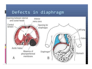
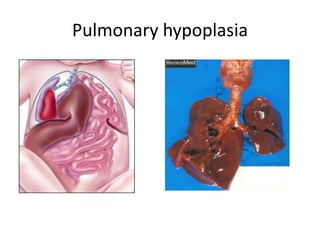
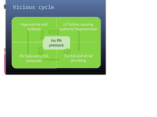
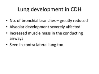
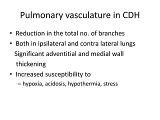
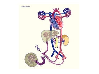
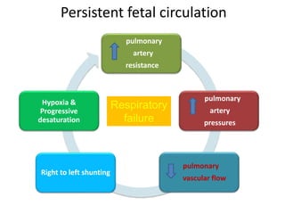
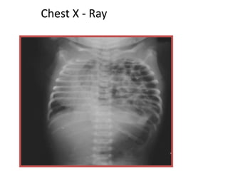
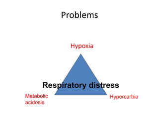
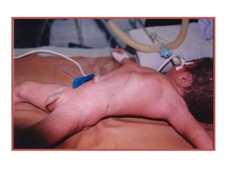
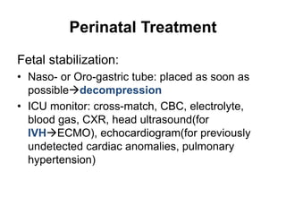
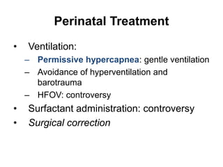
Congenital Diaphragmatic Hernia
Eyayalem M.
Introduction of CDH
• Incidence: about 1/2500
• Definition
– Failure of the pleural and peritoneal canal to
close at about GA 8 wk
– Herniation of the abdominal viscera into the
thoracic cavity and pulmonary hypoplasia
from compression by the viscera on the
developing lungs
• Cause: Unknown
Pathophysiology
• Diaphragmatic defect
• Failure of closure of pleuro-peritoneal canal
• Most common area is a postero-lateral defect
( Bochdalek )
• Left side more common
Pathophysiology
• Simple pathophysiology after herniation:
– Abdominal viscera fill the chest cavity
– Abdomen small & poorly developed
– Compressionpulmonary hypoplasia
– Dysfunction of the surfactant system
– Increased muscularization of the intra-acinar
pulmonary arteriespulmonary hypertension
– In very severe cases, left ventricular hypoplasia is
observed.
Pulmonary hypoplasia
Pathology of CDH
Following delivery
• Bowels fill with air
• Compression of ipsilateral lung
• Mediastinal shift
• Compression of contralateral lung(mechanical
compression of lung)
Lung development in CDH
• No. of bronchial branches – greatly reduced
• Alveolar development severely affected
• Increased muscle mass in the conducting
airways
• Seen in contra lateral lung too
Pulmonary vasculature in CDH
• Reduction in the total no. of branches
• Both in ipsilateral and contra lateral lungs
Significant adventitial and medial wall
thickening
• Increased susceptibility to
– hypoxia, acidosis, hypothermia, stress
Persistent fetal circulation
pulmonary
artery
resistance
pulmonary
artery
pressures
pulmonary
vascular flow
Right to left shunting
Hypoxia &
Progressive
desaturation
Respiratory
failure
Diagnosis
• CXR diagnostic
• Absence of diaphragm
• Scaphoid abdomen
• Bowel loops in chest
• Mediastinal shift
Prenatal Diagnosis
• Prenatal ultrasound: 59% detection rate, at
average GA 24.2 wk
• Predictors of outcome by prenatal ultrasound:
Polyhydramnios, Intrathoracic stomach or
liver, Abd circumference, lung-to-head
ratio(LHR), lung/transverse thorax ratio
• New predictors: PA diameter and LV mass (by
fetal echocardiography), MRI lung volumetry
• 1/3 P’t have associated malformations: CV, GU,
GI, CNS, chromosome anomalies
Chest X - Ray
Problems
Hypoxia
Respiratory distress
Metabolic
acidosis
Hypercarbia
Treatment
• Initial management Approach should be first medical
stabilization of the pt for improving respiratory and
general status and then proceeding for surgery after
stabilization
• GOALS
1. Reverse persistent pulm HTN to decrease rt to lt shunting.
2. Improvement in ventilation & oxygenation
3. Correction of acidosis
4. Correction of systemic hypotension
» Time taken to stabilize varies from 24-48 hrs to 7-10 days,
and upto 3 wks in some neonates
» Repair of hernia in emergency when the contents are
incarcerated
Perinatal Treatment
Fetal stabilization:
• Naso- or Oro-gastric tube: placed as soon as
possibledecompression
• ICU monitor: cross-match, CBC, electrolyte,
blood gas, CXR, head ultrasound(for
IVHECMO), echocardiogram(for previously
undetected cardiac anomalies, pulmonary
hypertension)
Perinatal Treatment
• Ventilation:
– Permissive hypercapnea: gentle ventilation
– Avoidance of hyperventilation and
barotrauma
– HFOV: controversy
• Surfactant administration: controversy
• Surgical correction

Chapter 6-2 Congenital diaphragmatic hernia.ppt

  • 1.
  • 2.
    Introduction of CDH •Incidence: about 1/2500 • Definition – Failure of the pleural and peritoneal canal to close at about GA 8 wk – Herniation of the abdominal viscera into the thoracic cavity and pulmonary hypoplasia from compression by the viscera on the developing lungs • Cause: Unknown
  • 3.
    Pathophysiology • Diaphragmatic defect •Failure of closure of pleuro-peritoneal canal • Most common area is a postero-lateral defect ( Bochdalek ) • Left side more common
  • 4.
    Pathophysiology • Simple pathophysiologyafter herniation: – Abdominal viscera fill the chest cavity – Abdomen small & poorly developed – Compressionpulmonary hypoplasia – Dysfunction of the surfactant system – Increased muscularization of the intra-acinar pulmonary arteriespulmonary hypertension – In very severe cases, left ventricular hypoplasia is observed.
  • 6.
  • 7.
    Pathology of CDH Followingdelivery • Bowels fill with air • Compression of ipsilateral lung • Mediastinal shift • Compression of contralateral lung(mechanical compression of lung)
  • 10.
    Lung development inCDH • No. of bronchial branches – greatly reduced • Alveolar development severely affected • Increased muscle mass in the conducting airways • Seen in contra lateral lung too
  • 11.
    Pulmonary vasculature inCDH • Reduction in the total no. of branches • Both in ipsilateral and contra lateral lungs Significant adventitial and medial wall thickening • Increased susceptibility to – hypoxia, acidosis, hypothermia, stress
  • 14.
    Persistent fetal circulation pulmonary artery resistance pulmonary artery pressures pulmonary vascularflow Right to left shunting Hypoxia & Progressive desaturation Respiratory failure
  • 15.
    Diagnosis • CXR diagnostic •Absence of diaphragm • Scaphoid abdomen • Bowel loops in chest • Mediastinal shift
  • 16.
    Prenatal Diagnosis • Prenatalultrasound: 59% detection rate, at average GA 24.2 wk • Predictors of outcome by prenatal ultrasound: Polyhydramnios, Intrathoracic stomach or liver, Abd circumference, lung-to-head ratio(LHR), lung/transverse thorax ratio • New predictors: PA diameter and LV mass (by fetal echocardiography), MRI lung volumetry • 1/3 P’t have associated malformations: CV, GU, GI, CNS, chromosome anomalies
  • 17.
  • 18.
  • 20.
    Treatment • Initial managementApproach should be first medical stabilization of the pt for improving respiratory and general status and then proceeding for surgery after stabilization • GOALS 1. Reverse persistent pulm HTN to decrease rt to lt shunting. 2. Improvement in ventilation & oxygenation 3. Correction of acidosis 4. Correction of systemic hypotension » Time taken to stabilize varies from 24-48 hrs to 7-10 days, and upto 3 wks in some neonates » Repair of hernia in emergency when the contents are incarcerated
  • 21.
    Perinatal Treatment Fetal stabilization: •Naso- or Oro-gastric tube: placed as soon as possibledecompression • ICU monitor: cross-match, CBC, electrolyte, blood gas, CXR, head ultrasound(for IVHECMO), echocardiogram(for previously undetected cardiac anomalies, pulmonary hypertension)
  • 22.
    Perinatal Treatment • Ventilation: –Permissive hypercapnea: gentle ventilation – Avoidance of hyperventilation and barotrauma – HFOV: controversy • Surfactant administration: controversy • Surgical correction