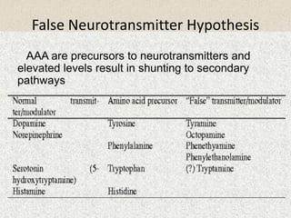
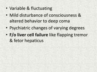
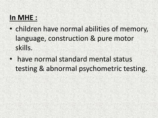
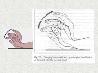
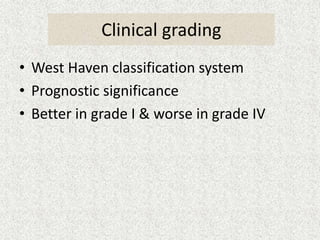
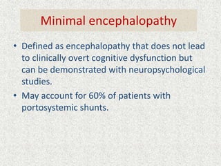
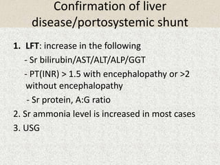
Hepatic Encephalopathy 
Dr Bikash Ranjan Praharaj 
Post Graduate, Dept of Pediatrics 
MKCG Medical College, Berhampur
• Definition 
• Etiology & classification 
• Pathogenesis 
• Precipitating factors 
• Clinical manifestation 
• Management 
• Outcome
Definition 
Hepatic encephalopathy (HE) is a complex 
metabolic mental state disorder with a 
spectrum of potentially reversible 
neuropsychiatric abnormalities seen in 
patients with severe acute or chronic liver 
dysfunction after exclusion of other brain 
diseases
Characterized by 
 Disturbances in consciousness & behaviour 
 Personality changes 
 Fluctuating neurologic signs, asterixis or 
flapping tremor 
 Distinctive EEG changes
Epidemiology 
 Exact data regarding incidence and prevalence is 
lacking 
 60-70% of patients with liver cirrhosis, while 
clinically unremarkable have pathologic changes on 
EEG and psychometric tests.(MHE) 
 Prevalence of minimal HE is about 53% in patients 
with extra hepatic portal vein obstruction 
 Approximately 50% of patients with liver cirrhosis 
develop HE after surgical portosystemic bypass 
procedures
Type Description Subcategory Subdivision 
A 
Encephalopathy associated with 
acute liver failure, typically 
associated with cerebral edema 
_____ ______ 
B 
Encephalopathy with Porto-systemic 
bypass and no 
intrinsic hepatocellular disease 
_____ ______ 
C 
Encephalopathy associated with 
cirrhosis or portal 
hypertension ⁄ Porto-systemic shunts 
Episodic 
Persistent 
Minimal 
•Percipated 
•Spontaneous 
•Recurrent 
•Mild 
•Severe 
•Treatment dependent 
Classification
Pathogenesis Theories 
–Ammonia hypothesis 
– False neurotransmitters & AA imbalance 
– Increase permeability of BBB 
–GABA hypothesis 
– Others
The Urea Cycle Aspartate Transaminase(AST) 
Alanine Transaminase (ALT)
Neurotoxic Action of Ammonia 
• Readily crosses blood-brain barrier 
• Ammonia reacts with α-ketoglutatrate to produce 
glutamate and glutamine 
• Consumption of α-ketoglutatrate, NADH and ATP, 
inhibition of pyruvate decarboxylase decrease 
TCA cycle activity which is vital for brain metabolism 
• Increased glutamine formation depletes glutamate 
stores which are needed by neural tissue l/t Irrepairable 
cell damage and neural cell death ensue. 
• Directly depress the cerebral blood flow & glucose 
metabolism 
• Direct toxic effect on the neuronal membrane
False neurotransmitters & Aminoacid imbalance 
• BCAA/AAA (N= 3-3.5, In hepatic coma=0.6- 
1.2) 
• BCAA : hyperinsulinemia  increased 
uptake & utilization by muscle & adipocytes 
• AAA : 
- insulin/glucagon --> catabolism of liver 
proteins & muscle --> AAA 
- Decrease hepatic deamination 
- Decrease gluconeogenesis
Which ultimately l/t 
Increase FNTs 
Decrease normal neurotransmitters 
Increase inhibitory neurotransmitters
False Neurotransmitter Hypothesis 
 AAA are precursors to neurotransmitters and 
elevated levels result in shunting to secondary 
pathways
Increase Permeability of Blood-Brain Barrier 
• Astrocyte (glial cell) volume is controlled by 
intracellular organic osmolyte which is glutamine 
• Increase glutamine levels in the brain result in 
increase volume of fluid within astrocytes resulting in 
cerebral edema (enlarged glial cells) 
• Neurological impairment 
“Alzheimer type II astrocytosis” 
– Pale, enlarged nuclei 
– characterisic of HE
GABA hypothesis 
• Major inhibitory neurotransmitter. 
• Evidence: increased GABAergic tone & 
Flumazenil improves clinical outcome 
• Cause 
- Decrease hepatic metabolism 
- Increase gut wall permeability
Some other theories 
• Dysregulation of serotonergic system 
(inversion of sleep rhythm) 
• Depletion of zinc & accumulation of Mn in 
globus pallidus. 
• Action of cytokines and bacterial LPS on 
astrocytes which are formed d/t inflmm. 
elsewhere in the body. 
• Neuronal NO synthase may increase c/t the 
altered cerebral perfusion.
Other neurotoxins 
• Mercaptans: Inhibit Na+-K+ ATPase 
• Short & medium chain fatty acids: 
inhibit Na+-K+ ATPase & Urea synthase 
• Phenol: a neurotoxin
Precipitating factors
CLINICAL MANIFESTATIONS
• Variable & fluctuating 
• Mild disturbance of consciousness & 
altered behavior to deep coma 
• Psychiatric changes of varying degrees 
• F/o liver cell failure like flapping tremor 
& fetor hepaticus
In MHE : 
• children have normal abilities of memory, 
language, construction & pure motor 
skills. 
• have normal standard mental status 
testing & abnormal psychometric testing.
Mild to moderate HE: 
• Decreased short term memory or 
forgetfulness 
• Loss of concentration & irritability 
• Asterixis, hyperventilation & 
hypothermia 
• Relative bradycardia (if ass. with increase 
ICP)
Clinical grading 
• West Haven classification system 
• Prognostic significance 
• Better in grade I & worse in grade IV
Minimal encephalopathy 
• Defined as encephalopathy that does not lead 
to clinically overt cognitive dysfunction but 
can be demonstrated with neuropsychological 
studies. 
• May account for 60% of patients with 
portosystemic shunts.
Clinical Manifestations & Diagnosis :MHE 
• Clinically normal 
• No mental deficit 
• Normal verbal ability 
• Deficit in attention ,visual perception, memory 
function, and learning 
• Impaired daily activities / driving 
• Only sophisticated tests such as 
EEG,CFF,ICT,NCT,DST, RBANS & PSE Syndrome 
test. 
• Neuroimaging : SPECT ,MRI,MRS.DWI
Manifestations & Diagnosis :MHE 
Number Connection Test (NCT) 
1 
2 
4 
3 
5 
6 
7 
8 
9 
10 
11 
12 
13 
14 
15 16 
17 
18 
19 20 
21 
25 
22 
23 
24 
Begin 
End 
Time to 
complete____________________ 
Draw a star 
SAMPLE HANDWRITING
Diagnosis of HE 
• No single laboratory test is sufficient to 
establish the diagnosis 
– No Gold Standard 
• Dx is mainly clinical on basis of history, clinical 
exam (includ mental status) & raised blood 
ammonia level
Diagnostic Criteria 
• Asterixis (“flapping tremor”) 
• Hx liver disease 
• Impaired performance on neuropsychological tests 
– Visual, sensory, brainstem auditory evoked potentials 
• Sleep disturbances 
• Fetor Hepaticus 
• EEG 
• PET scan 
– Changes of neurotransmission, astrocyte function 
• Elevated serum NH3 
– Stored blood contains ~30ug/L ammonia 
– Elevated levels seen in 90% pts with HE 
– Not needed for diagnosis
Investigations
Confirmation of liver 
disease/portosystemic shunt 
1. LFT: increase in the following 
- Sr bilirubin/AST/ALT/ALP/GGT 
- PT(INR) > 1.5 with encephalopathy or >2 
without encephalopathy 
- Sr protein, A:G ratio 
2. Sr ammonia level is increased in most cases 
3. USG
Detection of causative factors 
• Viral serologic markers: HBs Ag, HBe Ag, anti-HBc, 
HBV DNA increased in Hepatitis 
• TORCH screening 
• Autoimmune ab: ANA, ASMA, LKM1 
• Sr Cu, ceruloplasmin, urinary Cu : wilson’s disease 
• Urine for metabolic disorders 
• Sweat chloride & cystic fibrosis mutation studies 
• Alfa 1 antitrypsin levels : Alfa 1 antitrypsin def 
• Alfa feto protein : tyrosinemia type 1 
• Sr lactate & pyruvate : GSD & resp chain defects 
• Liver biopsy: cirrhosis
R/o other diseases with similar presentation 
• CT Scan: to r/o cerebral hemorrhage 
• EEG: r/o seizure disorder 
• CSF study: meningitis or encephalitis 
• Blood tests: metabolic causes of 
encephalopathy including hypoglycemia & 
uremia 
• Serum urea, Cr & electrolytes: renal failure
Detection of complications 
• ABG- hypoxia is common 
• CBC: to r/o infection 
• Hb,PCV,CPS 
• PT, aPTT 
• Pt count decreased in advanced cases & 
coagulopathy 
• Blood glucose: hypoglycemia 
• Sr ammonia 
• RFT
Differential Diagnosis 
Metabolic 
encephalopathies 
- Diabetes (hypoglycemia, 
ketoacidosis) 
- Hypoxia 
- Carbon dioxide narcosis 
Toxic 
encephalopathies 
- Alcohol (acute alcohol 
intoxication, delirium 
tremens, Wernicke- 
Korsakoff syndrome) 
- Drugs 
Intracranial events 
- Intracerebral 
bleeding or 
infarction 
-Tumor 
- Infections 
(abscess, 
meningitis) 
- Encephalitis 
Psychiatric diseases
Treatment of Hepatic Encephalopathy 
• Various measures in current treatment of HE 
– Strategies to lower ammonia production/absorption 
• Nutritional management 
– Protein restriction 
– BCAA supplementation 
• Medical management 
– Medications to counteract ammonia’s effect on brain cell 
function 
• Lactulose 
• Antibiotics 
– Devices to compensate for liver dysfunction 
– Liver transplantation
Proposed 
Complex 
Feedback 
Mechanisms 
In Treatment 
Of HE
Diet 
• Decreased protein intake with high 
carbohydrates 
• Calorie in the form of 10%D infusion 
• Protein restricted to 0.5-1 g/kg/day 
• Veg protein preferred as they are less 
amminogenic , contain less amount of 
methionine & AAA and more fibres 
• Dietary supplementation of BAA 
• 50% of non-protein calories should come from 
MCT
Lactulose/lactitol 
• Non absorbable synthetic diasachharide 
• Degraded by colonic bacteria to form lactic acid & acetic 
acid 
• Fecal acidity increase l/t decrease absorption of NH3 
• Favours growth of lactose fermenting bacteria & 
diminished growth of ammo producing bacteria like 
bacteroides 
• Detoxify short chain FAs produced in presence of blood 
& proteins 
Dose: 1-2 ml/kg per orally or as enema in higher doses 
N:B:- Alternatively, phosphate enema can be used
Actions Of Lactulose
Bowel sterilization 
• Neomycin : orally through NGT dose: 50- 
100mg/kg 
• Ampicillin 
• Rifaximin 
• metronidazole
Other measures 
• NGT aspiration 
• High colonic wash 
• Zn 
• L-Ornithine-L-Aspartate : oral/iv 
• Sodium Benzoate: 5g PO BD 
• H.Pylori eradication
Supportive care 
• Fluid & electrolyte balance: 
- Should contain 1meq/kg/d of glucose 
- Met acidosis: NaHco3 
- Hypokalemia: pot. Chloride 
• Early identification & T/t of GI bleeding, 
septicemia & hypoxia 
• Avoidance of ppt factors: drugs/paracentesis 
• Drugs: To improve sensorium e.g Flumazenil, l-dopa, 
bromocriptine
T/t in Resistant cases 
• Plasmapheresis/hemodialysis 
• exchange transfusion 
• Surgical shunt occlusion 
• Temporary hepatic support: 
- ELAD (Extracorporeal Liver Assist Devices) 
- MARS (Molecular Adsorbent Recirculating 
System) 
• Liver transplantation
T/t of complications 
1. CNS complications: 
• Cerebral edema: 
- Elevation of bed by 30 “,mannitol, 
hyperventilation & fluid restriction 
- Hypothermia & phenobarbitone 
• Seizures: phenytoin & gabapentin 
• Cerebral hypoxia: O2, N-acetylcysteine 
2. Hypotension: colloids/albumin infusion 
3. Bleeding: Inj Vit-k/ FFP/ Inj Ranitidine
4. Respiratory failure: 
- In Stage III & IV 
- Endotracheal Intubation 
5. Renal Failure: 
- Furosemide in a dose of 1-2 mg/kg in early 
stages if CVP > 8-10 cm of H2O 
- Hemodialysis in established cases 
- Urine output should be maintained 
- Dopamine: Improve renal perfusion 
6. Ascites: 5% albumin, bile acid binders
Monitoring Protocol 
Daily Once in 3 days Weekly 
•Blood glucose (2 hrly) 
•Sr electrolytes: Na, K, HCO3- 
•Hb, PCV, CPS 
-Renal function 
tests 
-PT 
-NEC 
-Sr amino acids 
-EEG
Minimal HE 
1.No established indication for treatment 
2.Consider changes in daily activities (avoid 
driving) 
3.In selected patients 
• Lactulose /lactitol 
• Dietary intervention vegetable based diet 
• Probiotics
Prophylaxis Of New Episodes 
1.Control of precipitating factors 
2.Nutritional support 
3.Adequate protein intake with dairy and 
vegetable based diets 
4.Vitamins 
5.Zinc supplementation 
6.Lactulose /lactitol as needed 
7. OLT evaluation
Course And Prognosis 
•Develops rapidly few hours – 1-2 days 
•Mortality in grade IV is 80% 
•Death usually due to brain herniation / edema ICH 
•Type C develops slowly – undulating course / 
recurrence 
•Neuropsychiatric manifestations are reversible 
•Can lead to permanent damage with dementia, extra 
pyramidal signs, cerebellar degeneration,myelopathy 
with spastic paraplegia, peripheral polyneuropthy 
•Liver TX can reverse all changes
Prognostic indicators 
FEATURES GOOD PROGNOSIS BAD PROGNOSIS 
AGE CHILDREN ADOLESCENTS 
ETIOLOGY PCM POISONING, HEP A HEP C 
DURATION OF 
ENCEPHALOPATHY 
< 7 DAYS > 7 DAYS 
COMA GRADE I & II III & IV 
LIVER SIZE ENLARGED SHRINKING/NON PALPABLE 
BLEEDING TENDENCY ABSENT PRESENT 
FLUID RETENTION ---- +++ 
SR ALBUMIN N 
PT N PROLONGED 
LIVER ENZYMES: AST/ALT N 
AFP 
ASS. COMPLICATIONS ABSENT PRESENT 
IMPROVEMENT OF 
RAPID 
SENSORIUM WITH T/t 
NO IMPROVEMENT AFTER 
48 HRS OF T/t
Take home points 
• Ammonia is the main culprit 
• Dx mainly by clinical exclusion 
• Bad prognostic indicators: 
- Liver span 
- Bilirubin level 
- Liver enzyme levels 
- Prothrombin time 
• T/t of precipitating causes & supportive care is 
the mainstay of t/t 
• Prognosis bad in type A & better in other types
Thanks

Hepatic encephalopathy

  • 1.
    Hepatic Encephalopathy DrBikash Ranjan Praharaj Post Graduate, Dept of Pediatrics MKCG Medical College, Berhampur
  • 2.
    • Definition •Etiology & classification • Pathogenesis • Precipitating factors • Clinical manifestation • Management • Outcome
  • 3.
    Definition Hepatic encephalopathy(HE) is a complex metabolic mental state disorder with a spectrum of potentially reversible neuropsychiatric abnormalities seen in patients with severe acute or chronic liver dysfunction after exclusion of other brain diseases
  • 4.
    Characterized by Disturbances in consciousness & behaviour  Personality changes  Fluctuating neurologic signs, asterixis or flapping tremor  Distinctive EEG changes
  • 5.
    Epidemiology  Exactdata regarding incidence and prevalence is lacking  60-70% of patients with liver cirrhosis, while clinically unremarkable have pathologic changes on EEG and psychometric tests.(MHE)  Prevalence of minimal HE is about 53% in patients with extra hepatic portal vein obstruction  Approximately 50% of patients with liver cirrhosis develop HE after surgical portosystemic bypass procedures
  • 6.
    Type Description SubcategorySubdivision A Encephalopathy associated with acute liver failure, typically associated with cerebral edema _____ ______ B Encephalopathy with Porto-systemic bypass and no intrinsic hepatocellular disease _____ ______ C Encephalopathy associated with cirrhosis or portal hypertension ⁄ Porto-systemic shunts Episodic Persistent Minimal •Percipated •Spontaneous •Recurrent •Mild •Severe •Treatment dependent Classification
  • 7.
    Pathogenesis Theories –Ammoniahypothesis – False neurotransmitters & AA imbalance – Increase permeability of BBB –GABA hypothesis – Others
  • 8.
    The Urea CycleAspartate Transaminase(AST) Alanine Transaminase (ALT)
  • 10.
    Neurotoxic Action ofAmmonia • Readily crosses blood-brain barrier • Ammonia reacts with α-ketoglutatrate to produce glutamate and glutamine • Consumption of α-ketoglutatrate, NADH and ATP, inhibition of pyruvate decarboxylase decrease TCA cycle activity which is vital for brain metabolism • Increased glutamine formation depletes glutamate stores which are needed by neural tissue l/t Irrepairable cell damage and neural cell death ensue. • Directly depress the cerebral blood flow & glucose metabolism • Direct toxic effect on the neuronal membrane
  • 11.
    False neurotransmitters &Aminoacid imbalance • BCAA/AAA (N= 3-3.5, In hepatic coma=0.6- 1.2) • BCAA : hyperinsulinemia  increased uptake & utilization by muscle & adipocytes • AAA : - insulin/glucagon --> catabolism of liver proteins & muscle --> AAA - Decrease hepatic deamination - Decrease gluconeogenesis
  • 12.
    Which ultimately l/t Increase FNTs Decrease normal neurotransmitters Increase inhibitory neurotransmitters
  • 13.
    False Neurotransmitter Hypothesis  AAA are precursors to neurotransmitters and elevated levels result in shunting to secondary pathways
  • 14.
    Increase Permeability ofBlood-Brain Barrier • Astrocyte (glial cell) volume is controlled by intracellular organic osmolyte which is glutamine • Increase glutamine levels in the brain result in increase volume of fluid within astrocytes resulting in cerebral edema (enlarged glial cells) • Neurological impairment “Alzheimer type II astrocytosis” – Pale, enlarged nuclei – characterisic of HE
  • 16.
    GABA hypothesis •Major inhibitory neurotransmitter. • Evidence: increased GABAergic tone & Flumazenil improves clinical outcome • Cause - Decrease hepatic metabolism - Increase gut wall permeability
  • 17.
    Some other theories • Dysregulation of serotonergic system (inversion of sleep rhythm) • Depletion of zinc & accumulation of Mn in globus pallidus. • Action of cytokines and bacterial LPS on astrocytes which are formed d/t inflmm. elsewhere in the body. • Neuronal NO synthase may increase c/t the altered cerebral perfusion.
  • 18.
    Other neurotoxins •Mercaptans: Inhibit Na+-K+ ATPase • Short & medium chain fatty acids: inhibit Na+-K+ ATPase & Urea synthase • Phenol: a neurotoxin
  • 19.
  • 20.
  • 21.
    • Variable &fluctuating • Mild disturbance of consciousness & altered behavior to deep coma • Psychiatric changes of varying degrees • F/o liver cell failure like flapping tremor & fetor hepaticus
  • 22.
    In MHE : • children have normal abilities of memory, language, construction & pure motor skills. • have normal standard mental status testing & abnormal psychometric testing.
  • 23.
    Mild to moderateHE: • Decreased short term memory or forgetfulness • Loss of concentration & irritability • Asterixis, hyperventilation & hypothermia • Relative bradycardia (if ass. with increase ICP)
  • 25.
    Clinical grading •West Haven classification system • Prognostic significance • Better in grade I & worse in grade IV
  • 27.
    Minimal encephalopathy •Defined as encephalopathy that does not lead to clinically overt cognitive dysfunction but can be demonstrated with neuropsychological studies. • May account for 60% of patients with portosystemic shunts.
  • 28.
    Clinical Manifestations &Diagnosis :MHE • Clinically normal • No mental deficit • Normal verbal ability • Deficit in attention ,visual perception, memory function, and learning • Impaired daily activities / driving • Only sophisticated tests such as EEG,CFF,ICT,NCT,DST, RBANS & PSE Syndrome test. • Neuroimaging : SPECT ,MRI,MRS.DWI
  • 29.
    Manifestations & Diagnosis:MHE Number Connection Test (NCT) 1 2 4 3 5 6 7 8 9 10 11 12 13 14 15 16 17 18 19 20 21 25 22 23 24 Begin End Time to complete____________________ Draw a star SAMPLE HANDWRITING
  • 30.
    Diagnosis of HE • No single laboratory test is sufficient to establish the diagnosis – No Gold Standard • Dx is mainly clinical on basis of history, clinical exam (includ mental status) & raised blood ammonia level
  • 31.
    Diagnostic Criteria •Asterixis (“flapping tremor”) • Hx liver disease • Impaired performance on neuropsychological tests – Visual, sensory, brainstem auditory evoked potentials • Sleep disturbances • Fetor Hepaticus • EEG • PET scan – Changes of neurotransmission, astrocyte function • Elevated serum NH3 – Stored blood contains ~30ug/L ammonia – Elevated levels seen in 90% pts with HE – Not needed for diagnosis
  • 32.
  • 33.
    Confirmation of liver disease/portosystemic shunt 1. LFT: increase in the following - Sr bilirubin/AST/ALT/ALP/GGT - PT(INR) > 1.5 with encephalopathy or >2 without encephalopathy - Sr protein, A:G ratio 2. Sr ammonia level is increased in most cases 3. USG
  • 34.
    Detection of causativefactors • Viral serologic markers: HBs Ag, HBe Ag, anti-HBc, HBV DNA increased in Hepatitis • TORCH screening • Autoimmune ab: ANA, ASMA, LKM1 • Sr Cu, ceruloplasmin, urinary Cu : wilson’s disease • Urine for metabolic disorders • Sweat chloride & cystic fibrosis mutation studies • Alfa 1 antitrypsin levels : Alfa 1 antitrypsin def • Alfa feto protein : tyrosinemia type 1 • Sr lactate & pyruvate : GSD & resp chain defects • Liver biopsy: cirrhosis
  • 35.
    R/o other diseaseswith similar presentation • CT Scan: to r/o cerebral hemorrhage • EEG: r/o seizure disorder • CSF study: meningitis or encephalitis • Blood tests: metabolic causes of encephalopathy including hypoglycemia & uremia • Serum urea, Cr & electrolytes: renal failure
  • 36.
    Detection of complications • ABG- hypoxia is common • CBC: to r/o infection • Hb,PCV,CPS • PT, aPTT • Pt count decreased in advanced cases & coagulopathy • Blood glucose: hypoglycemia • Sr ammonia • RFT
  • 37.
    Differential Diagnosis Metabolic encephalopathies - Diabetes (hypoglycemia, ketoacidosis) - Hypoxia - Carbon dioxide narcosis Toxic encephalopathies - Alcohol (acute alcohol intoxication, delirium tremens, Wernicke- Korsakoff syndrome) - Drugs Intracranial events - Intracerebral bleeding or infarction -Tumor - Infections (abscess, meningitis) - Encephalitis Psychiatric diseases
  • 38.
    Treatment of HepaticEncephalopathy • Various measures in current treatment of HE – Strategies to lower ammonia production/absorption • Nutritional management – Protein restriction – BCAA supplementation • Medical management – Medications to counteract ammonia’s effect on brain cell function • Lactulose • Antibiotics – Devices to compensate for liver dysfunction – Liver transplantation
  • 39.
    Proposed Complex Feedback Mechanisms In Treatment Of HE
  • 40.
    Diet • Decreasedprotein intake with high carbohydrates • Calorie in the form of 10%D infusion • Protein restricted to 0.5-1 g/kg/day • Veg protein preferred as they are less amminogenic , contain less amount of methionine & AAA and more fibres • Dietary supplementation of BAA • 50% of non-protein calories should come from MCT
  • 41.
    Lactulose/lactitol • Nonabsorbable synthetic diasachharide • Degraded by colonic bacteria to form lactic acid & acetic acid • Fecal acidity increase l/t decrease absorption of NH3 • Favours growth of lactose fermenting bacteria & diminished growth of ammo producing bacteria like bacteroides • Detoxify short chain FAs produced in presence of blood & proteins Dose: 1-2 ml/kg per orally or as enema in higher doses N:B:- Alternatively, phosphate enema can be used
  • 42.
  • 43.
    Bowel sterilization •Neomycin : orally through NGT dose: 50- 100mg/kg • Ampicillin • Rifaximin • metronidazole
  • 44.
    Other measures •NGT aspiration • High colonic wash • Zn • L-Ornithine-L-Aspartate : oral/iv • Sodium Benzoate: 5g PO BD • H.Pylori eradication
  • 45.
    Supportive care •Fluid & electrolyte balance: - Should contain 1meq/kg/d of glucose - Met acidosis: NaHco3 - Hypokalemia: pot. Chloride • Early identification & T/t of GI bleeding, septicemia & hypoxia • Avoidance of ppt factors: drugs/paracentesis • Drugs: To improve sensorium e.g Flumazenil, l-dopa, bromocriptine
  • 46.
    T/t in Resistantcases • Plasmapheresis/hemodialysis • exchange transfusion • Surgical shunt occlusion • Temporary hepatic support: - ELAD (Extracorporeal Liver Assist Devices) - MARS (Molecular Adsorbent Recirculating System) • Liver transplantation
  • 47.
    T/t of complications 1. CNS complications: • Cerebral edema: - Elevation of bed by 30 “,mannitol, hyperventilation & fluid restriction - Hypothermia & phenobarbitone • Seizures: phenytoin & gabapentin • Cerebral hypoxia: O2, N-acetylcysteine 2. Hypotension: colloids/albumin infusion 3. Bleeding: Inj Vit-k/ FFP/ Inj Ranitidine
  • 48.
    4. Respiratory failure: - In Stage III & IV - Endotracheal Intubation 5. Renal Failure: - Furosemide in a dose of 1-2 mg/kg in early stages if CVP > 8-10 cm of H2O - Hemodialysis in established cases - Urine output should be maintained - Dopamine: Improve renal perfusion 6. Ascites: 5% albumin, bile acid binders
  • 49.
    Monitoring Protocol DailyOnce in 3 days Weekly •Blood glucose (2 hrly) •Sr electrolytes: Na, K, HCO3- •Hb, PCV, CPS -Renal function tests -PT -NEC -Sr amino acids -EEG
  • 50.
    Minimal HE 1.Noestablished indication for treatment 2.Consider changes in daily activities (avoid driving) 3.In selected patients • Lactulose /lactitol • Dietary intervention vegetable based diet • Probiotics
  • 51.
    Prophylaxis Of NewEpisodes 1.Control of precipitating factors 2.Nutritional support 3.Adequate protein intake with dairy and vegetable based diets 4.Vitamins 5.Zinc supplementation 6.Lactulose /lactitol as needed 7. OLT evaluation
  • 52.
    Course And Prognosis •Develops rapidly few hours – 1-2 days •Mortality in grade IV is 80% •Death usually due to brain herniation / edema ICH •Type C develops slowly – undulating course / recurrence •Neuropsychiatric manifestations are reversible •Can lead to permanent damage with dementia, extra pyramidal signs, cerebellar degeneration,myelopathy with spastic paraplegia, peripheral polyneuropthy •Liver TX can reverse all changes
  • 53.
    Prognostic indicators FEATURESGOOD PROGNOSIS BAD PROGNOSIS AGE CHILDREN ADOLESCENTS ETIOLOGY PCM POISONING, HEP A HEP C DURATION OF ENCEPHALOPATHY < 7 DAYS > 7 DAYS COMA GRADE I & II III & IV LIVER SIZE ENLARGED SHRINKING/NON PALPABLE BLEEDING TENDENCY ABSENT PRESENT FLUID RETENTION ---- +++ SR ALBUMIN N PT N PROLONGED LIVER ENZYMES: AST/ALT N AFP ASS. COMPLICATIONS ABSENT PRESENT IMPROVEMENT OF RAPID SENSORIUM WITH T/t NO IMPROVEMENT AFTER 48 HRS OF T/t
  • 54.
    Take home points • Ammonia is the main culprit • Dx mainly by clinical exclusion • Bad prognostic indicators: - Liver span - Bilirubin level - Liver enzyme levels - Prothrombin time • T/t of precipitating causes & supportive care is the mainstay of t/t • Prognosis bad in type A & better in other types
  • 55.