
Recommended
More Related Content
Similar to CONGENITAL ESOPHAGEAL.pptx
Similar to CONGENITAL ESOPHAGEAL.pptx (20)
More from musayansa
More from musayansa (20)
Recently uploaded
Recently uploaded (20)
CONGENITAL ESOPHAGEAL.pptx
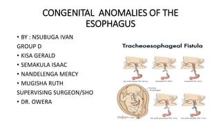
- 1. CONGENITAL ANOMALIES OF THE ESOPHAGUS • BY : NSUBUGA IVAN GROUP D • KISA GERALD • SEMAKULA ISAAC • NANDELENGA MERCY • MUGISHA RUTH SUPERVISING SURGEON/SHO • DR. OWERA
- 2. INTRODUCTION • Embryology: • Its part of foregut • Simple tube passing through to the hind end, its whole length supported by a dorsal mesentery attached to the midline in front of the aorta • Supplied by the coeliac artery running through the dorsal mesogastrium • At 4th week of gestation-appearance of the respiratory diverticulum at the ventral wall of the foregut, at border with the pharyngeal gut
- 3. Continua…… • Tracheal esophageal septum: divides foregut into • Ventral portion-respiratory primodium • Dorsal portion-esophagus • Its short at first, lengthens rapidly with descent of the heart and lungs • About 25cm long in adults Blood supply • Esophageal branches-left gastric artery-coelic trunk • Esophageal branch-inferior thyroid artery, right bronchial artery, descending thoracic aorta
- 11. Continua…. • Histology: • Mucosa-stratified squamous non-keratinized epithelium • Submucosa • Muscularis • adventitia
- 12. ESOPHAGEAL ATRESIA • Defin: an interruption in the continuity of the esophagus with/out fistula to the trachea • First documented case in 1670 by William Durston’s published description of “a narrative of a monstrous birth in polymouth…” he described a blind- ending upper esophageal pouch in the right infant of a set of female thoracopagus-conjoined twins PATHOGENESIS • The pathogenesis of congenital EA malformations remains unknown • The pathogenesis is heterogeneous and multifactorial and involves multiple genes and complex gene-environment interaction
- 13. Continua…. • Theories of foregut occlusion and failure of recanalization of the intestinal lumen • Another theory has been put forth by Cozzi and col- leagues and others in which it is suggested that EA is a component of cephalic neurocristopathy. • pathogenesis of EA may be related to defective pharyngeal arch development-DiGeorge syndrome
- 14. Epidemiology • The reported incidence of EA, with or without TEF (EA TEF), is 1:3500 live-born infants; • In Uganda mean incidence was 3.16 per 10,000 live birth according to N.kakembo et al 2020
- 15. Associated Anomalies • between 50% and 70% of infants with EA have at least one associated congenital malformation • Syndromic malformation: VACTERL(vertebral defects, anal atresia, cardiac defects, trachea-esophageal fistula, renal anomalies, and limb abnormalies), CHARGE(coloboma, choanal atresia, esophageal atresia), Fanconi anemia, Opitz G(affects midline structures), and Goldenhar syndrome.
- 16. Continua…..
- 17. Continua…. • Associated neurologic anomalies include neural tube defects (2.3%), hydrocephalus (5.2%), holoprosencephaly (2.3%), and anophthalmia or microphthalmos (3.7%) • Other reported associated anomalies include choanal atresia (5.2%), facial cleft (7.2%), abdominal wall defects (4.3%), and diaphragmatic hernia (2.9%)
- 18. Classification
- 19. Continua…. • In addition to anatomic classifications, in 1962 D. J. Waterston, R. E. Bonham Carter, and Eoin Aberdeen of the Hospital for Sick Children in London developed a classification system rela- ted to risk factors in infants with EA
- 20. Continua… • infants in the “good”-risk category (A) were typically treated with immediate operative repair, “moderate”-risk infants (B) were managed by delayed repair, and “high”-risk infants (C) were treated by staged repair.
- 21. Okamoto and colleagues revised Spitz classification
- 22. Diagnosis and Clinical Findings • Prenatal detection of EA by ultrasonography relies on the finding of a small or absent stomach bubble and associated maternal polyhydramnios. • The ultrasonographic finding of an esophageal pouch in the middle of the fetal neck in association with poly- hydramnios and a small stomach may increase the accuracy of prenatal diagnosis of EA. • Most infants with EA are symptomatic in the first few hours of life.
- 23. Continua…. • The earliest clinical sign of EA is usually excessive salivation that results from pooling of secretions in the pharynx. • Typically, the first feeding is followed by regurgitation, choking, and coughing. • Other features: cyanosis with or without feeding, respiratory distress, inability to swallow, and inability to pass a feeding or suction catheter through the mouth or nose into the stomach
- 24. Continua…. • Incase distal fistula is present: the abdomen distends as inspired air passes through the fistula into the stomach. • Pulmonary compromise can become significant as gastric fluid refluxes through the TEF, spills into the trachea and lungs, and leading to chemical pneumonitis • As the abdomen distends with air, the diaphragms (the principal muscles of respiration in the newborn) elevate and pulmonary status worsens. • Aspiration of saliva from the upper pouch into the trachea further exacerbates the pulmonary compromise
- 25. Continua… • The diagnosis of EA can be confirmed by passing a firm catheter through the mouth into the esophagus to the point at which resistance is met. • A few milliliters of air can be injected through the tube and used as a contrast agent to distend the upper esophageal pouch as frontal and lateral films are obtained
- 27. Continua… • A flexible catheter such as a feeding tube will often curl up in the upper pouch, also suggesting the diagnosis of EA
- 28. Continua…. • If necessary, 0.5 to 1.0 mL of diluted barium can be used as a contrast agent and injected into the upper pouch to confirm the diagnosis
- 29. Continua… • Air in the stomach and bowel confirms the presence of a distal TEF. • Absence of air in the abdomen typically represents isolated EA without distal TEF
- 30. Continua….. • The presence or absence of a proximal TEF must be confirmed by bronchoscopy. • The diagnosis of TEF without EA is more difficult and requires a high index of suspicion on the basis of clinical symptoms. • Investigations are triggered by repeated coughing or choking during feedings, often with pulmonary infiltrates on chest radiograph suggesting aspiration. • The dx can be made by barium esophagography in the prone position ; however, bronchoscopy with or without esophagoscopy is often required to confirm the diagnosis.
- 32. Continua… • The clinical evidence of associated anomalies should be considered in the diagnostic evaluation for EA. • physical examination focused on seeking known associated defects (such as those of the VACTERL association), • additional testing includes: echocardiography, renal ultrasonography, and chromosomal analysis.
- 33. Preoperative Treatment • Pneumonitis from aspiration of upper pouch secretions and reflux of gastric acid through the TEF causing worsening respiratory distress is the most critical preoperative problem for an infant with EA-TEF. • Immediate management: measures to prevent further aspiration and treatment of pneumonitis. A sump catheter should be positioned in the upper esophageal pouch to continuously aspirate saliva under low-pressure suction.(double-lumen Replogle type of catheter is best)
- 34. Continua…… • Infant positioned to minimize reflux of gastric fluid up through the TEF. -An upright sitting position or the head-up, prone position. • Broad-spectrum antibiotic coverage • Pulmonary physiotherapy • Intravenous fluid therapy with 10% dextrose and hypotonic saline-to maintain fluid, electrolyte, and glucose balance. • Vitamin K analogue should also be administered before surgery.
- 35. Continua…. • NOTE: Routine endotracheal intubation should be avoided bcoz of the risk for gastric perforation and worsening respiratory distress as the abdomen becomes distended from ventilation through the TEF.
- 36. Operative Repair • depends greatly on the specific type of anomaly present and the occurrence of associated anomalies. ESOPHAGEAL ATRESIA WITH DISTAL TRACHEOESOPHAGEAL FISTULA • Emergent operation for EA with distal TEF is seldom necessary, • Period of 24 to 48 hours between diagnosis and operation • Full assessment of the infant and treatment of pulmonary insufficiency including atelectasis and pneumonitis.
- 37. Continua….. • Open thoracotomy or thoracoscopic division of the fistula with primary anastomosis of the esophagus is the operative procedure of choice-if possible • For the open approach, the infant is usually positioned for a standard right posterolateral thoracotomy, with the right arm extended above the head and the head slightly flexed.
- 38. Continua…. • Through the 4th intercostal space • Most pediatric surgeons continue to advocate the extrapleural approach for EA-TEF repair bcoz a substantial anastomotic leak does not result in empyema but merely causes an esophagocutaneous fistula, which typically closes in 1 to 2 weeks. • Surgeons who use the transpleural route argue that the operative time is shorter and, with current antibiotic options, the risk for empyema after a leak is minimal.
- 40. Continua…. • Effort is made to preserve the vagal fibers that supply the distal portion of the esophagus. • Close the fistula with absorbable suture such as polydioxanone-Bcoz occasional intratracheal granuloma has occurred with permanent sutures. • It is important to leave 1 to 2 mm of esophagus on the tracheal end of the fistula to minimize the risk for postoperative tracheal stricture, while not leaving an excessive amount that would act as a tracheal diverticulum.
- 41. Continua…. • The air-tightness of the tracheal closure can be tested by filling the thoracic cavity with warm saline and watching for bubbles with positive-pressure ventilation. • A small tube should be passed through the distal end of the esophagus to ensure that the lumen is adequate and open and to aspirate air distending the stomach.-ensures distal esophageal patency and the potential for early postoperative enteral feeding if desired.
- 42. Continua…. • In infants with EA and distal TEF, another technique for handling the fistula and uniting the esophageal ends is the end-to-side anastomosis developed by Sulamaa and colleague and championed by Duhamel and others: -a single ligature is used to ligate the fistula; the upper pouch is mobilized down to the lower esophageal segment, and a wide, oblique, end-to-side anastomosis is performed.→risk for recurrent TEF formation.
- 43. LONG-GAP ESOPHAGEAL ATRESIA • Occasionally in infants with EA-TEF, the upper pouch is high and the distance between the upper and lower esophageal segments limits the ability to easily complete end-to-end esophagoesophagostomy with acceptable tension. • In the case of isolated EA, in which a “long gap” is expected,most pediatric surgeons would proceed with the operative placement of a gastrostomy tube, a period of observation,and an attempted delayed primary repair.
- 44. Continua…. • During the first several months of life, the gap btwn the two ends of the esophagus tends to lessen bcoz of spontaneous growth, possibly related in part to reflux of bolus gastric feedings into the lower esophagus, which makes primary repair more feasible. OR • Upper pouch bougienage-weighted bougie is passed through the mouth into the upper pouch, with forward pressure applied daily or twice daily for 6 to 12 weeks before attempting delayed primaryrepair.
- 45. Continua….. • Collis gastroplasty with Nissen fundoplication has also been described as a means of lengthening the distal end of the esophagus to avoid esophageal replacement.
- 46. ESOPHAGEAL REPLACEMENT • Colon replacement, or ileocolon, has been widely practiced for many years as a method of esophageal replacement. • The jejunum has also been used for esophageal replacement both in a Roux-en-Y fashion and as a free graft with microvascular anastomosis. • Gastric transposition has more recently become a well established and successful esophageal substitute in infants with EA.
- 47. ESOPHAGEAL ATRESIA WITHOUT TRACHEOESOPHAGEAL FISTULA • Infants born with isolated EA have almost no esophagus in their thorax. • There is 52% incidence of prematurity, an 11% incidence of Down syndrome, and a 10% incidence of duodenal atresia; 19% cardiac, 16% urogenital, 14% vertebral, • The clinical manifestations of an infant with isolated EA are similar to those of an infant with EA-TEF
- 48. Continua….. • A gastrostomy tube should be placed within the first 24 to 48 hours of life, and delayed primary repair,-Placement of the gastrostomy tube→ early institution of enteral feedings, →subsequent enlargement of the diminutive stomach and stretching of the distal esophageal pouch.
- 49. ISOLATED (H-TYPE) TRACHEOESOPHAGEAL FISTULA • Congenital TEF without EA or the “H” type of TEF (perhaps more anatomically accurately called the “N” type) occurs in approx 4% of esophageal anomalies reported. • Usually manifested in the first few days of life when the neonate repeatedly chokes on attempting to feed or has unexplained cyanotic spells. • Intermittent abdominal distention during coughing or crying can occur as air passes through the fistula into the stomach.
- 50. Continua…. • Recurrent bouts of pneumonia typically involving the right upper lobe, in older infants and children • A high index of suspicion • Dx of isolated TEF can be suspected if plain radiographs of the chest show evidence of aspiration pneumonitis with gastric distention. • A reliable way to establish the diagnosis is by tube video esophagography performed while the infant is prone;
- 52. ESOPHAGEAL ATRESIA WITH AN UPPER POUCH FISTULA • There are two versions of this anomaly: (1) proximal pouch fistula in association with distal TEF (double fistula) (2) proximal pouch fistula without distal TEF. • Dx made by a preoperative proximal pouch contrast study→a small upper pouch in addition to identifying the fistula. • Preoperative bronchoscopy to make or confirm the diagnosis of a proximal pouch fistula
- 54. Outcomes • Survival rates for infants who have EA with or without TEF have improved dramatically in the past 50 years.
- 55. Complications • Risk factors for complications twin birth, preoperative intubation, birth weight less than 2500 g, long-gap atresia anastomotic leak postoperative intubation longer than 4 days, inability to feed at the end of 1 month.
- 56. EARLY COMPLICATIONS • Anastomotic Leak ; -Occurs in approximately 13% to 16% of patients with EA. -Most leaks are clinically insignificant and can be managed with adequate drainage and nutritional support -A patent mediastinal drain in place, if a retropleural approach is undertaken -Typically recognized early (24 to 48 hours) after the initial repair. -The infant frequently deteriorates from tension pneumothorax or mediastinitis uncontrolled by drainage and antibiotics.
- 57. Continua…. • Leading factors; -Poor surgical technique, ischemia of the esophageal ends, use of myotomy, and excessive tension at the anastomotic site. • Reoperation for control of sepsis with adequate drainage and attempted repair of the anastomotic leak are warranted.
- 58. Esophageal Stricture • Common complication • Spitz and Hitchcock proposed that stricture be defined as the presence of symptoms (dysphagia and recurrent respiratory problems from aspiration or foreign body obstruction) and narrowing noted on endoscopy or contrast esophagography
- 59. Contributing factors • Poor anastomotic technique(excessive tension, two-layered anastomosis, and silksuture material), • Long gap, ischemia at the ends of the esophagus, • GERD, • Anastomotic leak. • Treated by dilation performed via antegrade or retrograde bougienage.-Savary-gilliard type dilators prograde, endoscopic balloon dilators
- 60. Continua….. • NOTE: (1) A recalcitrant stricture resistant to repeated dilations will require resection and reanastomosis or even esophageal replacement. (2) Application of mitomycin c to the stricture under endoscopic control has been reported to reduce stricture formation after dilation. (3) Prescribe H2 blockers incase of GERD, or PPI
- 61. Recurrent Tracheoesophageal Fistula • occurs in 3% to 14% of patients after initial operative division or ligation. • anastomotic leak with local inflammation and erosion through the previous site of TEF repair. • Symptoms : -coughing and choking or cyanosis with feedings -recurrent pulmonary infections • The diagnosis may be suggested by: -an air-filled esophagus on plain radiographs of the chest.
- 62. Continua…. NOTE: Can be minimized by the use of a pleural flap, vascularized pericardial flap, and azygos vein flap, interposed between the esophageal and tracheal suture lines • typically requires surgical repair-Thoracotomy with fistula ligation and division is the operation of choice.
- 63. LATE COMPLICATIONS • Gastroesophageal Reflux Disease -GERD occurs in 30% to 70% of patients after repair of EA. -Due to shortening of the intra-abdominal portion of the esophagus because of anastomotic tension and/or esophageal motor dysfunction, either acquired as a result of operative manipulation or intrinsic to the congenital anomaly itself. -results to esophagitis later in life -vomiting, dysphagia, and recurrent anastomotic stenosis,
- 64. Continua…. • Txt- H2 blockers, PPI, Collis-Nissen fundoplication to gain intra- abdominal esophageal length.
- 65. Tracheomalacia • “brassy” or “barking” cough in mild cases to recurrent pneumonia or acute, life-threatening apneic spells.
- 66. CONGENITAL STENOSIS OF THE ESOPHAGUS • Incomplete separation of the respiratory tract from the primitive foregut at 25th day of life. • Highest in japan • Nihoul-Fekete classification: (1) a membranous web or diaphragm-rarest (2)fibromuscular thickening, (3) stricture secondary to tracheobronchial remnants in the wall of the esophagus.-most common
- 67. Continua…. • Diagnosis: -Symptoms of CES usually begin in infancy with progressive dysphagia and vomiting, generally after the introduction of semisolid or solid foods around the age of 6 months. -Regurgitation and respiratory distress in the newborn. -Contrast esophagography typically reveals an abrupt distal esophageal narrowing, high-frequency endoscopic ultrasonography
- 69. Continua…. • Differentiating CES from achalasia and strictures caused by GERD; esophageal manometry and pH monitoring. TREATMENT • Bougienage-CES secondary to esophageal webs and fibromuscular hyperplasia. • Serial hydrostatic balloon dilation • Modified Hill gastropexy and Nissen fundoplication,with or without pyloroplasty/Collis gastroplasty in combination with Nissen fundoplication
- 70. Continua….. • Complications : • GERD • Reported complications from treatment -esophageal perforation and failure of therapy(balloon) • Complications of resection and anastomosis include -esophageal leak with mediastinitis, which can be successfully treated by mediastinal drainage.
- 71. LARYNGOTRACHEOESOPHAGEAL CLEFT • a rare congenital anomaly consisting of a midline communication between the larynx, trachea, and esophagus. • an arrest of the cranial extension of the tracheoesophageal septum that permits the persistence of an esophagotrachea. • Male:female 3:5
- 72. Continua….. • Associated congenital anomalies: • GIT-anal defects (21%), anomalies of rotation and fixation (13%), and meconium ileus (8%). • Genitourinary anomalies: hypospadias, inguinal hernias, undescended testes, and renal agenesis • Cardiovascular anomalies: ventricular septal defects, coarctation of the aorta, and transposition of the great vessels
- 73. Pettersson’s Classification Described three types of clefts: • type I, limited to the larynx and involving part or all of the cricoid plate; • type II, extending beyond the cricoid lamina to the cervical trachea; • type III, involving the entire trachea down to the carina. • type IV in which the cleft extends beyond the carina to involve one or both mainstem bronchi.(suggested by Ryan et al)
- 74. Continua…. • Symptoms of LTEC vary depending on the extent of the cleft, • immediately after birth exhibit respiratory distress aggravated by feeding. • a characteristic toneless or hoarse cry, cyanosis, • choking, increased secretions, and recurrent aspiration pneumonia.
- 75. Management • minimize aspiration and stabilize the airway. • Asymptomatic type I clefts may require no operative intervention, • Endoscopic therapy in other symptomatic types • Type II LTEC can be approached through a lateral pharyngotomy, posterior pharyngotomy, or anterior laryngofissure. • Operative management for types III and IV LTEC requires a combined cervical and thoracic approach.
- 76. REFERENCE • Arnold G. Coran, Thomas M. Krummel (2012) pediatric surgery ch69, 893-918
- 77. END THE PYTHOGAREAN, ALWAYS SEEK KNOWLEDGE EXCELLENCE IS NOT A MONOPOLY GRATIAS TIBI VALDE