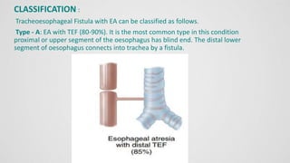
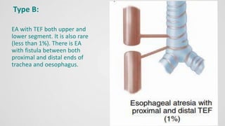
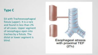
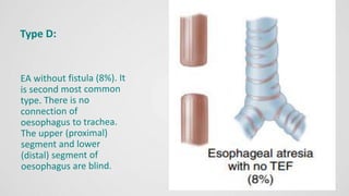
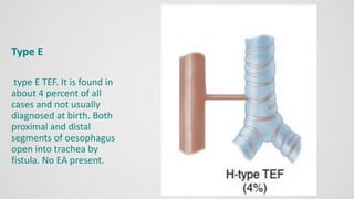
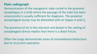
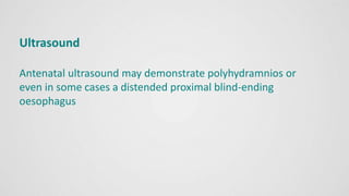
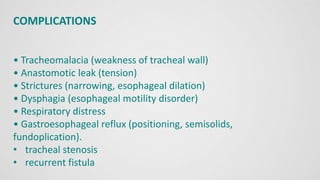
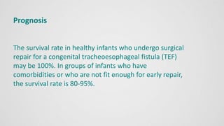
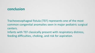
TRACHEOSPHAGEAL
FISTULA
Presented by
Group D
GROUP D MEMBERS
Obot, Edidiong Raymond
Akpaetuk Righteousness A
Nelson, Henry Edet
Etim Eastsunsam Elerius
CONTENTS
Introduction
Epidemiology
Classification
Aethiopathogenesis
Clinical manifestations
Diagnosis
Management
Complications
Prognosis
Conclusion
OBJECTIVES
• Objective 1: To define Tracheoesophageal Fistula
(TEF) and explain its causes and prevalence.
Objective 2: To review the various types of TEF and
their associated clinical presentations
• Objective 3: To describe the common symptoms of
TEF in infants and the importance of seeking prompt
medical attention.
• Objective 4: To outline the diagnostic procedures
used to identify TEF and explain the role of surgical
intervention.
DEFINITION
Tracheoesophageal Fistula (TEF)
is an abnormal connection between trachea and
oesophagus which is usually associated with Oesophageal
Atresia (EA).
EA is failure of the oesophagus to form a continuous
passage from the pharynx to the stomach during fourth
week of gestation. Incidence:
EPIDEMIOLOGY
Tracheoesophageal Fistula (TEF) occurs 1 in 3,500 live births
across the world.
In India 18,000 cases are recorded per year.
They appear to be equal sex incidence.
A history of maternal polyhydramnios is present in
approximately 50% of infants with the defects
CLASSIFICATION :
Tracheoesophageal Fistula with EA can be classified as follows.
Type - A: EA with TEF (80-90%). It is the most common type in this condition
proximal or upper segment of the oesophagus has blind end. The distal lower
segment of oesophagus connects into trachea by a fistula.
Type B:
EA with TEF both upper and
lower segment. It is also rare
(less than 1%). There is EA
with fistula between both
proximal and distal ends of
trachea and oesophagus.
Type C
EA with Tracheoesophageal
fistula (upper). It is a rare
and found in less than 2%
of all cases. Upper segment
of oesophagus open into
trachea by a fistula. The
distal or lower segment is
blind.
Type E
type E TEF. It is found in
about 4 percent of all
cases and not usually
diagnosed at birth. Both
proximal and distal
segments of oesophagus
open into trachea by
fistula. No EA present.
AETIOPATHOGENESIS
 Embryology
 Acquired TEF
 Association
 Etiology
Embryology
Knowledge of embryology is essential to understand the
pathogenesis of congenital TEFs.
The esophagus and trachea both develop from the primitive
foregut. In a 4- to 6-week-old embryo, the caudal part of the
foregut forms a ventral diverticulum that evolves into the trachea.
The longitudinal tracheoesophageal fold fuses to form a septum
that divides the foregut into a ventral laryngotracheal tube and a
dorsal esophagus. The posterior deviation of the
tracheoesophageal septum causes incomplete separation of the
esophagus from the laryngotracheal tube and results in a TEF.
Incomplete formation of the esophagus is known as
esophageal atresia, which may be associated with TEFs.
Many anatomic variations of esophageal atresia with or
without a TEF may occur.
The most common anomaly consists of a blind esophageal
pouch and a distal TEF.
Pure esophageal atresia without a TEF is the second most
common form. The third most common anomaly is the H-type
fistula, which consists of a TEF without esophageal atresia
Acquired TEF
 Traumatic TEFs occur secondary to either blunt trauma or
open avulsion injury to the neck and thorax. In blunt
traumatic injuries, the TEF is intrathoracic and is usually
located at the carina level. The TEF appears several days later
as a result of tracheal wall necrosis
 TEFs occur uncommonly at the time of
tracheostomy or secondary to improper
positioning of the tracheal tube because of
improper tracheal incision. The malpositioned
tracheostomy tube exerts posterior pressure
against the esophagus, resulting in tissue damage
and a TEF.
 In tuberculosis (TB) involvement of the esophagus
and mediastinal lymph nodes may result in an
acquired tubercular TEF
 Malignancy
ASSOCIATION
Approximately 17-70% of children with tracheoesophageal fistulas (TEFs)
have associated developmental anomalies. These include:
cardiac anomalies (15-19%)
PDA
ASD
VSD
right sided aortic arch
VATER/VACTERL
gastrointestinal anomalies (22%)
imperforate anus
malrotation
aneuploidic chromosomal abnormalities
trisomy 21
trisomy 18
trisomy 13
non-aneuploidic syndromic associations 5
Feingold syndrome
CHARGE syndrome
Pallister-Hall syndrome
ETIOLOGY
Although no definite cause exists for congenital
tracheoesophageal fistulas (TEFs)
 Association with trisomies 18, 21, and 13 has been reported.
 use of decongestants that contain imidazoline derivatives by
women during the first trimester of pregnancy has been
linked to an increased risk for congenital TEFs.
 There is only a minimal hereditary/genetic component with an
incidence in twins and those with family history being only
approximately 1% .
Causes of acquired TEFs include:
iatrogenic injury
blunt chest
neck trauma
prolonged mechanical ventilation via endotracheal or
tracheostomy tube,
excessive tube cuff pressure in patients ventilated for
lung disease
small-cell and non-small-cell lung cancer.
CLINICAL MANIFESTATION
EXTRAUTERINE
A single-artery umbilical cord
exhibit drooling
mucus and saliva bubbling from the nose and
mouth.
Excessive salivation
Apnoea Increased respiratory distress
Abdominal distention after feeding
Three C’s of Tracheoesophageal fistula are:
Coughing Choking Cyanosis
INTRA UTERINE
polyhydramnios (increased fluid in the uterus)
Absent stomach
Dilated esophageal pouch
When TEF is suspected, the first feeding
should be delayed until a diagnostic study is
performed.
DIAGNOSIS
The simplest test for TEF is to gently attempt to place a 10F
(french replogle tube) or larger tube via the mouth into the
stomach. The passage
of the tube is blocked at the level of the atresia. A chest x-
ray: reveals the tube coiled in the esophageal pouch. Air
can be injected through the tube to outline the atretic
pouch.
Barium should not be used because of extreme risk of
aspiration, but a tiny amount of dilute water-soluble
contrast agent can be given carefully, then aspirated when
the defect is clearly shown.
Antenatal diagnosis
can be done by: ultrasonography
Postnatal diagnostic procedures include
Ultrasonography
plain X-ray abdomen
bronchoscopy
karyotyping for asssociated syndromes
ECG and Echocardiogram can be done to detect
associated cardiac anomalies.
Plain radiograph
Demonstration of the nasogastric tube curled in the proximal
oesophagus in a child where the passage of the tube has been
unsuccessful is usually sufficient for diagnosis. The proximal
oesophageal stump may be distended with air (types A and C).
The presence of air in the stomach and bowel in the setting of
oesophageal atresia implies that there is a distal fistula.
Often the lungs demonstrate areas of consolidation/atelectasis
due to recurrent aspiration.
Ultrasound
Antenatal ultrasound may demonstrate polyhydramnios or
even in some cases a distended proximal blind-ending
oesophagus
Differential diagnosis of TEF/EA
includes:
esophageal stricture or diverticulum
pharyngeal pseudodiverticulum
severe GERD
vascular ring
iatrogenic esophageal perforation
laryngo-tracheo-esophageal cleft
esophageal webs
esophageal duplication congenital shortened esophagus
tracheal agenesis or atresia.
Medical Management
• The infant is immediately deprived of oral intake (NPO)
• Start IV fluids.
• Place infant in the position least likely to cause aspiration of
either mouth or stomach secretions i.e. supine with head end
raised to 45° or head turned to one side.
• Removal of secretions from the mouth and upper pouch
requires frequent or continuous suction with Replogle’s catheter
every 5 min gently with pressure of 50 cm of H₂O.
• Broad spectrum antibiotic therapy is often instituted
Nasogastric tube aspiration
Nasogastric tube to be kept in situ and suctioning to be done
frequently to prevent aspiration. The blind pouch to be washed
with normal saline to prevent blocking of tube with thick
mucus. Gastrostomy is done to decompress the stomach and to
prevent aspiration and afterwards to feed the infant
Supportive care should include
maintenance of nutritional requirement and warmth,
prevention of infection, antibiotic therapy, respiratory support,
detection and treatment of complications, continuous
monitoring of patient condition, chest physiotherapy and
postural drainage.
Surgical Management:
The treatment of TEF is surgical. The fistula is divided and
ligated. The esophageal ends are approximated and
anastomosed. In some cases, primary anastomosis cannot be
performed because of a long gap between the proximal and
distal
esophagus. Various techniques have been described to treat
this problem, including pulling up the stomach, elongating the
esophagus by myotomy, and simply delaying esophageal
anastomosis and providing continuous suction to the upper
pouch while allowing for growth.
Immediate primary repair: indications are
no pulmonary complications
weight of the child >2 to 2.5kgs
no major congenital malformations
healthy baby
gap between distal and proximal esophagus < 2cm
Delayed surgical intervention: indications are
pneumonia
sepsis
congenital malformations
severe prematurity
SURGICAL MANAGEMENT STAGES OF OPERATION:
Surgical correction can be done in stages with division of
fistula.
Gastrostomy is performed in initial stage followed by
esophageal or colonic transplant after 1year.
Other surgical intervention includes
Cervical esophagostomy
esophago- coloplasty
esophago-gastroplasty
NURSING INTERVENTION
Preoperative intervention
• Preventing aspiration by positioning, suctioning and nothing by
mouth, thus reducing chance of respiratory infections.
Postoperative intervention
• Maintaining clear airway
• Providing adequate feeding by I/V fluid and gastrostomy
feeding
• Reducing pain by analgesics and comfort measures
• Maintaining chest tube drainage with necessary precautions.
Nursing interventions contd...
Preventing infection by
general cleanlines
hygienic measures
administrating antibiotics.
COMPLICATIONS
• Tracheomalacia (weakness of tracheal wall)
• Anastomotic leak (tension)
• Strictures (narrowing, esophageal dilation)
• Dysphagia (esophageal motility disorder)
• Respiratory distress
• Gastroesophageal reflux (positioning, semisolids,
fundoplication).
• tracheal stenosis
• recurrent fistula
Prognosis
The survival rate in healthy infants who undergo surgical
repair for a congenital tracheoesophageal fistula (TEF)
may be 100%. In groups of infants who have
comorbidities or who are not fit enough for early repair,
the survival rate is 80-95%.
conclusion
Tracheoesophageal fistula (TEF) represents one of the most
common congenital anomalies seen in major pediatric surgical
centers.
Infants with TEF classically present with respiratory distress,
feeding difficulties, choking, and risk for aspiration.
THANK YOU
TRACHEOSPHAGEAL FISTULA.pptx an abnormalities

TRACHEOSPHAGEAL FISTULA.pptx an abnormalities

  • 1.
  • 2.
    GROUP D MEMBERS Obot,Edidiong Raymond Akpaetuk Righteousness A Nelson, Henry Edet Etim Eastsunsam Elerius
  • 3.
  • 4.
    OBJECTIVES • Objective 1:To define Tracheoesophageal Fistula (TEF) and explain its causes and prevalence. Objective 2: To review the various types of TEF and their associated clinical presentations • Objective 3: To describe the common symptoms of TEF in infants and the importance of seeking prompt medical attention. • Objective 4: To outline the diagnostic procedures used to identify TEF and explain the role of surgical intervention.
  • 5.
    DEFINITION Tracheoesophageal Fistula (TEF) isan abnormal connection between trachea and oesophagus which is usually associated with Oesophageal Atresia (EA). EA is failure of the oesophagus to form a continuous passage from the pharynx to the stomach during fourth week of gestation. Incidence:
  • 6.
    EPIDEMIOLOGY Tracheoesophageal Fistula (TEF)occurs 1 in 3,500 live births across the world. In India 18,000 cases are recorded per year. They appear to be equal sex incidence. A history of maternal polyhydramnios is present in approximately 50% of infants with the defects
  • 8.
    CLASSIFICATION : Tracheoesophageal Fistulawith EA can be classified as follows. Type - A: EA with TEF (80-90%). It is the most common type in this condition proximal or upper segment of the oesophagus has blind end. The distal lower segment of oesophagus connects into trachea by a fistula.
  • 9.
    Type B: EA withTEF both upper and lower segment. It is also rare (less than 1%). There is EA with fistula between both proximal and distal ends of trachea and oesophagus.
  • 10.
    Type C EA withTracheoesophageal fistula (upper). It is a rare and found in less than 2% of all cases. Upper segment of oesophagus open into trachea by a fistula. The distal or lower segment is blind.
  • 12.
    Type E type ETEF. It is found in about 4 percent of all cases and not usually diagnosed at birth. Both proximal and distal segments of oesophagus open into trachea by fistula. No EA present.
  • 13.
    AETIOPATHOGENESIS  Embryology  AcquiredTEF  Association  Etiology
  • 14.
    Embryology Knowledge of embryologyis essential to understand the pathogenesis of congenital TEFs. The esophagus and trachea both develop from the primitive foregut. In a 4- to 6-week-old embryo, the caudal part of the foregut forms a ventral diverticulum that evolves into the trachea. The longitudinal tracheoesophageal fold fuses to form a septum that divides the foregut into a ventral laryngotracheal tube and a dorsal esophagus. The posterior deviation of the tracheoesophageal septum causes incomplete separation of the esophagus from the laryngotracheal tube and results in a TEF.
  • 17.
    Incomplete formation ofthe esophagus is known as esophageal atresia, which may be associated with TEFs. Many anatomic variations of esophageal atresia with or without a TEF may occur. The most common anomaly consists of a blind esophageal pouch and a distal TEF. Pure esophageal atresia without a TEF is the second most common form. The third most common anomaly is the H-type fistula, which consists of a TEF without esophageal atresia
  • 18.
    Acquired TEF  TraumaticTEFs occur secondary to either blunt trauma or open avulsion injury to the neck and thorax. In blunt traumatic injuries, the TEF is intrathoracic and is usually located at the carina level. The TEF appears several days later as a result of tracheal wall necrosis
  • 19.
     TEFs occuruncommonly at the time of tracheostomy or secondary to improper positioning of the tracheal tube because of improper tracheal incision. The malpositioned tracheostomy tube exerts posterior pressure against the esophagus, resulting in tissue damage and a TEF.  In tuberculosis (TB) involvement of the esophagus and mediastinal lymph nodes may result in an acquired tubercular TEF  Malignancy
  • 20.
    ASSOCIATION Approximately 17-70% ofchildren with tracheoesophageal fistulas (TEFs) have associated developmental anomalies. These include: cardiac anomalies (15-19%) PDA ASD VSD right sided aortic arch VATER/VACTERL gastrointestinal anomalies (22%) imperforate anus malrotation
  • 21.
    aneuploidic chromosomal abnormalities trisomy21 trisomy 18 trisomy 13 non-aneuploidic syndromic associations 5 Feingold syndrome CHARGE syndrome Pallister-Hall syndrome
  • 22.
    ETIOLOGY Although no definitecause exists for congenital tracheoesophageal fistulas (TEFs)  Association with trisomies 18, 21, and 13 has been reported.  use of decongestants that contain imidazoline derivatives by women during the first trimester of pregnancy has been linked to an increased risk for congenital TEFs.  There is only a minimal hereditary/genetic component with an incidence in twins and those with family history being only approximately 1% .
  • 23.
    Causes of acquiredTEFs include: iatrogenic injury blunt chest neck trauma prolonged mechanical ventilation via endotracheal or tracheostomy tube, excessive tube cuff pressure in patients ventilated for lung disease small-cell and non-small-cell lung cancer.
  • 24.
    CLINICAL MANIFESTATION EXTRAUTERINE A single-arteryumbilical cord exhibit drooling mucus and saliva bubbling from the nose and mouth. Excessive salivation Apnoea Increased respiratory distress Abdominal distention after feeding Three C’s of Tracheoesophageal fistula are: Coughing Choking Cyanosis
  • 25.
    INTRA UTERINE polyhydramnios (increasedfluid in the uterus) Absent stomach Dilated esophageal pouch When TEF is suspected, the first feeding should be delayed until a diagnostic study is performed.
  • 26.
    DIAGNOSIS The simplest testfor TEF is to gently attempt to place a 10F (french replogle tube) or larger tube via the mouth into the stomach. The passage of the tube is blocked at the level of the atresia. A chest x- ray: reveals the tube coiled in the esophageal pouch. Air can be injected through the tube to outline the atretic pouch. Barium should not be used because of extreme risk of aspiration, but a tiny amount of dilute water-soluble contrast agent can be given carefully, then aspirated when the defect is clearly shown.
  • 28.
    Antenatal diagnosis can bedone by: ultrasonography Postnatal diagnostic procedures include Ultrasonography plain X-ray abdomen bronchoscopy karyotyping for asssociated syndromes ECG and Echocardiogram can be done to detect associated cardiac anomalies.
  • 29.
    Plain radiograph Demonstration ofthe nasogastric tube curled in the proximal oesophagus in a child where the passage of the tube has been unsuccessful is usually sufficient for diagnosis. The proximal oesophageal stump may be distended with air (types A and C). The presence of air in the stomach and bowel in the setting of oesophageal atresia implies that there is a distal fistula. Often the lungs demonstrate areas of consolidation/atelectasis due to recurrent aspiration.
  • 30.
    Ultrasound Antenatal ultrasound maydemonstrate polyhydramnios or even in some cases a distended proximal blind-ending oesophagus
  • 31.
    Differential diagnosis ofTEF/EA includes: esophageal stricture or diverticulum pharyngeal pseudodiverticulum severe GERD vascular ring iatrogenic esophageal perforation laryngo-tracheo-esophageal cleft esophageal webs esophageal duplication congenital shortened esophagus tracheal agenesis or atresia.
  • 32.
    Medical Management • Theinfant is immediately deprived of oral intake (NPO) • Start IV fluids. • Place infant in the position least likely to cause aspiration of either mouth or stomach secretions i.e. supine with head end raised to 45° or head turned to one side. • Removal of secretions from the mouth and upper pouch requires frequent or continuous suction with Replogle’s catheter every 5 min gently with pressure of 50 cm of H₂O. • Broad spectrum antibiotic therapy is often instituted
  • 33.
    Nasogastric tube aspiration Nasogastrictube to be kept in situ and suctioning to be done frequently to prevent aspiration. The blind pouch to be washed with normal saline to prevent blocking of tube with thick mucus. Gastrostomy is done to decompress the stomach and to prevent aspiration and afterwards to feed the infant Supportive care should include maintenance of nutritional requirement and warmth, prevention of infection, antibiotic therapy, respiratory support, detection and treatment of complications, continuous monitoring of patient condition, chest physiotherapy and postural drainage.
  • 34.
    Surgical Management: The treatmentof TEF is surgical. The fistula is divided and ligated. The esophageal ends are approximated and anastomosed. In some cases, primary anastomosis cannot be performed because of a long gap between the proximal and distal esophagus. Various techniques have been described to treat this problem, including pulling up the stomach, elongating the esophagus by myotomy, and simply delaying esophageal anastomosis and providing continuous suction to the upper pouch while allowing for growth.
  • 35.
    Immediate primary repair:indications are no pulmonary complications weight of the child >2 to 2.5kgs no major congenital malformations healthy baby gap between distal and proximal esophagus < 2cm Delayed surgical intervention: indications are pneumonia sepsis congenital malformations severe prematurity
  • 36.
    SURGICAL MANAGEMENT STAGESOF OPERATION: Surgical correction can be done in stages with division of fistula. Gastrostomy is performed in initial stage followed by esophageal or colonic transplant after 1year. Other surgical intervention includes Cervical esophagostomy esophago- coloplasty esophago-gastroplasty
  • 37.
    NURSING INTERVENTION Preoperative intervention •Preventing aspiration by positioning, suctioning and nothing by mouth, thus reducing chance of respiratory infections. Postoperative intervention • Maintaining clear airway • Providing adequate feeding by I/V fluid and gastrostomy feeding • Reducing pain by analgesics and comfort measures • Maintaining chest tube drainage with necessary precautions.
  • 38.
    Nursing interventions contd... Preventinginfection by general cleanlines hygienic measures administrating antibiotics.
  • 39.
    COMPLICATIONS • Tracheomalacia (weaknessof tracheal wall) • Anastomotic leak (tension) • Strictures (narrowing, esophageal dilation) • Dysphagia (esophageal motility disorder) • Respiratory distress • Gastroesophageal reflux (positioning, semisolids, fundoplication). • tracheal stenosis • recurrent fistula
  • 40.
    Prognosis The survival ratein healthy infants who undergo surgical repair for a congenital tracheoesophageal fistula (TEF) may be 100%. In groups of infants who have comorbidities or who are not fit enough for early repair, the survival rate is 80-95%.
  • 41.
    conclusion Tracheoesophageal fistula (TEF)represents one of the most common congenital anomalies seen in major pediatric surgical centers. Infants with TEF classically present with respiratory distress, feeding difficulties, choking, and risk for aspiration.
  • 42.