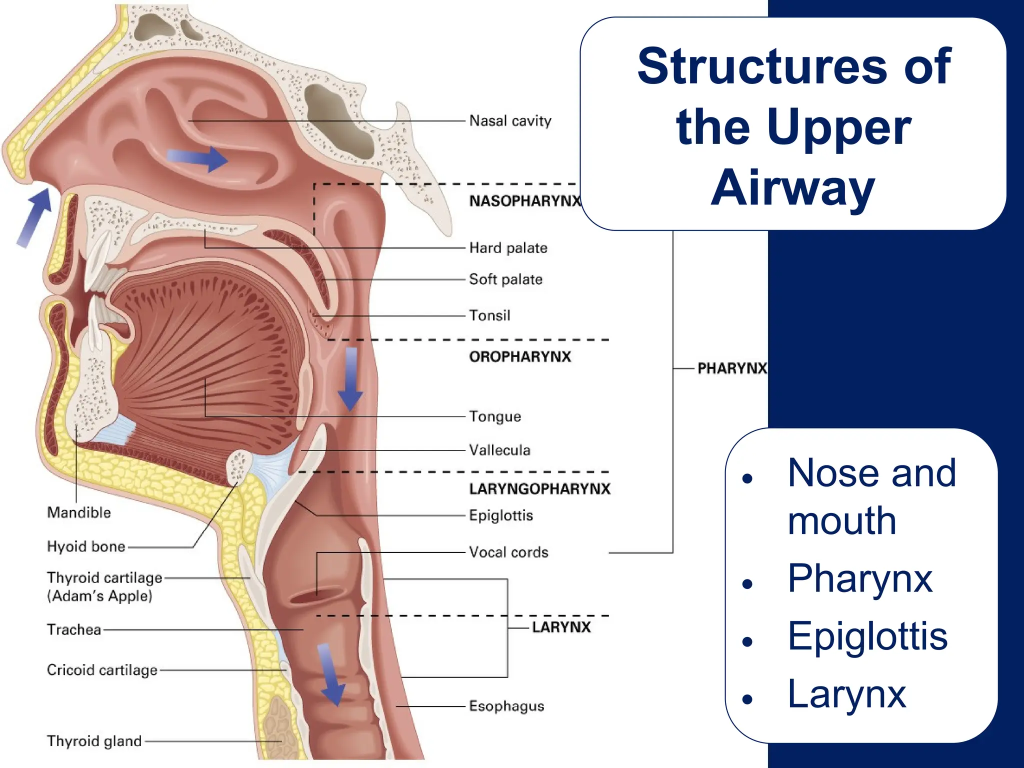
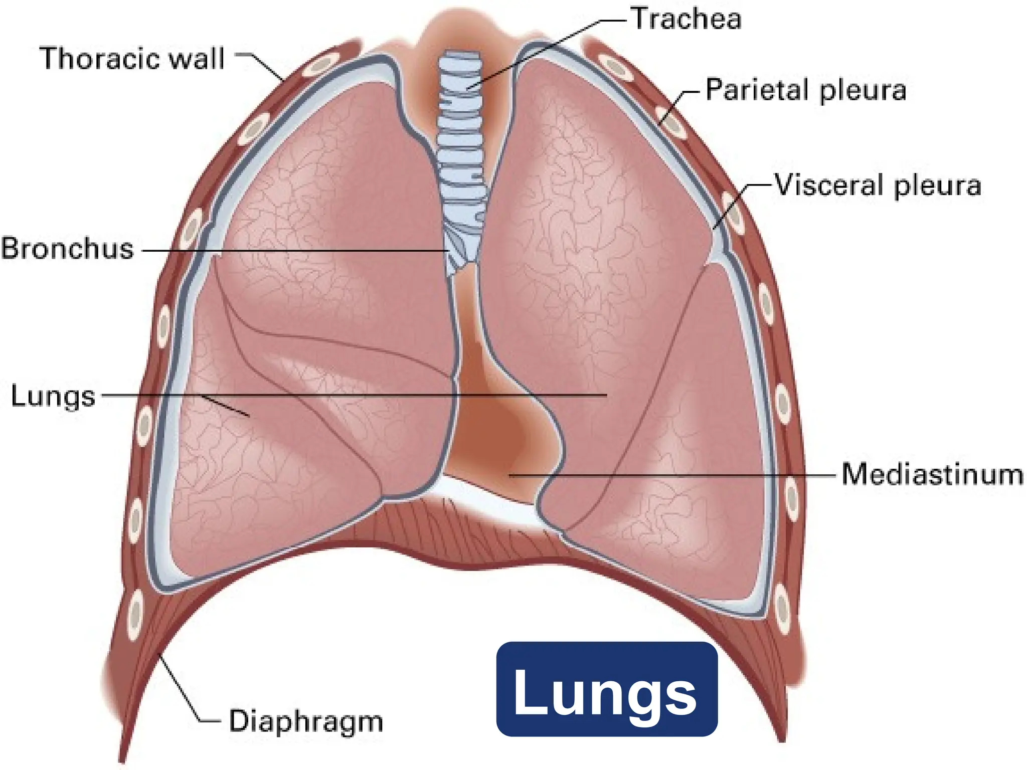
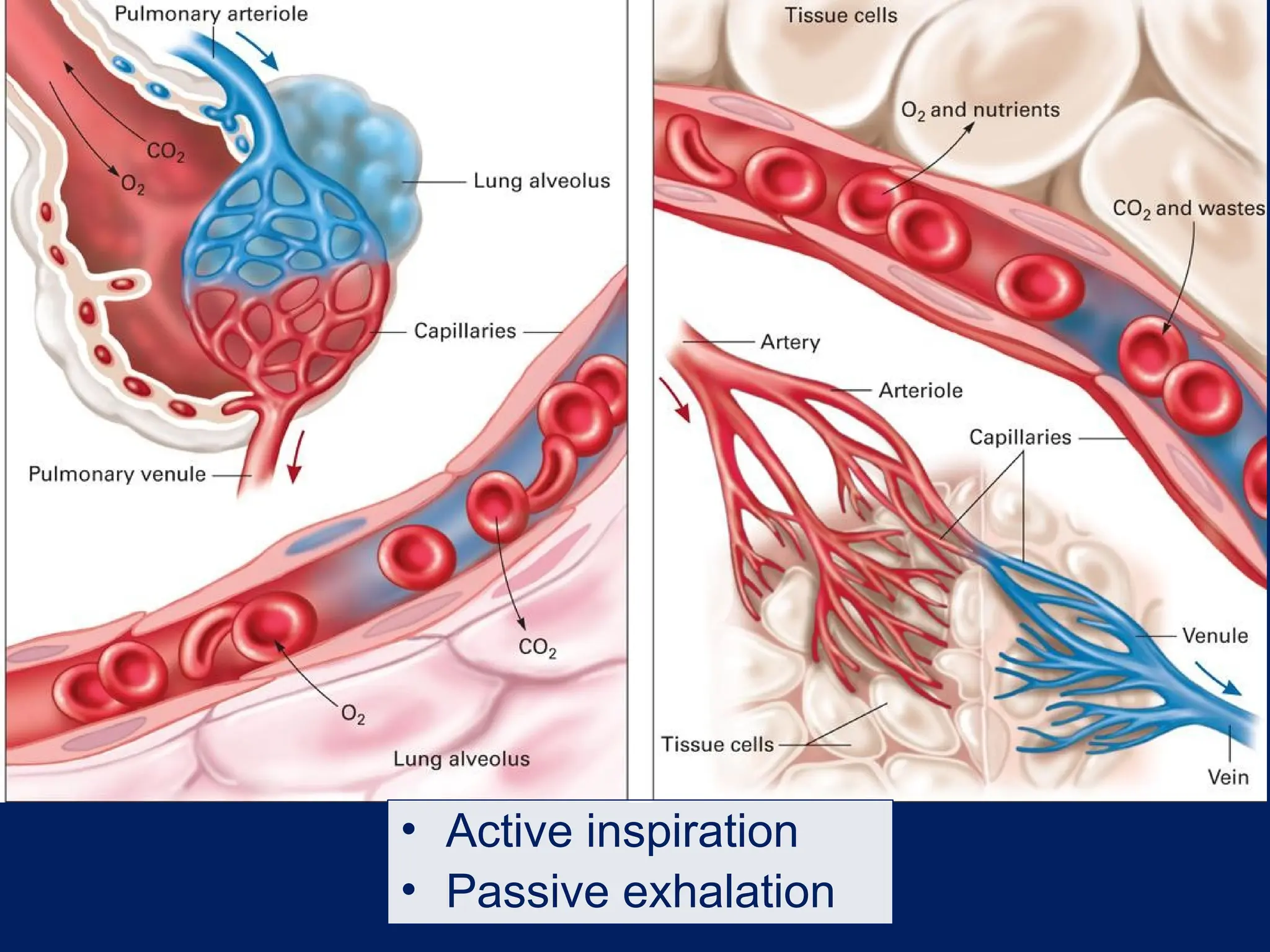
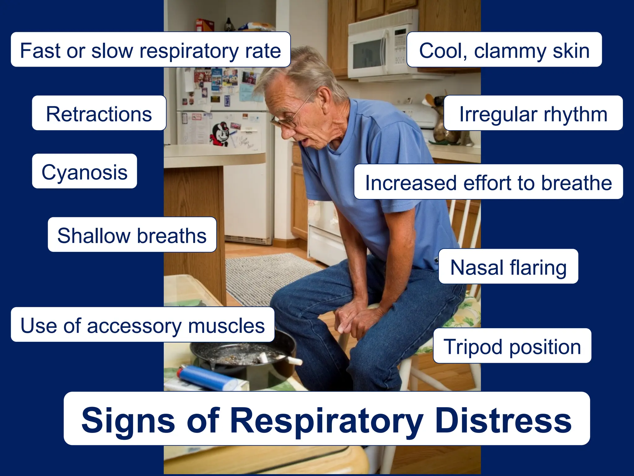
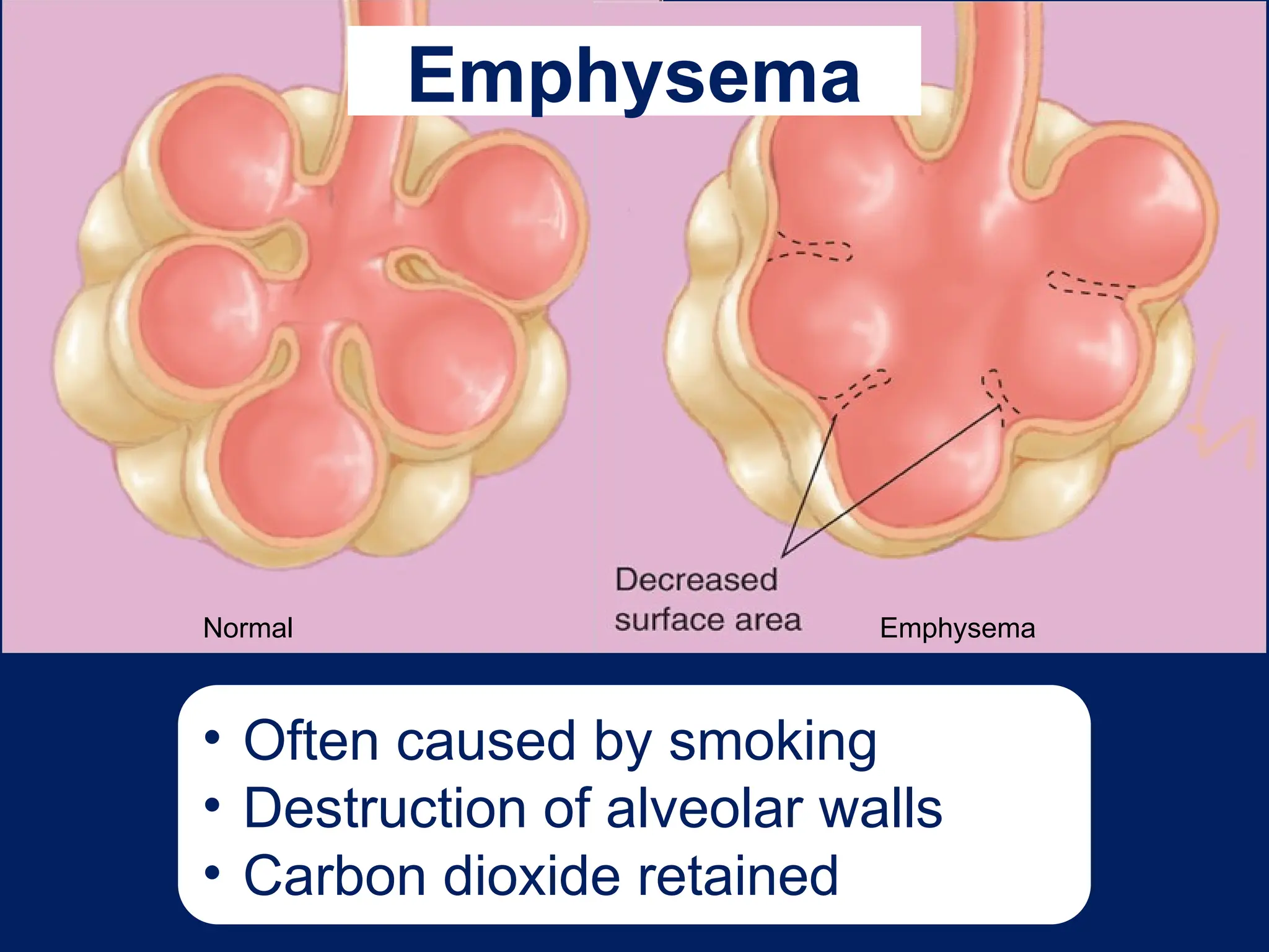
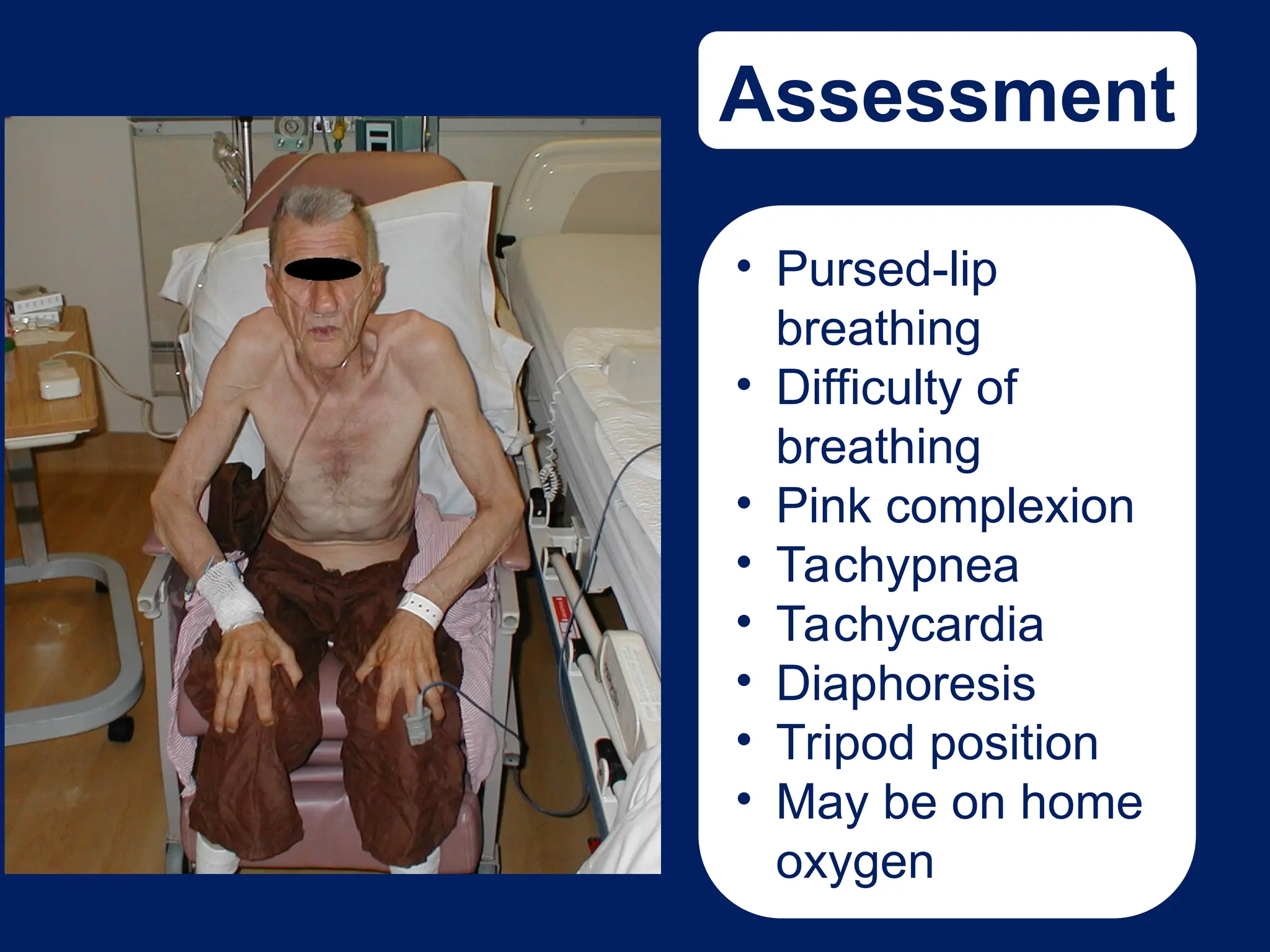
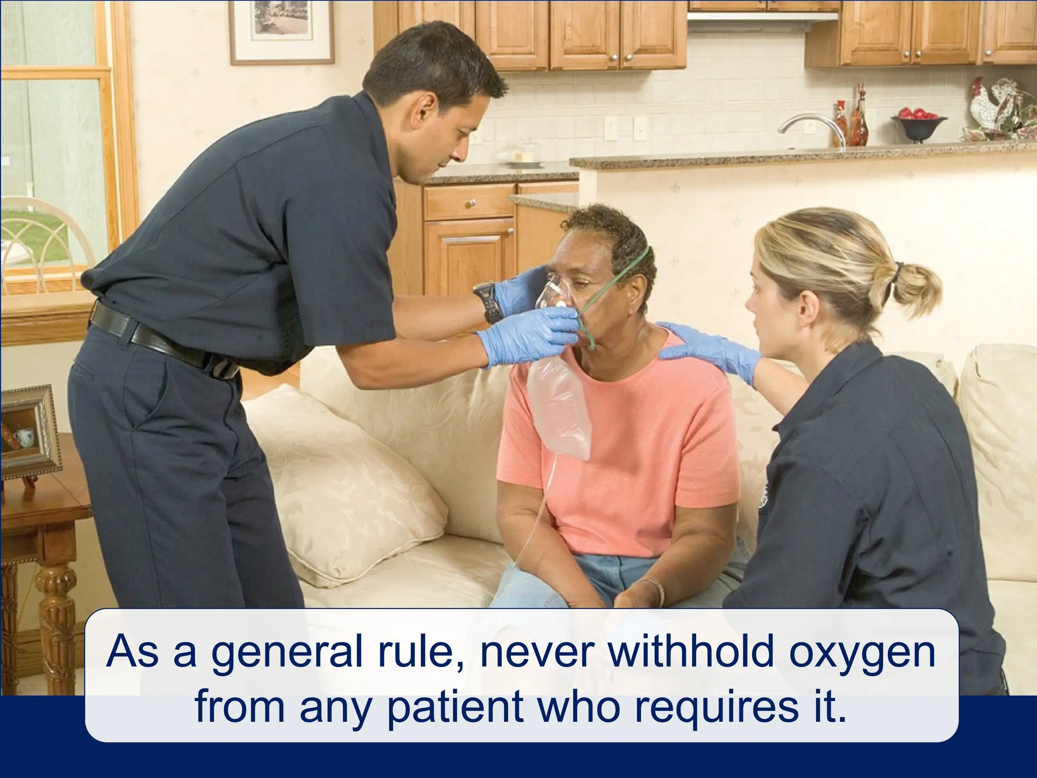
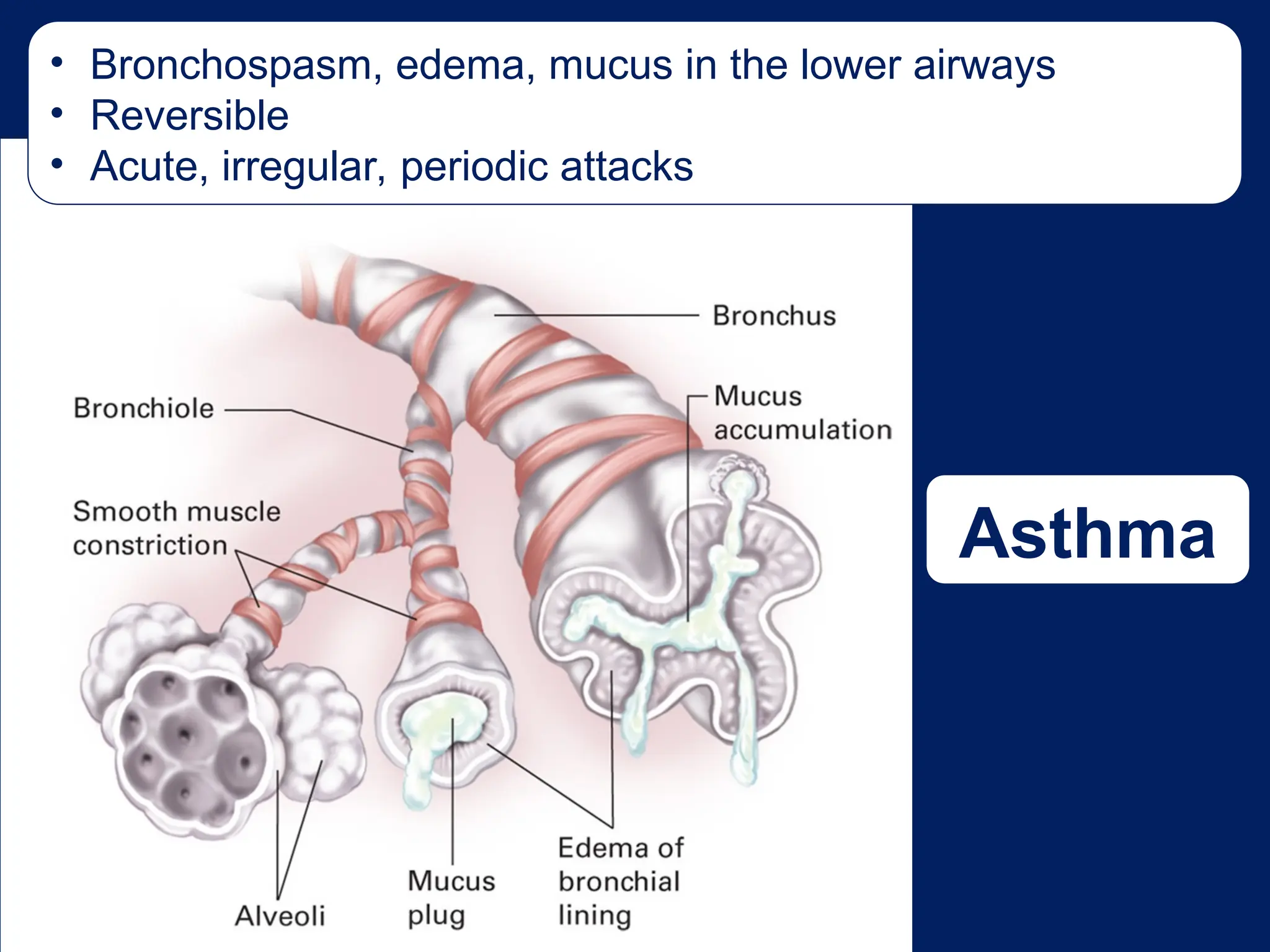
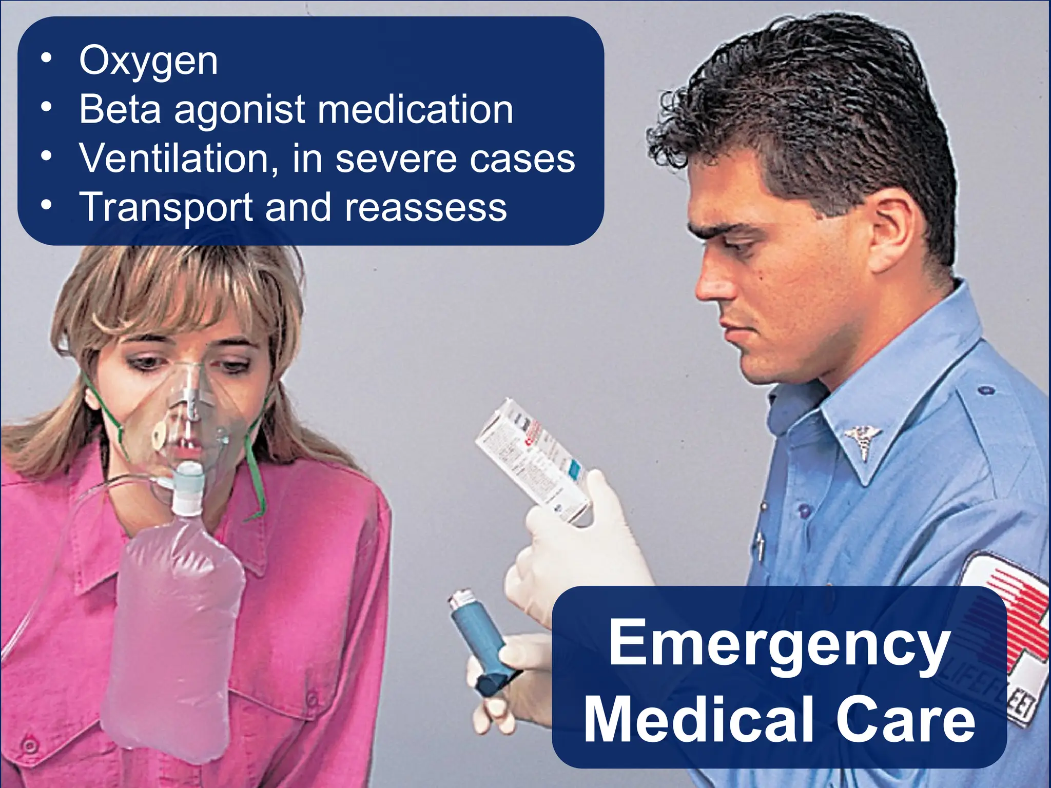
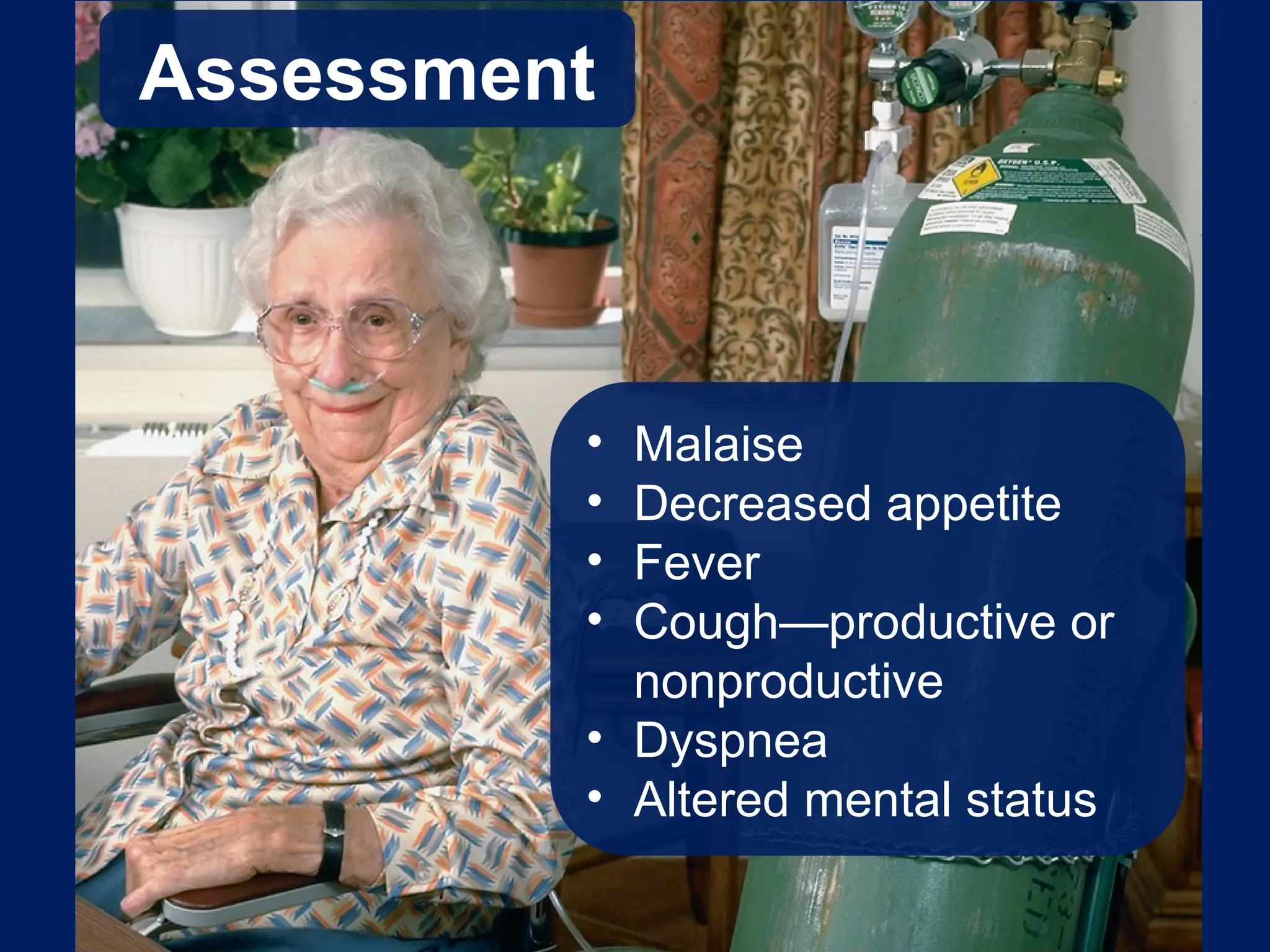
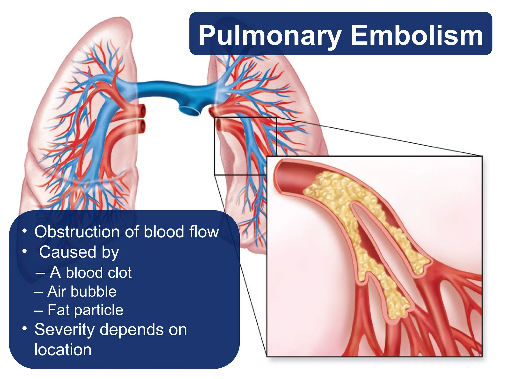
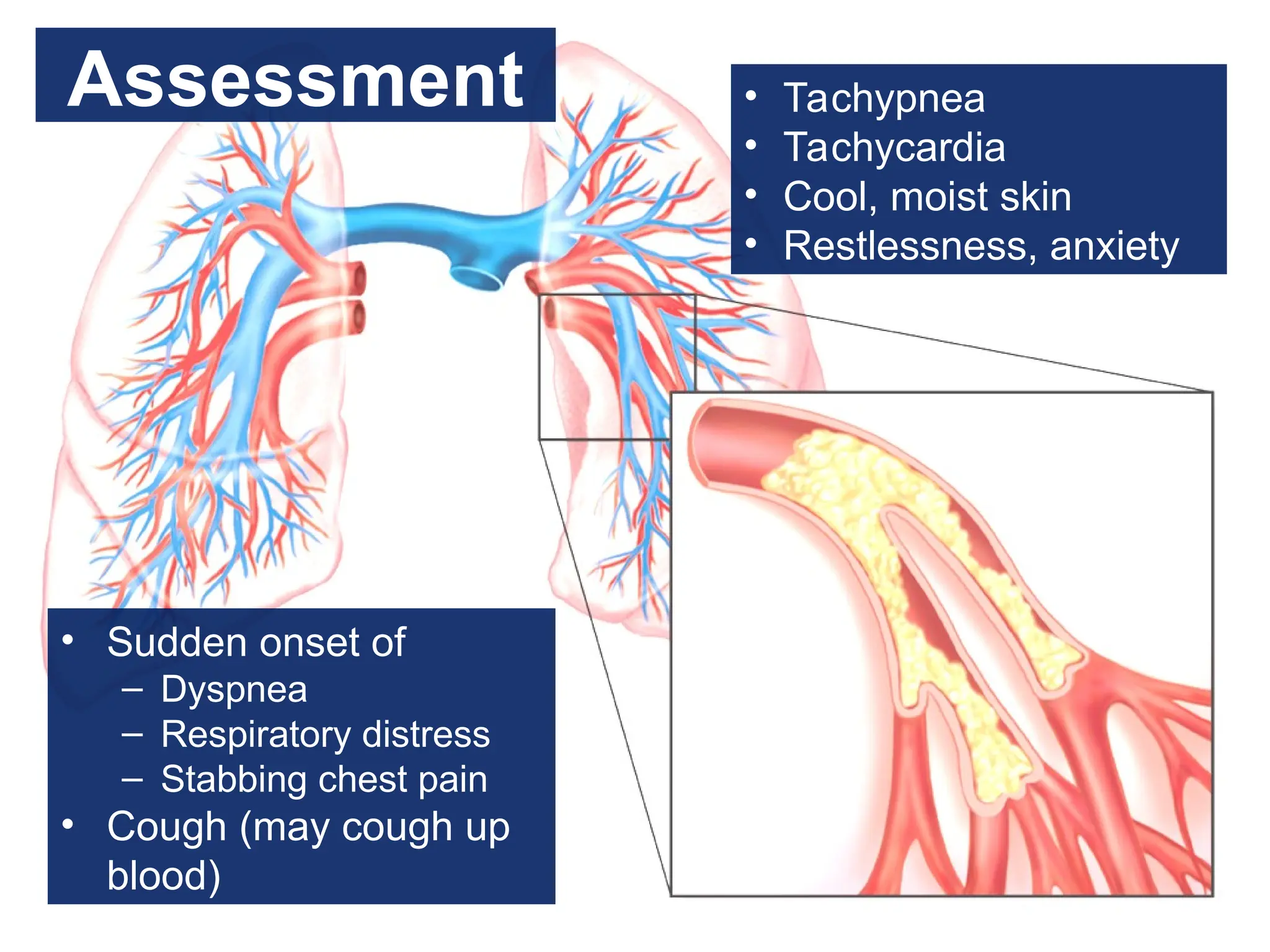
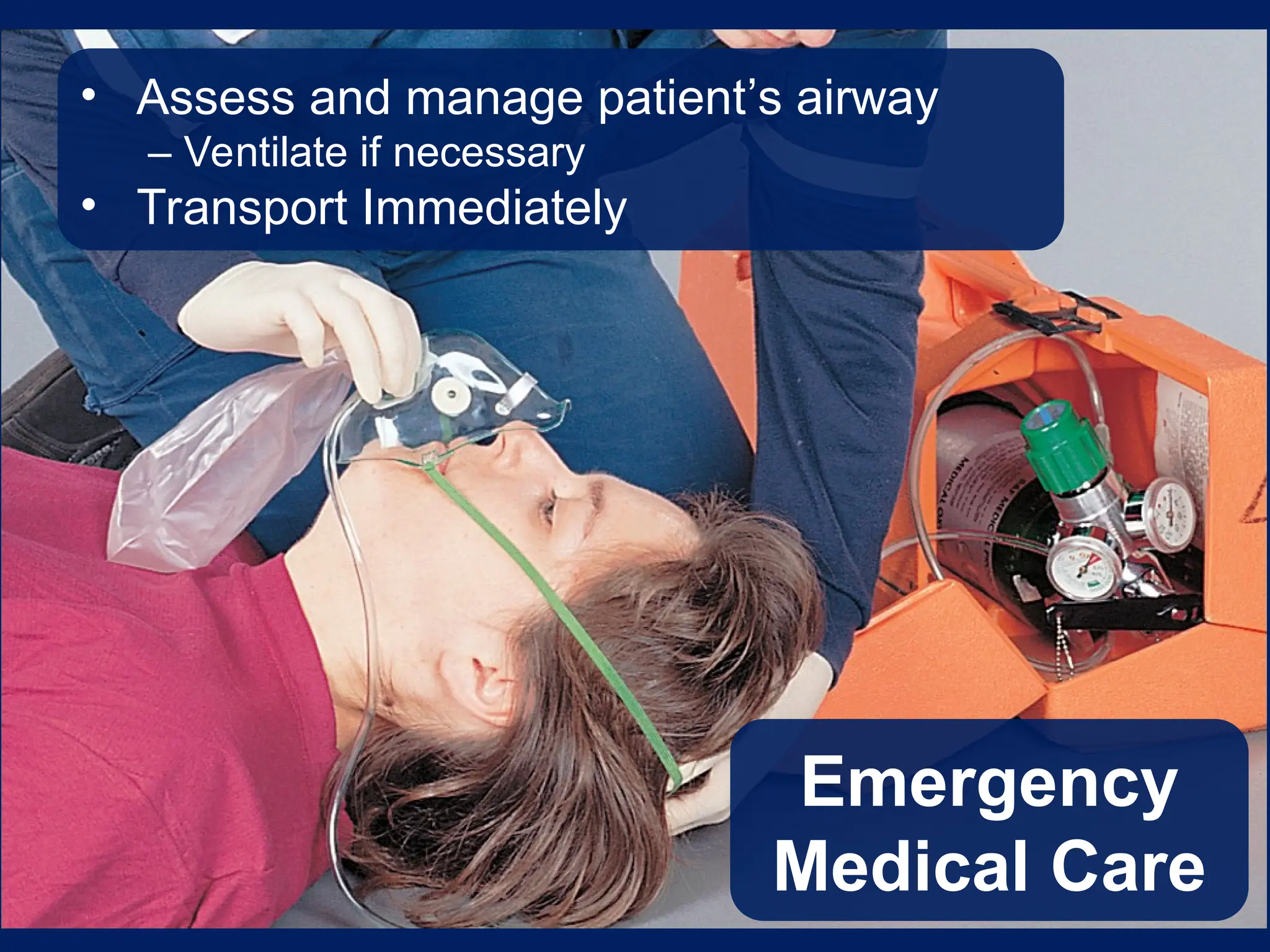
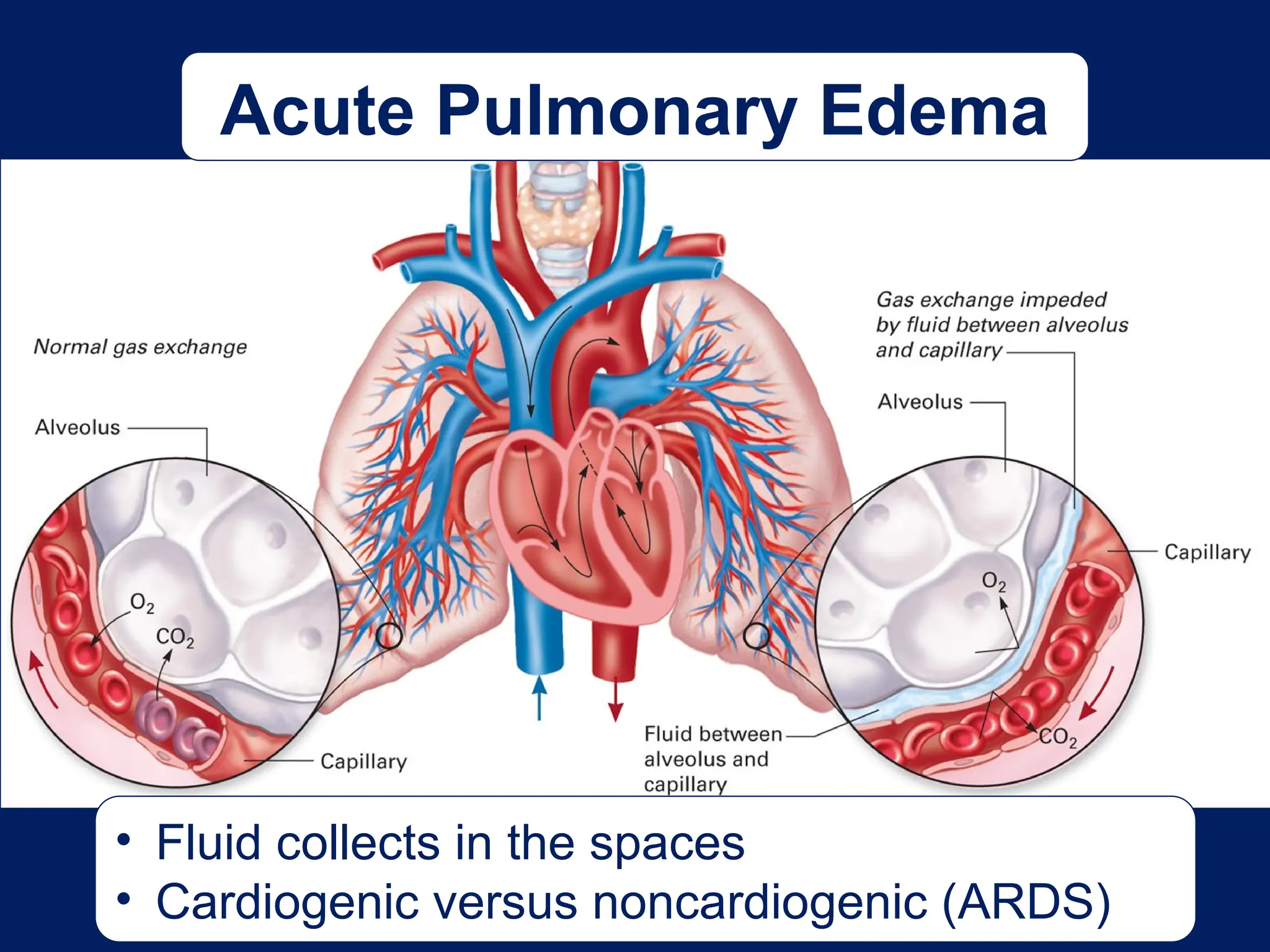
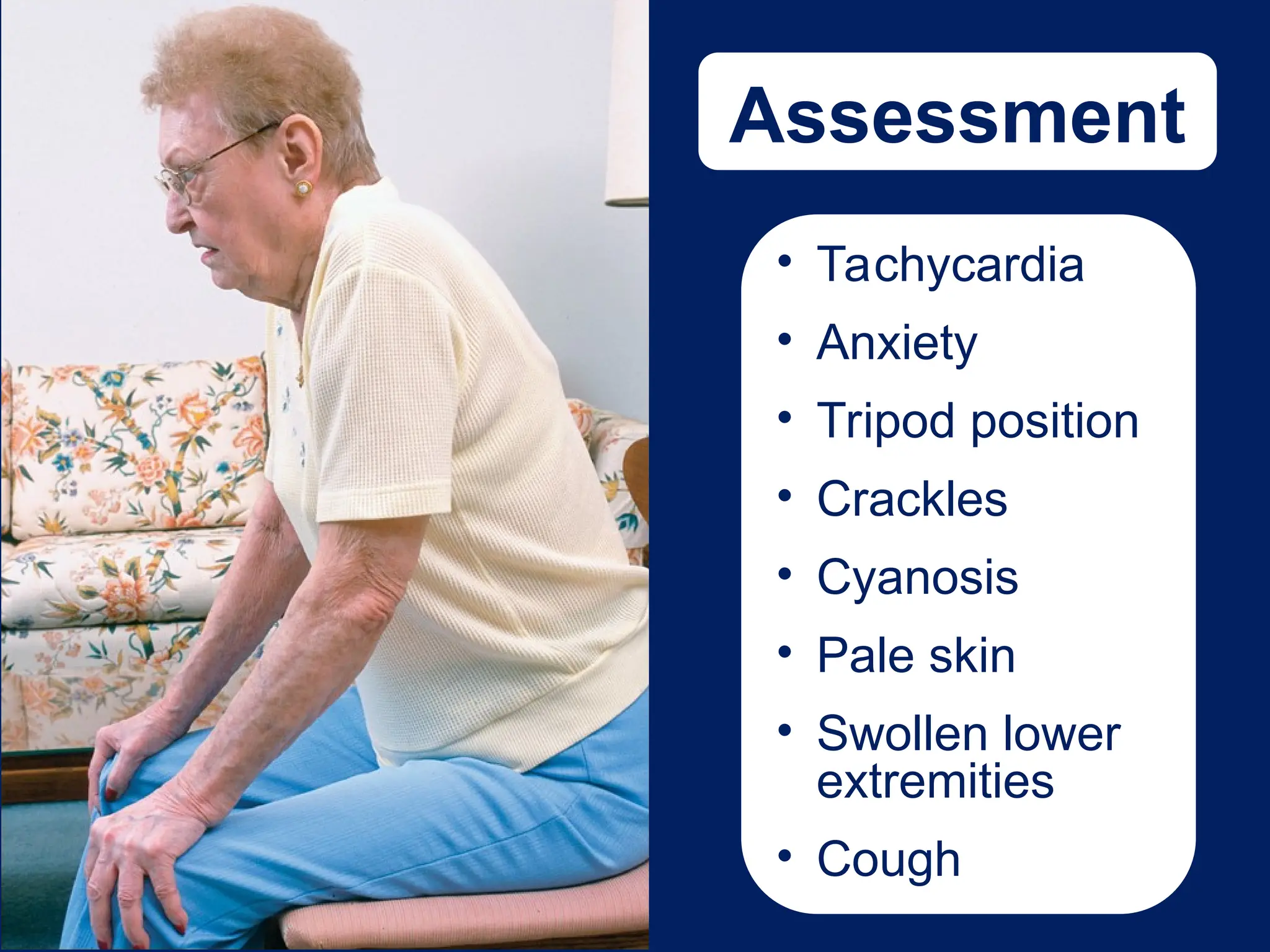
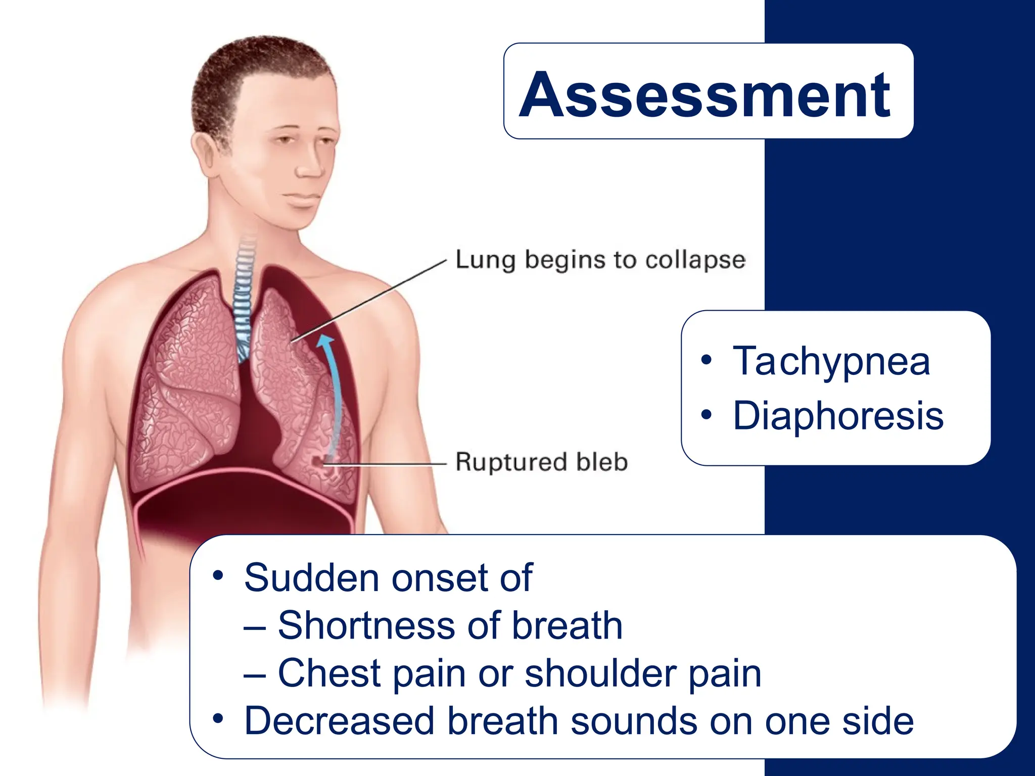
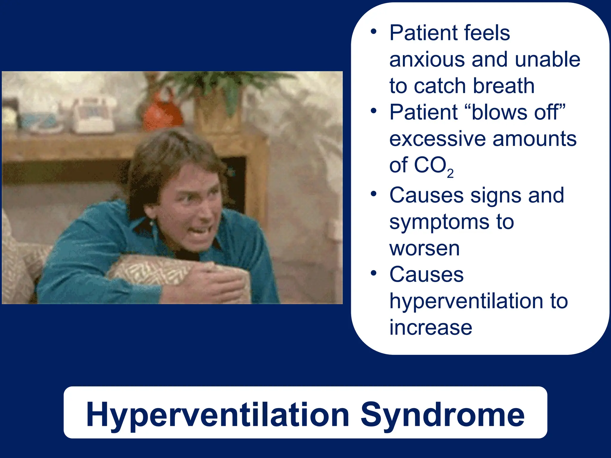
Chapter 16
Respiratory Emergencies
Copyright ©2010 by Pearson Education, Inc.
All rights reserved.
Prehospital Emergency Care, Ninth Edition
Joseph J. Mistovich • Keith J. Karren
Anatomy
Structures of
the Upper
Airway
• Nose and
mouth
• Pharynx
• Epiglottis
• Larynx
Structures of
the Lower
Airway
• Trachea
• Bronchi
• Bronchioles
• Lungs
Lungs
Normal Breathing
• Active inspiration
• Passive exhalation
Abnormal Breathing
As a result of several
possibilities
• Increased width
• Lack of perfusion
• Filling of the alveoli
• Bronchoconstriction
Abnormal Breathing
Assessing Breath
Sounds
• High-pitched,
musical,
whistling
• Constriction of
bronchioles
Wheezing
Rhonchi
• Snoring or rattling noises
• Caused by thick mucous secretions
Crackles
• Bubbly or crackling
sounds
• Associated with
fluid around the
alveoli
Respiratory Distress
Terms to Know…
• Hypoxemia
– Decreased oxygen in blood stream
• Hypoxia
– Decreased oxygen in the tissues
• Dyspnea
– Shortness of breath
• Apnea
– Respiratory arrest
Signs of Respiratory Distress
Fast or slow respiratory rate
Retractions
Cool, clammy skin
Irregular rhythm
Increased effort to breathe
Shallow breaths
Cyanosis
Nasal flaring
Use of accessory muscles
Tripod position
• Can quickly lead to cardiac arrest
• Managed with rapid oxygen delivery
Respiratory Arrest
Pathophysiology of Conditions
That Cause Respiratory Distress
Emphysema
Obstructive Pulmonary Diseases
Emphysema
Normal
• Often caused by smoking
• Destruction of alveolar walls
• Carbon dioxide retained
Emphysema
Assessment
• Thin, barrel-chest appearance
• Coughing, nonproductive
• Prolonged exhalation
• Diminished breath sounds
• Wheezing and rhonchi
Assessment
• Pursed-lip
breathing
• Difficulty of
breathing
• Pink complexion
• Tachypnea
• Tachycardia
• Diaphoresis
• Tripod position
• May be on home
oxygen
Obstructive Pulmonary Diseases
Chronic Bronchitis
Chronic Bronchitis
• Caused by smoking
• Inflammation, swelling, and thickening of the bronchi
and bronchioles
• Excessive mucous production
• Typically overweight
• Chronic cyanotic complexion
• Difficulty in breathing
• Productive chronic cough
• Coarse rhonchi
• Wheezes and crackles
Assessment
Emergency
Medical Care
• Treat the same as any patient experiencing
shortness of breath
• Hypoxic drive a rare complication
As a general rule, never withhold oxygen
from any patient who requires it.
Obstructive Pulmonary Diseases
Asthma
• Bronchospasm, edema, mucus in the lower airways
• Reversible
• Acute, irregular, periodic attacks
Asthma
Assessment
• Dyspnea
• Nonproductive
cough
• Wheezing
• Tachypnea
• Tachycardia
• Anxiety and
apprehension
• Chest tightness
• SpO2 < 95
percent
• Extreme fatigue or exhaustion
• Inability to speak
• Quiet or absent breath sounds
• SpO2 < 90 percent with patient on oxygen
Symptoms that Require Ventilation
Emergency
Medical Care
• Oxygen
• Beta agonist medication
• Ventilation, in severe cases
• Transport and reassess
Other Diseases That Cause
Respiratory Distress
Pneumonia
Pneumonia • Common disease of the
elderly and those with
suppressed immune
systems
• Acute infectious disease
• Caused by bacteria or virus
Need image 16-05
here – we don’t
have it - WDS
Assessment
• Malaise
• Decreased appetite
• Fever
• Cough—productive or
nonproductive
• Dyspnea
• Altered mental status
Emergency
Medical Care
• Treat the same as any patient experiencing
shortness of breath
• May administer metered-dose inhaler or
small-volume nebulizer
Other Diseases That Cause
Respiratory Distress
Pulmonary Embolism
Pulmonary Embolism
• Obstruction of blood flow
• Caused by
– A blood clot
– Air bubble
– Fat particle
• Severity depends on
location
Assessment
• Sudden onset of
– Dyspnea
– Respiratory distress
– Stabbing chest pain
• Cough (may cough up
blood)
• Tachypnea
• Tachycardia
• Cool, moist skin
• Restlessness, anxiety
Emergency
Medical Care
• Assess and manage patient’s airway
– Ventilate if necessary
• Transport Immediately
Pulmonary
Embolism
https://www.youtube.com/watch?v=SzsQWIMYbN8
Other Diseases That Cause
Respiratory Distress
Acute Pulmonary
Edema
Acute Pulmonary Edema
• Fluid collects in the spaces
• Cardiogenic versus noncardiogenic (ARDS)
Assessment
• Tachycardia
• Anxiety
• Tripod position
• Crackles
• Cyanosis
• Pale skin
• Swollen lower
extremities
• Cough
Emergency
Medical Care
Manage the patient’s airway
• Positive pressure ventilation may be necessary
• May improve the patient’s status
Other Diseases That Cause
Respiratory Distress
Spontaneous Pneumothorax
Spontaneous
Pneumothorax
• Sudden rupture of the visceral lining
• More likely in tall, thin males
• Change in pressure causes lung to
collapse
Assessment
• Sudden onset of
– Shortness of breath
– Chest pain or shoulder pain
• Decreased breath sounds on one side
• Tachypnea
• Diaphoresis
Emergency Medical Care
• Treat shortness of breath
• Be prepared to support ventilations
• Monitor for signs of a tension pneumothorax
Other Diseases That Cause
Respiratory Distress
Hyperventilation
Syndrome
Hyperventilation Syndrome
• Patient feels
anxious and unable
to catch breath
• Patient “blows off”
excessive amounts
of CO2
• Causes signs and
symptoms to
worsen
• Causes
hyperventilation to
increase
Assessment
• Nervousness and anxiety
• Dizziness
• Shortness of breath
• Chest tightness
• Numbness and tingling around the mouth, hands, and feet
• Tachypnea
• Carpopedal spasm
Emergency
Medical Care
Emergency
Medical Care
• Coach patient to slow down breathing
– NEVER use a paper bag
– NEVER withhold oxygen
• If possible, remove the source of anxiety
Other Diseases That Cause
Respiratory Distress
Epiglottitis
Epiglottitis
• Inflammation of
upper airway
• Becoming more
common in adults
• Can be life
threatening
Assessment
• High fever
• Sore throat
• Drooling
• Anxiety and
apprehension
• Tripod position
• Fatigue
• Inspiratory stridor
Emergency
Medical Care
• Administer O2
• Keep patient calm
• Rapid transport
• Consider ALS
intercept
• BVM if necessary
Other Diseases That Cause
Respiratory Distress
Pertussis
Pertussis
• Whooping cough
• Similar to cold at
start
• Rapid coughing
• Leads to
complications
Assessment
• History of upper
respiratory
infection
• Coughing fits
• “Whoop” heard at
end of cough
• Dyspnea during
cough
Emergency
Medical Care
• Calm patient
• Administer oxygen
• Expedite transport
• Consider ALS
• Decontaminate
ambulance afterward
Other Diseases That Cause
Respiratory Distress
Cystic Fibrosis
Cystic
Fibrosis
• Hereditary disease
• Diagnosed early in life
• No cure
• Causes pulmonary failure
Assessment
Bronchiole • Known history of
cystic fibrosis
• Recurrent coughing
• Fever
• Expectoration of thick
mucus
• Recurrent infections
• Trouble speaking and
breathing
Emergency
Medical Care
• Provide O2 via nonrebreather mask
• Consider saline nebulizer (per protocol)
• Move patient into position of comfort
• Consider ALS intercept
Other Diseases That Cause
Respiratory Distress
Poisonous Exposures
Poisonous Exposures
Any type of inhalation injury that
occurs secondary to exposure to a
toxic substance(s)
(© Brendan McDermid/Reuters/Corbis)
Assessment
• History consistent with an inhalation injury
• Presence of chemicals about the face from
the exposure
• Findings of respiratory distress
Emergency
Medical Care
• EMS safety first
• Rescue patients
• ABCs
• Provide O2
• Gather information regarding exposure
• Consider ALS intercept
Other Diseases That Cause
Respiratory Distress
Viral Respiratory
Infections
Viral Respiratory Infections
• Common to all age groups; most serious in children
• Caused by many viruses
• Can lead to more serious infections
Assessment
• Nasal congestion
• Sore throat
• Mild respiratory distress
• Fever
• Malaise
• Poor feeding habits
Emergency Medical Care
• Treat symptoms
• Put patient in
position of comfort
• Monitor for changes
Metered-Dose Inhalers and
Small-Volume Nebulizers
Inhalers and
Nebulizers
Medication is deposited on site of bronchoconstriction.
See skill slides for proper administration.
(© Carl Leet, YSU)
Advair
Not for emergency use
Age-Related Variations:
Pediatrics and Geriatrics
Pediatric Patients
Pediatrics
Primary assessment
• Rule out trauma first
• Spot signs in general impression
Respiratory Distress in the
Pediatric Patient:
Assessment and Care
Signs of Respiratory
Distress
Monitor
for
worsening
condition.
Respiratory
Failure
• Altered mental status
• Bradycardia
• Hypotension
• Irregular breathing pattern
Begin immediate positive
pressure ventilations.
Geriatric Patients
Geriatrics
Primary assessment
• Rule out trauma first
• Spot signs in primary assessment
• Patient will deteriorate rapidly
Respiratory Distress in the
Geriatric Patient:
Assessment and Care
Respiratory Distress
• Retractions
• Accessory muscle use
• Tachypnea
• Tachycardia
• Nasal flaring
Respiratory Failure
• Diminished or absent lung
sounds
• Altered mental status
• Irregular breathing pattern
• Cyanosis
Assessment and Care:
General Guidelines
Assessment-Based
Approach: Respiratory
Distress
Scene size-up
Primary assessment
ABCs
Transport priority
Secondary assessment
Emergency care
Reassessment
Chapter 16 Respiratory Emergencies for EMT

Chapter 16 Respiratory Emergencies for EMT

Editor's Notes

  • #1 Advance Preparation Review local protocols for administration of respiratory drugs and treatment of respiratory emergencies. Bring MDI samples or demonstration units and small-volume nebulizers for demonstration and student practice. Bring enough stethoscopes to class for pairs of students to practice auscultation of breath sounds. Bring enough small drinking straws or coffee stirrers to class for each student. (See slide 22.) Arrange for lab instructors and equipment for skills practice. A ratio of one instructor for every four students is recommended.
  • #3 Talking Points The primary purpose of the upper and lower airways is the conduction of air into and out of the lungs. The third portion of the respiratory system is the lungs and accessory structures, which work in concert with the upper and lower airways to allow the oxygenation of body cells and the elimination of carbon dioxide from the blood stream.
  • #6 Discussion Question What are the characteristics of normal breathing?
  • #7 Talking Points The following should occur in a patient who is breathing adequately, providing no other condition or injury is involved: Normal mental status Normal muscle tone Normal pulse oximeter reading Normal skin condition findings Point to Emphasize Normal breathing requires an intact airway and is characterized by normal rate, rise and fall of the chest, and rhythm; bilateral clear breath sounds; and no use of accessory muscles.
  • #8 Point to Emphasize Lung conditions can lead to decreased efficiency of gas exchange across the alveolar-capillary membrane.
  • #9 Talking Points Abnormal breathing is a result of several possibilities: Increased width of the space between the alveoli and blood vessels Lack of perfusion of the pulmonary capillaries from the right ventricle of the heart Filling of the alveoli with fluid, blood, or pus Bronchoconstriction of bronchioles 2’ to reactive airway problems Discussion Questions How does the body respond to decreases in oxygen and increases in carbon dioxide levels? Why is increased respiratory rate an indication of respiratory distress?
  • #10 Point to Emphasize Three basic types of abnormal breath sounds are wheezing, rhonchi, and crackles. Teaching Tip Demonstrate the correct locations for assessing breath sounds. Discussion Questions What conditions are associated with wheezing? What conditions are associated with rhonchi? What conditions are associated with crackles in the lungs?
  • #11 Talking Points Wheezing is a high-pitched, musical, whistling sound that is best heard initially on exhalation but may also be heard during inhalation in more severe cases. It is an indication of swelling and constriction of the inner lining of the bronchioles. Wheezing that is diffused (heard over all the lung fields) is a primary indication for the administration of a beta agonist medication by metered-dose inhaler or by small-volume nebulizer. Wheezing is usually heard in asthma, emphysema, and chronic bronchitis. It may also be heard in pneumonia, congestive heart failure, and other conditions when they cause bronchoconstriction.
  • #12 Talking Points Rhonchi are very often heard in chronic bronchitis, emphysema, aspiration, and pneumonia as snoring or rattling noises. They indicate obstruction of the larger conducting airways of the respiratory tract by thick secretions of mucus. Once characteristic of rhonchi is that the quality of sound changes if the person coughs or sometimes even when the person changes position.
  • #13 Talking Points Crackles are associated with fluid that has surrounded or filled the alveoli or very small bronchioles. The crackling sound is commonly associated with the alveoli and terminal bronchioles “popping” open with each inhalation. The bases of the lungs posteriorly will reveal crackles first because of the natural tendency of fluid to be pulled downward by gravity. Crackles may indicate pulmonary edema or pneumonia. This type of breath sound typically does not change with coughing or movement. Class Activity Have pairs of students practice listening to each other’s breath sounds in the proper locations. Knowledge Application Given several descriptions of breath sounds, students should be able to suggest general causes of abnormal sounds.
  • #14 Teaching Time 20 minutes Class Activity Have students attempt to breathe normally through drinking straws or coffee stirrers to demonstrate the increased respiratory effort and decreased airflow associated with severe bronchoconstriction. Critical Thinking Discussion What is the significance of the patient’s experience of anxiety in respiratory distress?
  • #15 Talking Points Failing to breathe adequately, even for a short time, will result in hypoxemia and cellular death, which will lead to all the other bodily systems starting to falter as well. Symptoms of respiratory distress include shortness of breath, abnormal upper airway sounds, faster- or slower-than-normal breathing rates, and poor chest rise and fall. These may be indications that the cells of the body are not getting an adequate supply of oxygen, a condition known as hypoxia. Dyspnea and apnea can result from a large number of causes, but most typically they involve the respiratory tract or the lungs. Because quick intervention and appropriate emergency care can truly be life saving in a respiratory emergency, it is important for you to understand the anatomy and basic physiology of the respiratory tract and lungs and the techniques of airway management and artificial ventilation. Knowledge Application Give several examples of conditions leading to hypoxia, such as constricted airways, a blood clot in the pulmonary artery, and ask students to explain how each leads to hypoxia and respiratory distress.
  • #16 Talking Points The common findings a patient with respiratory distress may display: Subjective complaint of shortness of breath Restlessness Increased (early distress) or decreased (late distress) pulse rate Changes to the rate of depth of breathing Skin color changes Abnormal breathing, lung, or airway sounds Difficulty or inability to speak Retractions (suprasternal, supraclavicular, subclavicular, intercostal) Altered mental status Abdominal breathing (excessive abdominal muscle utilization) Excessive coughing (with or without expectorating material) Tripod positioning Decrease in pulse oximetry reading, especially below 95 percent
  • #17 Talking Points Respiratory arrest is when the breathing effort ceases completely. Respiratory arrest can lead to cardiac arrest in minutes, if not properly managed, because of a lack of oxygen delivery to the brain and heart. Whether breathing difficulty is caused by trauma or a medical condition, your first priority will be to determine if the patient is in respiratory distress and only in need of oxygen therapy, or if he is in respiratory failure or respiratory arrest where he will need immediate ventilation with a bag-valve-mask or other ventilation device and supplemental oxygen.
  • #18 Teaching Time 120 minutes Points to Emphasize The goals of managing patients with respiratory distress are the same, but the way in which the goals are achieved in each patient may be different. Not all signs and symptoms of a given disorder must be present for the patient to have the disorder. Class Activity Before lecturing on this section, assign small groups of students to each disorder in this section. Give students 15 minutes to read about and do more research on their topic before presenting their information to the class. Be prepared to correct any misunderstandings and fill in any gaps. Critical Thinking Discussion What is the relative importance of determining the exact cause of the patient’s problem?
  • #19 Point to Emphasize Obstructive pulmonary diseases result in reduced airflow through the respiratory tract, which can lead to hypoxia.
  • #20 Talking Points Emphysema is more common in men than in women and the primary causation factor is cigarette smoking. Persons who are exposed continuously to environmental toxins are also predisposed to developing emphysema. In emphysema, the lung tissue loses its elasticity, the alveoli become distended with trapped air, and the walls of the alveoli are destroyed. Loss of the alveolar wall reduces the surface area in contact with pulmonary capillaries. Therefore, a drastic disruption in gas exchange occurs, and the patient becomes hypoxic and begins to retain carbon dioxide. Breathing is extremely difficult for the emphysema patient. Exhaling becomes an active rather than a passive process, requiring muscular contraction, and the patient uses most of his energy to breathe. The loss of lung elasticity and trapping of air cause the chest to increase in diameter, producing the barrel-chest appearance.
  • #21 Talking Points Many of the signs and symptoms of emphysema are similar to those listed earlier for respiratory distress and may include the following: Thin, barrel-chest appearance Coughing, but with little sputum Prolonged exhalation Diminished breath sounds Wheezing and rhonchi on auscultation (Signs and symptoms continue on the next slide.)
  • #22 Talking Points Additional signs and symptoms of emphysema include: Pursed-lip breathing Extreme difficulty of breathing on minimal exertion Pink complexion (These patients are often called “pink puffers.”) Tachypnea (breathing rate usually greater than 20 per minute at rest) Tachycardia Diaphoresis Tripod position May be on home oxygen
  • #24 Talking Points In chronic bronchitis, the alveoli remain unaffected by the disease, but inflamed and swollen bronchioles and thick mucus restrict airflow to the alveoli so that they do not expand fully, causing respiratory distress and possible hypoxia. Recurrent infections leave scar tissue that further narrows the airway. A major problem with chronic bronchitis is the swelling and thickening of the lining of the lower airways and an increase in mucus production. The airways become very narrow, causing a high resistance to air movement and chronic difficulty in breathing.
  • #25 Talking Points Signs and symptoms of chronic bronchitis are: Typically overweight Chronic cyanotic complexion (They are often called “blue bloaters”.) Difficulty in breathing, but less prominent than with emphysema Vigorous productive chronic cough with sputum Coarse rhonchi usually heard upon auscultation of the lungs Wheezes and possibly crackles at the bases of the lungs Respiratory infections that lead to more acute episodes
  • #27 Talking Points Ensuring an open airway and adequate breathing, moving the patient into a position of comfort, and administrating supplemental oxygen are key elements in managing the patient. The patient may also have a prescribed metered-dose inhaler or small-volume nebulizer. COPD patients may develop a hypoxic drive. Normally, the body’s respiratory receptors respond to rising carbon dioxide levels to stimulate breathing. In COPD patients, constantly high carbon dioxide levels in the blood from poor gas exchange cause the respiratory receptors to respond to low levels of oxygen. Theoretically, if high concentrations of oxygen are administered to the patient, the receptors pick up the increased oxygen level in the blood and send signals to the respiratory control center to reduce or even stop breathing. In the prehospital setting, this is a rare event and is not a major concern. Oxygen administration should take precedence over a concern about whether the hypoxic drive is going to be lost and cause the patient to stop breathing.
  • #28 Talking Points If you have categorized the COPD patient as being a high priority patient, deliver high-flow, high-concentration oxygen via a nonrebreather mask at 15 lpm. If the patient is not in significant distress or is not a priority patient, medical direction may order you to place the patient on a nasal cannula at two to three liters per minute. Since many COPD patients are on home oxygen, medical direction may advise you to apply a nasal cannula at the same liter flow or possibly one lpm higher than the home oxygen setting. Follow local protocol or medical direction’s order for oxygen administration in the COPD patient. Point to Emphasize Never withhold oxygen from a patient who needs it.
  • #29 Point to Emphasize Status asthmaticus is a life-threatening emergency.
  • #30 Talking Points The following conditions in the asthma patient contribute to the increasing resistance to air flow and difficulty in breathing: bronchospasm, edema, and increased secretion of mucus that causes plugging of the smaller airways. Asthma patients usually suffer acute, irregular, periodic attacks but between the attacks usually have either no or very few signs or symptoms. Status asthmaticus is a prolonged life-threatening attack producing inadequate breathing and severe signs and symptoms. It does not respond to either oxygen or medication and requires immediate and rapid transport to the hospital. Consider requesting ALS backup. Discussion Question What are the major differences among asthma, emphysema, and chronic bronchitis?
  • #31 Talking Points Signs and symptoms of asthma are: Dyspnea that may progressively worsen Nonproductive cough Wheezing on auscultation (typically expiratory) Tachypnea Tachycardia Anxiety and apprehension Possible fever Typical allergic signs and symptoms: runny nose, sneezing, red or bloodshot eyes, stuffy nose Chest tightness Inability to sleep SpO2 less than 95 percent before oxygen administration
  • #32 Talking Points Symptoms that require ventilation include: Extreme fatigue or exhaustion Inability to speak Quiet or absent breath sounds SpO2 less than 90 percent with patient on oxygen Excessive diaphoresis Accessory muscle use of the neck, chest, and/or abdomen Confusion Cyanosis to the core of the body Heart rate greater than 150 per minute or a slow rate Tachypnea
  • #33 Talking Points Establish and maintain an airway, apply oxygen or begin positive pressure ventilation with supplemental oxygen, and assess the adequacy of circulation. If the patient has a prescribed metered-dose inhaler, administration of the beta agonist should provide some relief of the breathing difficulty. When providing positive pressure ventilation to a patient suffering a severe asthma attack, the increase in resistance in the bronchioles will make ventilation more difficult to perform. Watch for chest rise when providing ventilation to determine the necessary volume and pressure needed to effectively ventilate the patient. Calm the patient to reduce work of breathing. Transport and continuously reassess the breathing status during the ongoing assessment. Discussion Question What are the similarities in treatment of all patients with obstructive pulmonary diseases?
  • #34 Point to Emphasize Pneumonia is an infection in the lungs that impairs gas exchange. Discussion Question What are some risk factors for pneumonia?
  • #35 Talking Points Pneumonia is a common cause of death in the United States, especially in the elderly. Patients with suppressed immune systems and others who are on immunosuppressive drugs, such as transplant patients, are also very prone to pneumonia. Pneumonia is primarily an acute infectious disease, caused by bacterium or a virus that affects the lower respiratory tract and causes lung inflammation and fluid- or pus-filled alveoli. This leads to poor gas exchange and eventual hypoxia. Pneumonia can also be caused by inhalation of toxic irritants or aspiration of vomitus and other substances. Additional risk factors include cigarette smoking, alcoholism, and exposure to cold temperatures.
  • #36 Talking Points Pneumonia patients generally appear ill and may complain of fever and severe chills. Signs and symptoms vary with the cause and patient age: Malaise and decreased appetite Fever (may not occur in the elderly) Cough—may be productive or nonproductive Dyspnea (less frequent in the elderly) Tachypnea and tachycardia Chest pain—sharp and localized and usually made worse when breathing deeply or coughing Decreased chest wall movement and shallow respirations The patient may splint his thorax with his arm. Crackles and rhonchi on auscultation Altered mental status, especially in the elderly Diaphoresis Cyanosis SpO2 less than 95 percent
  • #37 Talking Points Manage the pneumonia patient no differently than any patient having difficulty in breathing. Ensure adequate oxygenation and breathing. Pneumonia is an acute infectious disease process that is not usually associated with severe bronchoconstriction unless it occurs as a complication of asthma or COPD. Therefore, you would not expect the patient to have a metered-dose inhaler or small-volume nebulizer for this condition, nor would you necessarily consider their use unless indications of bronchoconstriction are present. Consult medical direction and follow your local protocol for the use of the metered-dose inhaler or small-volume nebulizer.
  • #38 Point to Emphasize A pulmonary embolism is a sudden obstruction of blood flow from the heart to the lungs, preventing oxygenation of the blood. Talking Point Patients at risk for suffering a pulmonary embolism are those who experience long periods of immobility (such as bedridden individuals, those who travel for a long period confined in one position, and those with splints to extremities); those with heart disease, recent surgery, long-bone fractures, venous pooling associated with pregnancy, cancer, deep vein thrombosis (development of clots in the veins, most commonly in the legs); those taking estrogen therapy; and those who smoke.
  • #39 Talking Points Pulmonary embolism (PE) is a sudden blockage of blood flow through a pulmonary artery or one of its branches. The embolism is usually caused by a blood clot, but it may also be caused by an air bubble, a fat particle, a foreign body, or amniotic fluid. The embolism prevents blood from flowing to the lung. As a result, some areas of the lung have oxygen in the alveoli but are not receiving any blood flow. This leads to a decrease in gas exchange and subsequent hypoxia, the severity of which depends on the size of the embolism or the number of alveoli affected. Discussion Question What are risk factors for pulmonary embolism?
  • #40 Talking Points Not all signs and symptoms will always be present with PE. The three most common are chest pain, dyspnea, and tachypnea. Signs and symptoms of PE depend on the size of the obstruction: – Sudden onset of unexplained dyspnea – Signs of difficulty in breathing or respiratory distress; rapid breathing – Sudden onset of sharp, stabbing chest pain – Cough (may cough up blood) – Tachypnea – Tachycardia – Syncope (fainting) – Cool, moist skin – Restlessness, anxiety, or sense of doom – Decrease in blood pressure (late sign) – Cyanosis (may be severe) (late sign) – Distended neck veins (late sign) – Crackles – SpO2 less than 95 percent
  • #41 Talking Points Open the airway and apply oxygen by nonrebreather mask or initiate positive pressure ventilation with supplemental oxygen. Begin oxygen administration early on and continuously monitor the patient for signs of respiratory arrest. Immediately transport the patient.
  • #42 Talking Points Open the airway and apply oxygen by nonrebreather mask or initiate positive pressure ventilation with supplemental oxygen. Begin oxygen administration early on and continuously monitor the patient for signs of respiratory arrest. Immediately transport the patient.
  • #43 Point to Emphasize Pulmonary edema is the presence of fluid in the space between the alveoli and the capillaries that surround them, interfering with gas exchange.
  • #44 Talking Points Pulmonary edema is most frequently seen in patients with cardiac dysfunction leading to congestive heart failure. The most significant problem associated with pulmonary edema is hypoxia. Cardiogenic pulmonary edema is typically related to an inadequate pumping function of the heart that drastically increases the pressure in the pulmonary capillaries. This forces fluid to leak into the space between the alveoli and capillaries and, eventually, into the alveoli themselves. Noncardiogenic pulmonary edema, also known as acute respiratory distress syndrome (ARDS), results from destruction of the capillary bed that allows fluid to leak out. Common causes of noncardiogenic pulmonary edema are severe pneumonia, aspiration of vomit, submersion, narcotic overdose, inhalation of smoke or other toxic gases, ascent to a high altitude, and trauma. The causes may differ, but the signs and symptoms are the same.
  • #45 Talking Points Symptoms of acute pulmonary edema include: Dyspnea, especially on exertion Difficulty in breathing when lying flat (orthopnea) Frothy sputum Tachycardia Anxiety, apprehension, combativeness, confusion Tripod position with legs dangling Fatigue Crackles and possibly wheezing on auscultation Cyanosis or dusky-color skin Pale, moist skin Distended neck veins Swollen lower extremities Cough Symptoms of cardiac compromise SpO2 less than 95 percent
  • #46 Talking Points If there is any evidence of inadequate breathing, begin positive pressure ventilation with supplemental oxygen. Even if the patient is still awake and able to talk to you, if the breathing status is inadequate, you should begin ventilation. Positive pressure ventilation will force the oxygen across the alveoli and into the capillaries, which will improve oxygenation and the patient’s condition. Explain to the patient what you are doing and why. If the breathing is adequate, administer oxygen via a nonrebreather mask at 15 lpm, and closely monitor the breathing status. Keep the patient in an upright sitting position and transport without delay. Discussion Question What are the treatment needs of patients with pulmonary edema?
  • #48 Talking Points In spontaneous pneumothorax, a portion of the visceral pleura ruptures without any trauma having been applied to the chest. This allows air to enter the pleural cavity, disrupting its normally negative pressure and causing the lung to collapse. This condition most often occurs in males who are tall, thin, and lanky and between the ages of 20 and 40. The visceral pleura stretches within the chest cavity beyond its normal limit when the patient experiences an increase in intrathoracic pressure from an activity such as coughing, lifting a heavy object, or straining. The lung collapse causes a disturbance in gas exchange and can lead to hypoxia. Point to Emphasize A key finding in spontaneous pneumothorax is a sudden onset of shortness of breath without any evidence of trauma to the chest and with decreased breath sounds upon assessment.
  • #49 Talking Points The signs and symptoms of a spontaneous pneumothorax are: Sudden onset of shortness of breath Sudden onset of sharp chest pain or shoulder pain Decreased breath sounds to one side of the chest (most often heard first at the apex, or top, of lung) Subcutaneous emphysema Tachypnea Diaphoresis Pallor Cyanosis may be seen late and in a large pneumothorax SpO2 less than 95 percent Discussion Question What would you expect to find in the history and assessment of a patient with a spontaneous pneumothorax?
  • #50 Talking Points The patient with a spontaneous pneumothorax requires supplemental oxygen. If the breathing is adequate, apply a nonrebreather mask at 15 lpm. If inadequate breathing is present, provide positive pressure ventilation. Perform positive pressure ventilation in a patient suffering from a pneumothorax with great care since the pneumothorax could easily be converted into a tension pneumothorax (air entering the pleural cavity that cannot escape, eventually causing lung collapse). Use the most minimal tidal volume necessary to ventilate the patient effectively. If cyanosis, hypotension, and significant resistance to ventilation occur, suspect a tension pneumothorax. The pulse oximeter reading will also decline severely with the development of a tension pneumothorax. Contact ALS backup if a tension pneumothorax is suspected.
  • #51 Point to Emphasize It is not always possible to distinguish between hyperventilation syndrome due to anxiety and respiratory distress due to a medical problem. Never withhold oxygen from the patient.
  • #52 Talking Points The hyperventilation syndrome patient is often anxious and feels unable to catch his breath. The patient then begins to breathe faster and deeper, causing many of the signs and symptoms of hyperventilation to occur. The true hyperventilation syndrome patient will “blow off” excessive amounts of carbon dioxide, which further worsens the signs and symptoms. The patient becomes more anxious because of the symptoms and breathes even faster. One result is that the amount of calcium in the body decreases, causing the muscles of the feet and hands to cramp. Point to Emphasize Patients with true hyperventilation syndrome are often in an emotionally charged situation that is producing an anxious state. Calming measures often go a long way in helping relieve hyperventilation syndrome.
  • #53 Talking Points Signs and symptoms of hyperventilation syndrome are: Nervousness and anxiety Dizziness Shortness of breath Chest tightness Numbness and tingling around the mouth, hands, and feet Tachypnea Tachycardia Spasms of the fingers and feet causing them to cramp (carpopedal spasm) Possible seizures in a patient with a seizure disorder
  • #54 Talking Points The primary management is to get the patient to calm down and slow his breathing. Remove the patient from the source of anxiety or remove the source of anxiety from the scene. Instruct the patient to consciously slow down his rate of breathing and the amount of air he is breathing. Have the patient close his mouth and breathe through his nose, but coach the patient to assist his breathing. Do not have the patient breathe into a paper bag or oxygen mask not connected to oxygen to allow him to rebreathe carbon dioxide. These techniques can be fatal if the patient has a true underlying medical condition that is causing the hyperventilation syndrome. Only use a carbon dioxide rebreathing technique if no underlying medical conditions exist and you are specifically instructed by medical direction. In the prehospital setting, it is common to administer oxygen. Apply the pulse oximeter to measure the oxygen content of the blood. Never withhold oxygen from a patient. Discussion Question Why is breathing into a paper bag not an appropriate treatment for patients who are hyperventilating?
  • #55 Talking Points The primary management is to get the patient to calm down and slow his breathing. Remove the patient from the source of anxiety or remove the source of anxiety from the scene. Instruct the patient to consciously slow down his rate of breathing and the amount of air he is breathing. Have the patient close his mouth and breathe through his nose, but coach the patient to assist his breathing. Do not have the patient breathe into a paper bag or oxygen mask not connected to oxygen to allow him to rebreathe carbon dioxide. These techniques can be fatal if the patient has a true underlying medical condition that is causing the hyperventilation syndrome. Only use a carbon dioxide rebreathing technique if no underlying medical conditions exist and you are specifically instructed by medical direction. In the prehospital setting, it is common to administer oxygen. Apply the pulse oximeter to measure the oxygen content of the blood. Never withhold oxygen from a patient. Discussion Question Why is breathing into a paper bag not an appropriate treatment for patients who are hyperventilating?
  • #57 Talking Points Epiglottitis is an inflammation affecting the upper airway. Conditions that can cause epiglottitis include infectious, chemical, and traumatic agents. In previous years, the most common cause for epiglottitis was H influenzae Type B, and this emergency was almost exclusive to children; however, with the advent of the Hib vaccination, the incidence in children has dropped dramatically. Currently, other organisms such as viruses, fungi, and bacteria are the causes, especially among adults. It can be an acute, severe, and life-threatening condition if left untreated. Point to Emphasize Do not inspect or place anything in the mouth of a patient with suspected epiglottitis.
  • #58 Talking Points Signs and symptoms of epiglottitis are: Dyspnea, usually with a more rapid onset High fever (although it can occur with only mild fevers) Sore throat Inability to swallow with drooling Anxiety and apprehension Tripod position, usually with jaw jutted forward Fatigue High pitched inspiratory stridor Cyanosis or dusky-color skin Trouble speaking SpO2 less than 95 percent
  • #59 Talking Points Ensure oxygenation and prevent airway obstruction. If the patient’s breathing is still adequate, administer high-flow, high-concentration oxygen at 15 lpm. Maintain a calm and quiet environment to help the patient remain calm and reduce his respiratory distress. Keep the patient in a position that is comfortable to him, and expedite transport with ALS intercept if possible. Never force an inspection of the airway so long as the patient is adequately exchanging air. Airway maneuvers in a patient with epiglottitis are only warranted in extreme cases of respiratory occlusion. If the patient deteriorates and requires assisted ventilations with a bag-valve-mask device, squeeze the bag slowly. This helps direct air past the obstruction and into the lungs rather than into the esophagus.
  • #60 Talking Points Pertussis (also known as “whooping cough”) is a respiratory disease characterized by uncontrolled coughing. It is a highly contagious disease that affects the respiratory system and is caused by bacteria that reside in the upper airway of an infected person. It is spread by respiratory droplets that are discharged from the nose and mouth during coughing. Pertussis has been found to occur in all age brackets, but it is mostly reported in children. The younger the patient, the more severe the clinical condition that develops.
  • #61 Talking Points Pertussis often starts out seeming very similar to a cold or a mild upper respiratory infection. Because of this, an older patient or the parents of an infant or child may try “waiting it out” before seeking medical care. Thus by the time the patient presents to EMS, the condition may be severe. Within two weeks or so of onset, the patient will develop episodes of rapid coughing (15 to 24 episodes in close sequence) as the body attempts to expel thick mucus from the airway, followed by a “crowing” or “whooping” sound made during inhalation as the patient breathes in deeply. Complications of pertussis include pneumonia, dehydration, seizures, brain injuries, ear infections, and even death.
  • #62 Talking Points Signs and symptoms of pertussis are: History of upper respiratory infection Sneezing, runny nose, low grade fever General malaise (weakness, fatigue, not feeling well) Fever Increase in frequency and severity of coughing Sore throat Coughing fits, usually more common at night Vomiting Inspiratory “whoop” heard at the end of coughing burst Cyanosis during coughing burst Diminishing pulse oximetry finding Exhaustion from expending energy during coughing burst Trouble speaking and breathing (dyspnea) during burst
  • #63 Talking Points The patient will likely be anxious and scared, so ensure a quiet and calm environment. Keep the patient in a position of comfort. Administer high-flow, high-concentration oxygen at 15 lpm via nonrebreather mask. Administering humidified oxygen may help mucus become less viscous and be expelled more easily. Encourage the patient to expectorate any mucus that is brought up with the coughing. Expedite transport and consider an ALS intercept. Because it is a very contagious disease, take all Standard Precautions to prevent cross-contamination (to include putting a mask on the patient), so long as it does not impede the patient’s breathing. Following transport of a patient with known or suspected pertussis, consider totally disinfecting the patient compartment of the ambulance.
  • #64 Point to Emphasize Cystic fibrosis is a chronic disease, and often patients are very young.
  • #65 Talking Points Cystic fibrosis (CF) is a hereditary disease. Although it commonly causes pulmonary dysfunction as a result of changes in the mucus secreting glands of the lungs, it also affects the sweat glands, the pancreas, the liver, and the intestines. The pulmonary complications are the most common cause for a patient with this affliction to summon EMS. There is not yet a cure for CF, and many individuals with this disease die at a young age (20s to 30s) as a result of pulmonary failure. In CF, an abnormal gene alters the functioning of the mucous glands lining the respiratory system, producing an overabundant amount of thick, sticky mucus. This thick mucus layer blocks the airways as well as increases the incidence of lung infections since bacteria can readily grow in the thick mucus. Repeated lung infections cause scarring and pulmonary damage leading to pulmonary failure and death. Discussion Question How is cystic fibrosis different from other respiratory diseases?
  • #66 Talking Points Signs and symptoms of cystic fibrosis are: Commonly a known history of the disease Recurrent coughing General malaise (weakness, fatigue, not feeling well) Fever Expectoration of thick mucus during coughing Sore throat Recurrent episodes or history of pneumonia, bronchitis, and sinusitis Gastrointestinal complaints that may include diarrhea and greasy and/or foul smelling bowel movements Abdominal pain from intestinal gas Malnutrition or low weight despite a healthy appetite Dehydration Clubbing of the digits Trouble speaking and breathing (dyspnea) with mucus buildup
  • #67 Talking Points Emergency treatment of a patient suffering from exacerbation of CF is geared toward symptomatic relief of the respiratory distress. If the patient is breathing adequately, provide high-flow, high-concentration oxygen at 15 lpm via a nonrebreather mask. If the patient is expectorating thick mucus, provide humidified oxygen to help thin the secretions so that expelling the mucus with coughing is easier. Consider administering normal saline through a small-volume nebulizer to aid in this. (Follow local protocol or medical direction). Place the patient in a sitting position for comfort and to aid breathing. Establish ongoing pulse oximetry and, in severe cases, attempt to rendezvous with an ALS unit. If the patient’s condition is severe enough that breathing becomes inadequate, deliver oxygen via positive pressure ventilation.
  • #68 Point to Emphasize Symptoms of respiratory distress are among the most common initial findings consistent with toxic inhalation injuries. Discussion Question What are some sources of toxic inhalation exposures?
  • #69 Talking Points “Poisonous inhalation injury” is an umbrella label for any type of inhalation injury that occurs secondary to exposure to a toxic substance(s) that can cause airway occlusion and/or pulmonary dysfunction by inhibiting the normal exchange of gases at the cellular level. These substances can have many effects on the body as they all lead to cellular hypoxia by some mechanism. Some cause the soft tissue that lines the upper airway to swell to the point that airway occlusion occurs. Some cause the displacement of oxygen in the atmosphere and, hence, hypoxia. Chemicals may be caustic to the point that the alveolar lining is damaged, severely hampering gas exchange. In all of these situations, without immediate and effective treatment, inadequate cellular respiration, cellular death, and the death of the patient may occur.
  • #70 Talking Points Signs and symptoms of poisonous inhalation injuries are: History consistent with a inhalation injury (house fire, industrial accident, and so on). The presence of chemicals about the face from the exposure Findings of respiratory distress (consistent with either adequate or inadequate breathing) Fever Cough, stridor, wheezing, or crackles Sore throat Oral or pharyngeal burns, possible hoarseness Dizziness or feelings of malaise Headache, confusion, or altered mental status Seizures Cyanosis or other skin color changes Nausea, vomiting, or abdominal distress/pain Copious secretions Vital sign changes
  • #71 Talking Points  After ensuring that there is no threat to you, the most important primary treatment is to limit the exposure if the patient is still in the toxic environment. Be sure, however, that you can rescue the patient in a manner that is not dangerous to you. If you cannot effect a rescue safely, remain at a safe distance until properly trained providers can do so. Ensure an open airway and provide oxygenation for the adequately breathing patient by providing oxygen at 15 lpm via a nonrebreather mask. If the patient is breathing inadequately, provide positive pressure ventilation at a rate of 15 per minute with supplemental oxygen. Prior to transport, try to ascertain as much information as possible about the inhaled poison for the receiving facility. Because of the criticality of the patient or the potential for rapid deterioration, try to arrange an ALS intercept en route to the receiving facility. Provide early notification to the receiving facility so personnel can prepare adequately for your arrival.
  • #72 Point to Emphasize Croup generally worsens at night.
  • #73 Talking Points Viral respiratory infection (VRI) is a condition of the respiratory system caused by a virus. Common VRIs include bronchiolitis, colds, and flu. In most adults, VRIs are fairly mild, self-limiting, and confined to the upper respiratory system. Viral respiratory infections are often referred to as upper respiratory infections (URIs) by the medical community because the majority of symptoms are found in the nose and throat. In children, the infection has a greater propensity to spread into the lower airways where more significant infections can occur, resulting in patient deterioration. When an infection involves lower airway structures, the diagnosis is often croup, bronchiolitis, or pneumonia. The VRI typically runs its course in about 14 days and, unless extenuating circumstances are present (a secondary respiratory infection of the lower airways or severe respiratory distress), rarely requires medical attention.
  • #74 Talking Points Signs and symptoms of viral respiratory infections include: Nasal congestion Sore or scratchy throat Mild respiratory distress, coughing Fever (usually around 101–102 degrees Fahrenheit) Malaise Headaches and body aches Irritability in infants and poor feeding habits Tachypnea Exacerbation of asthma if patient is asthmatic Knowledge Application Have students explain how each disorder can lead to dyspnea and hypoxia. Given several respiratory patient descriptions, students should be able to defend their opinion about what is wrong with the patient.
  • #75 Talking Points In all but the most severe (and uncommon) of cases, supportive treatment of positioning, oxygen therapy, emotional support, and gentle transport to the hospital is all that is necessary. If the infection is allowed to persist without medical attention and it develops into a more serious viral infection (especially for the very young or very old), high-flow, high-concentration oxygen and, occasionally, mechanical ventilation may become warranted. As needed, contact ALS for potential medication administration in patients with obvious or potential deterioration. Teaching Tips Draw a grid on the board with a respiratory disorder at the top of each column. Label two rows at the left “similar” and “different.” Have students state what is similar about all respiratory emergencies and what is unique or distinguishing about each disorder to help them learn about the disorders. Discuss specific protocols for your area.
  • #76 Teaching Time 30 minutes Talking Points Beta 2 agonists can be administered by a metered-dose inhaler (MDI) or a small-volume nebulizer (SVN) to relax the smooth muscle in the bronchioles. MDIs are more convenient, but some patients use SVNs at home. Many respiratory medications are administered by MDI or SVN. Not all are appropriate for emergency situations. Therefore, only administer bronchodilators by physician order (on-line or off-line). Even if a patient states he knows how to use an MDI, instruct the patient to ensure delivery of the medication. If oxygen is used to administer medication via SVN, the flow rate should be set at eight to ten lpm. Teaching Tip Pass MDI demonstrators and SVN equipment around the classroom so students can inspect and handle the devices. Discussion Question Why does the administration of a bronchodilator require a physician’s order?
  • #77 Talking Points The metered-dose inhaler (MDI) delivers a precise dose of medication directly on the bronchioles at the site of bronchoconstriction. Some MDIs connect to a spacer that prevents loss of medication to the outside and a greater amount of the medication being delivered. During administration, coach the patient to breathe in slowly and deeply, to hold his breath as long as he comfortably can, and to breathe out slowly through pursed lips. Some patients use a small-volume nebulizer (SVN), which is a device that has a drug reservoir into which the patient places the beta 2 medication in liquid form. The device is then attached to air or oxygen. Class Activity Have pairs of students instruct each other as if they were instructing a patient on the use of an MDI or SVN. Knowledge Application Have students role play with a partner, with one student acting as a patient who asks how his MDI medication works. The other student should explain the medication to his classmate.
  • #78 Talking Points A drug commonly prescribed for patients with uncontrolled asthma is the Advair Diskus. Unlike the short-acting beta 2 agonists (albuterol, Xopenex, Bronkosol) delivered via an MDI or SVN, Advair is a long-acting beta 2-specific drug that also contains a steroid. The drug comes in a rotodisk or discus delivery device that requires a different method of administration than the MDI or SVN. Even though Advair is used to treat asthma, it is not to be used as a rescue inhaler for the patient experiencing an acute asthma attack. Only a short-acting beta 2-agonist drug should be used in the emergency care of the patient experiencing an acute asthma attack.
  • #79 Teaching Time 45 minutes Point to Emphasize Age-related changes in anatomy and physiology affect the types of respiratory problems of concern, the way the disease presents, and the way patients respond to treatment.
  • #80 Points to Emphasize Form a general impression of the pediatric patient’s level of distress. Respiratory distress can progress quickly to respiratory failure in pediatric patients. Allow the pediatric patient with a respiratory emergency to stay with his parents or caregiver to reduce stress and prevent an increase in oxygen demand. Consider foreign body airway obstruction as a cause of respiratory distress or respiratory arrest in the pediatric patient.
  • #81 Talking Points During scene size-up with the infant or child, as with the adult patient, look for clues to help rule out trauma as a cause of the problem. You can spot many of the signs and symptoms of breathing difficulty as you form your general impression during the primary assessment. You can detect “from the doorway” labored or noisy breathing or a child who is sitting up in a tripod position, lying limply, or unresponsive even before you approach the patient. You will discover additional signs and symptoms as you contact the infant or child to assess mental status, airway, breathing, and circulation. Signs and symptoms of respiratory distress will precede respiratory failure in the infant or child. These signs and symptoms will be indications that the body is attempting to compensate for the poor oxygen and carbon dioxide exchange by increasing work of breathing.
  • #82 Talking Points The chest wall is extremely pliable in infants and young children, and the intercostal muscles (muscles between the ribs) are not well developed. Therefore, the child relies heavily on the diaphragm and abdominal muscles to breathe and does not rely very much on the intercostal muscles. Obvious abdominal movement is expected during normal breathing in an infant or young child. In a sense, the intercostal muscles in the infant and young child are viewed more as accessory muscles to breathing. Thus, in the infant and young child, retractions appear to be more prominent early in respiratory distress when they begin to use the intercostal muscles to assist in breathing.
  • #83 Talking Points Because respiratory distress may quickly proceed to respiratory failure in the infant or child, you must recognize any of these early signs of breathing difficulty: Increased use of accessory muscles to breathe Sternal and intercostal retractions during inspiration Tachypnea (increased breathing rate) Tachycardia (increased heart rate) Nasal flaring Prolonged exhalation Frequent coughing—may be present rather than wheezing in some children Cyanosis to the extremities Anxiety Teaching Tip Ask any students with children or young siblings to describe the signs and symptoms of croup.
  • #84 Talking Points Respiratory failure indicates that the cells are not receiving an adequate oxygen supply. The signs listed here occur late in a respiratory emergency and are an indication that you must immediately intervene and begin positive pressure ventilations: Altered mental status Bradycardia Hypotension Extremely fast, slow, or irregular breathing pattern Cyanosis to the mucous membranes and body core (late sign) Loss of muscle tone (limp appearance) Diminished or absent breath sounds Head bobbing Grunting See-saw or rocky breathing Decreased response to pain Inadequate tidal volume
  • #85 Point to Emphasize The chest wall is more compliant in pediatric patients and less compliant in geriatric patients.
  • #86 Talking Points Look for clues to help rule out trauma as a cause of the problem. You can spot many of the signs and symptoms of breathing difficulty as you form your general impression during the primary assessment. You can detect labored or noisy breathing, a patient in the tripod position, a patient lying in bed with multiple pillows behind his head, or unresponsiveness even before you approach the patient. You will discover additional signs and symptoms as you contact the patient to assess mental status, airway, breathing, and circulation. Only briefly will signs and symptoms of respiratory distress usually precede respiratory failure in the geriatrics. Geriatric patients do not have the compensatory mechanisms a younger adult has, and they typically decompensate much more rapidly. Whenever you see indications of respiratory distress, remember that these indicate that the body is attempting to compensate for the poor oxygen and carbon dioxide exchange by increasing the work of breathing, but this compensatory mechanism may be short lived.
  • #87 Discussion Question Why is rapid progression from respiratory distress to respiratory failure of high concern in pediatric and geriatric patients? Knowledge Application Ask students how to adapt their assessment to patients of various ages. Critical Thinking Discussion How can you help reduce a patient’s stress associated with a respiratory emergency?
  • #88 Talking Points Because respiratory distress may quickly proceed to respiratory failure in the geriatric patient, you must recognize these early signs of breathing difficulty: Increased use of accessory muscles to breathe Sternal and intercostal retractions during inspiration Tachypnea Tachycardia Nasal flaring, breathing with the mouth open Prolonged exhalation (with pursed lips) Frequent coughing Cyanosis Anxiety Inability to speak in full sentences
  • #89 Talking Points Respiratory failure indicates that the cells are not receiving an adequate oxygen supply. The signs listed here occur late in a respiratory emergency and are an indication that you must immediately intervene and begin positive pressure ventilations: Diminished or absent breath sounds Altered mental status Vital sign changes Extremely fast, slow, or irregular breathing pattern Cyanosis to the core of the body and mucous membranes Loss of muscle tone Decreased response to pain Inadequate tidal volume (poor chest rise and fall) Retractions (suprasternal, supraclavicular, subclavicular, intercostals)
  • #90 Teaching Time 90 minutes Talking Points Gain a general impression in the primary assessment by looking at the patient’s position, facial expression, ability to speak, and mental status. Use of accessory muscles, pallor, cyanosis, and diaphoresis are indications of respiratory distress and poor perfusion. Quickly determine the adequacy of breathing and intervene to improve inadequate breathing and oxygenation. Difficulty in breathing makes the patient a high priority for transport. Perform a secondary assessment and obtain a history. Determine if the patient uses an MDI or SVN for his condition. Determine if the patient has used his medication and how many times it has been used. Report this to medical direction, and consider the side effects of the medication in your assessment. Auscultation of the breath sounds is a critical step in assessing patients with respiratory emergencies. Pulsus paradoxus is a significant finding. Do not hesitate to give oxygen or assist ventilations as needed. Continuously reassess the patient for response to treatment.
  • #91 Teaching Tips Emphasize that patients do not need to have all signs and symptoms of respiratory distress to be in respiratory distress. Role play the assessment and management of a patient with respiratory distress. Discussion Questions What should you look for in the scene size-up? What should you look for in the primary assessment? Class Activity Give students ample opportunity to practice assessment and management of patients with respiratory emergencies. Knowledge Application Given a number of scenarios, students should be able to recognize respiratory distress and intervene appropriately. Critical Thinking Discussion How do you think signs and symptoms of respiratory distress can be missed by EMTs? What is the key to recognizing the patient’s problem?
  • #92 Talking Points Seek clues to determine whether the breathing difficulty is due to trauma or to a medical condition. Scan the scene for possible mechanisms of injury. At the same time, don’t forget to look for oxygen tanks, oxygen tubing, or oxygen concentrators, or medication inhalers or nebulizers at the scene. Continue with the primary assessment and check mental status, airway, breathing, and circulation. Several clues can help you form an impression of a patient who is suffering respiratory distress. These include the patient’s position, the patient’s face, the patient’s speech, the patient’s mental status, accessory muscle use, skin signs, nasal flaring, and pursed lips. Determine the transport priority.
  • #93 Talking Points If the patient is responsive, obtain a history using the OPQRST questions to evaluate the history of the present illness. If the patient is unresponsive, perform a rapid physical exam and collect as much information as possible from any family or bystanders at the scene. Establish an open airway. If the breathing is adequate but the patient complains of difficulty in breathing, administer oxygen at 15 liters per minute via a nonrebreather mask. If breathing is inadequate, begin positive pressure ventilation with supplemental oxygen. Determine if the patient has a prescribed beta 2 metered-dose inhaler (MDI). If so, contact medical direction for permission to administer the medication. Assist the patient with the administration of the medication. Be sure to comply with local protocols Place the patient in a position of comfort, most typically in a Fowler’s or semi-Fowler’s (sitting-up) position, and begin transport.