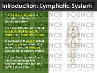
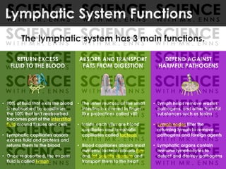
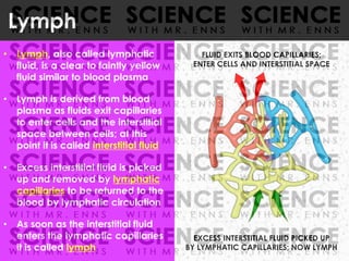
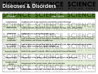
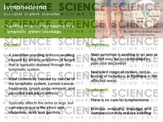
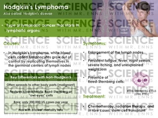
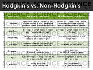
A Note About This Preview
THESE SAMPLE SLIDES ARE NOT THE ENTIRE PPT.
THE FULL PPT IS TOO LARGE TO UPLOAD AS A PREVIEW.
THESE SLIDES GIVE A BRIEF LOOK AT THE CONTENT
AND FORMAT OF EACH SECTION OF THE PPT..
SAMPLE SLIDES
The Lymphatic System
Part 1: Lymph and Lymphatic Vessels
• The lymphatic system is a
subsystem of the body’s
circulatory system
• It is a sophisticated network of
lymphatic fluid, lymphatic
vessels, and lymphatic organs
• Its main job is to collect a fluid
called lymph from intercellular
tissue spaces and transport it
back to the circulatory system
• The lymphatic system plays a
role in maintaining fluid
balance, absorbing fats, and
fighting pathogens
Introduction: Lymphatic System
RETURN EXCESS
FLUID TO THE BLOOD
• 90% of fluid that exits the blood
is reabsorbed by capillaries;
the 10% that isn’t reabsorbed
becomes part of the interstitial
fluid around tissues and cells
• Lymphatic capillaries absorb
excess fluid and proteins and
returns them to the blood
• Once reabsorbed, the excess
fluid is called lymph
ABSORB AND TRANSPORT
FATS FROM DIGESTION
• The inner mucosa of the small
intestine is covered in finger-
like projections called villi;
• Inside each vilus are blood
capillaries and lymphatic
capillaries called lacteals
• Blood capillaries absorb most
nutrients; lacteals absorb fats
and fat-soluble vitamins and
transport them to the heart
DEFEND AGAINST
HARMFUL PATHOGENS
• Lymph helps remove wastes,
pathogens, and other harmful
substances such as toxins
• Lymph nodes filter the
returning lymph to remove
pathogens and foreign agents
• Lymphatic organs contain
immune lymphocytes to
detect and destroy pathogens
Lymphatic System Functions
The lymphatic system has 3 main functions.
• Lymph, also called lymphatic
fluid, is a clear to faintly yellow
fluid similar to blood plasma
• Lymph is derived from blood
plasma as fluids exit capillaries
to enter cells and the interstitial
space between cells; at this
point it is called interstitial fluid
• Excess interstitial fluid is picked
up and removed by lymphatic
capillaries to be returned to the
blood by lymphatic circulation
• As soon as the interstitial fluid
enters the lymphatic capillaries
it is called lymph
Lymph
EXCESS INTERSTITIAL FLUID PICKED UP
BY LYMPHATIC CAPILLARIES; NOW LYMPH
FLUID EXITS BLOOD CAPILLARIES;
ENTER CELLS AND INTERSTITIAL SPACE
• Lymphatic capillaries are thin,
microscopic tubes that lay side
by side with blood capillaries;
they are found in all body
regions that have blood vessels
• Their walls are made of a single
layer of simple squamous cells
that overlap to form a simple
one-way valve that permits
excess fluid to enter but
prevents lymph from exiting
• Lymphatic capillaries are more
permeable than blood
capillaries to solvents, proteins,
and roving immune cells
Lymphatic Capillaries
ARTERIOLE VENULE
LYMPHATIC
CAPILLARIES
LYMPHATIC
VESSELS
BLOOD CAPILLARY
FLUID
ENTERS
NOW
LYMPH
LYMPHATIC CAPILLARY
SIMPLE SQUAMOUS ENDOTHELIUM
• Lymphatic vessels are thin
walled tubes distributed
throughout the body
• They form tubular networks
within most tissues and are
especially numerous in the skin
• Lymphatic vessels only flow one
way: from tissues to the heart;
they have valves to encourage
lymph to move in one direction
• There are 3 main types (in order
of size): lymphatic capillaries,
lymphatic vessels, and
lymphatic ducts
Lymphatic Vessels
LYMPHATIC
DUCTS
LYMPHATIC
VESSELS
LYMPHATIC
CAPILLARIES
• The lymphatic capillaries merge
with others to form lymphatic
vessels which are similar to veins
• Walls made of 3 layers:
• Tunica intima (endothelium)
• Tunica media (sm. muscle)
• Tunica externa (con. tissue)
• Have numerous 1-way valves
that prevent backflow of lymph
• Like veins, there is no pump: the
movement of lymph is assisted
by pressure exerted on vessel
walls by contractions of the
surrounding skeletal muscles
Lymphatic Vessels
3-LAYERED
WALLS
LYMPHATIC
CAPILLARY
LYMPHATIC
VESSEL
FLOW TOWARDS LYMPHATIC DUCTS
ONE-WAY
VALVE
• Lymphatic vessels merge with
others into one of two lymphatic
ducts which drain large regions
• The right lymphatic duct drains
lymph from the upper right
quadrant of the body, including
the right arm and right sides of
the head, neck, and thorax; it
arises in the neck and empties
into the right subclavian vein
• The thoracic duct drains the rest
of the body; it starts in the
abdominal cavity, rises upward
to the neck and empties into
the left subclavian vein
Lymphatic Ducts
REGION
DRAINED BY
R. LYMPHATIC
DUCT
RIGHT
LYMPHATIC DUCT
RIGHT
SUBCLAVIAN
VEIN
REGION
DRAINED BY
THORACIC
DUCT
THORACIC
DUCT
LEFT
SUBCLAVIAN
VEIN
CISTERNA
CHYLI
THORACIC
DUCT
RIGHT
LYMPHATIC
DUCT
The Lymphatic System
Part 2: Lymphatic Organs
• The organs of the immune and
lymphatic systems are
collectively called lymphoid
organs; there are two types:
• Primary lymphoid organs are
the site of production of new
lymphocytes from stem cells
• Include: bone marrow, thymus
• Secondary lymphoid organs
are sites where lymphocytes
mature and are exposed to
foreign substances
• Include: lymph nodes, thymus,
tonsils, spleen, appendix
Lymphoid Organs
Primary Lymphoid Organs
Sites of production of new lymphocytes from
stem cells and their maturation
bone marrow thymus
Secondary Lymphoid Organs
Sites of maturation of lymphocytes and
exposure to foreign substances
lymph
nodes
thymus tonsils spleen
appen
-dix
Lymph Node Anatomy
• Lymphatic vessels enter each node
on the convex side; these vessels
are called afferent lymphatic vessels
• Lymph vessels exit each node from
an indentation on the convex side
called the hilum; these vessels are
called efferent lymphatic vessels
• Each node is surrounded by a fibrous
connective tissue capsule that
features many inward extensions
called trabeculae that divide the
node into compartments called
lymph nodules or lobules
• Internally, each lymph node is
divided into two distinct regions: an
outer cortex and inner medulla
CORTEX
NODULES
HILUM
AFFERENT LYMPHATIC VESSELS
EFFERENT LYMPHATIC VESSELS
TRABECULAE
MEDULLA
CAPSULE
MALT
• Small clusters of lymphoid tissue
exist in various mucosal
membranes of the body, including
the digestive and respiratory tracts
• Collectively, the clusters are called
mucosa-associated lymphatic
tissue or MALT; which make up 50%
of all lymphoid tissue in the body
• MALT contains lymphocytes such as
T cells, B cells and macrophages,
which protect the body from
pathogens or other harmful
substances that may enter the body
through mucous membranes
• MALT is often subdivided into more
specific categories, including GALT,
BALT, NALT, SALT, and CALT
MALT
MUCOSA ASSOCIATED LYMPHATIC TISSUE
Location: mucous membranes in the body
MALT SUBDIVISIONS LOCATION
GALT
Gut Associated
Lymphatic Tissue
Intestine
BALT
Bronchus Associated
Lymphatic Tissue
Lungs
NALT
Nasal Associated
Lymphatic Tissue
Nose
SALT
Skin Associated
Lymphatic Tissue
Epidermis
CALT
Conjunctiva Associated
Lymphatic Tissue
Eye
• Tonsils are clusters of lymphoid tissue
found under mucous membranes
lining the nose, mouth, and throat
• As a component of MALT, their job is
to protect the body from pathogens
or harmful substances that may
enter via the nose or mouth
• There are 3 types of tonsils:
• Pharyngeal tonsils are are found in
the nasal cavity; they are called
adenoids when inflamed
• Palatine tonsils are found in the
throat behind the oral cavity
• Lingual tonsils are found at the base
of the tongue near the back
TONSILS
Tonsils MALT
PHARYNGEAL
TONSIL
PALANTINE
TONSIL
LINGUAL
TONSIL
Peyer’s Patches
• Peyer’s patches are small egg-
shaped nodules of lymphoid
tissue found under the mucous
membranes of the small
intestine, particularly in the lower
region called the ileum
• As a component of MALT (in this
case GALT) their job is to protect
the body from pathogens or
harmful substances that may
enter through the small intestine PEYER’S
PATCHES
PEYER’S
PATCHES
MALT
Spleen Anatomy
• The concave region of the spleen
where the splenic artery enters and
the splenic vein exits is the hilus
• The spleen in surrounded by a
connective tissue capsule with
inward extensions (trabeculae) that
divide it into lobules, each of which
features two types of tissues
• White pulp consists of lymphocytes
surrounding small arterioles
• Red pulp consists of sinuses filled
with blood and cords of lymphatic
cells, mainly macrophages
• Blood enters the spleen, moves
through the sinuses where it is filtered
and monitored, and then exits
HILUS
RED
PULP
CAPSULE
SPLENIC
ARTERY
SPLENIC
VEIN
WHITE
PULP
SINUS
TRABECULAE LOBULES
The Lymphatic System
Part 3: Lymphatic System Disorders
Disorder Description
Lymphedema Swelling in arm or leg caused by a lymphatic system blockage
Elephantiasis Tropical parasitic disease that causes swelling in the legs, arms, and genitalia
Lymphoma Malignant cancer of the lymphatic system
Hodgkin’s
Lymphoma
Type of lymphatic cancer that starts in lymphatic organs; can spread to the rest of the body;
less common with a lower mortality rate
Non-Hodgkin’s
Lymphoma
Type of lymphatic cancer that starts in B-cells and T-cells; does not spread beyond lymph
nodes. more common with higher mortality rate
Ruptured Spleen Break in the spleen’s capsule, often causing severe internal bleeding
Splenomegaly Enlargement of the spleen; often associated with blood cell destruction
Swollen Lymph
Nodes Enlargement of the lymph nodes, often due to infection
Tonsillitis Inflammation of the tonsils, most often due to a bacterial or viral infection
Diseases & Disorders
Symptoms:
• Main symptom is swelling in an arm or
leg that may be accompanied by
pain and discomfort
• Restricted range of motion, and a
feeling of heaviness or tightness in the
affected areas
Treatment:
• There is no cure for lymphedema
• Exercise, wrapping, massage, and
compression may reduce swelling
Lymphedema
Causes:
• A condition resulting in tissue swelling
caused by an accumulation of lymph
that is typically drained through the
lymphatic system
• Most commonly caused by cancer of
the lymphatic system, certain cancer
treatments, lymph node removal, and
parasites (see elephantiasis)
• Typically affects the arms or legs, but
can also occur in the chest wall,
abdomen, neck and genitals
• Swelling in arm or leg caused by a
lymphatic system blockage
Symptoms:
• Enlargement of the lymph nodes
• Persistent fatigue, fever, night sweats,
severe itching, and unexplained
weight loss
• Presence of
Reed-Sternberg cells:
Treatment:
• Chemotherapy, radiation therapy, and
in rare cases, stem-cell transplant
Hodgkin’s Lymphoma
Causes:
• In Hodgkin's lymphoma, white blood
cells called lymphocytes grow out of
control by replicating themselves in
the germinal centers of lymph nodes
• Type of lymphatic cancer that starts in
lymphatic organs
Key Differences with Non-Hodgkin’s
Often spreads to other body parts (malignant)
Features characteristic Reed-Sternberg or
“owl-eyes” cells
Rare; only 200,000 US cases per year
Has with a lower mortality rate
Hodgkin’s vs. Non-Hodgkin’s
Comparison Hodgkin’s Lymphoma Non-Hodgkin’s Lymphoma
Definition
Lymphatic cancer caused by the
overproduction of B or T cells in the
germinal center of the lymph nodes
Lymphatic cancer caused by the
overproduction of B or T cells
outside of the germinal centers
Location
Develops in lymph nodes in upper
body (armpits, chest, neck)
Develops in lymph nodes anywhere
in the body
Malignancy
Malignant; spreads from lymph
nodes to other body regions
Less malignant; remains in the
lymph nodes
Histology
Presence of Reed-Sternberg cells
(bi-nucleated B-cells – owl eyes)
No Reed-Sternberg cells
Frequency
Rare;
8,500 U.S. cases per year
More common;
80,000 U.S. cases per year
Age
Typically diagnosed in people 15-40
years of age; median age is 29
Typically diagnosed in adults over
60 years of age
Mortality
Mostly treatable;
90% 5-year survival rate
Not as treatable;
73% 5-year survival rate
Any Questions?
Name: Class: Date:
SUMMARY NOTES – LYMPHATIC SYSTEM
Part 1: Lymph and Lymphatic Vessels
1. What is the lymphatic system and what 3 things is it composed of?
2. Summarize the 3 main roles of the lymphatic system by filling in the table below.
3 Main Functions of the Lymphatic System
3. What is lymph and where does it come from?
4. Fill in the table below to state the 3 types of lymphatic vessels according to size.
3 Types of Lymphatic Vessels
Smallest Largest
5. Examine the diagram of a typical capillary bed and write the name for each labeled part.
Basic Anatomy of a Capillary Bed
a.
b.
c.
d.
e.
6. What are lacteals? Where are they located and what is their main function?
7. What are 3 ways that lymphatic vessels are similar to the veins of the circulatory system?
8. Contrast the right lymphatic duct and thoracic duct by filling in the summary table below.
Comparison Right Lymphatic Duct Thoracic Duct
What Body
Region It Drains
Where It Arises
Where It Empties
Part 2: Lymphatic Organs
9. Contrast primary and secondary lymphoid organs by filling in the summary table below.
Comparison Primary Lymphoid Organs Secondary Lymphoid Organs
Definition
Organs
10. Lymph nodes are the most numerous lymphoid organs of the body. What are their 3 main jobs?
3 Main Jobs of Lymph Nodes
11. Examine the diagram of a lymph node and write the name for each labeled part.
Internal Anatomy of a Lymph Node
a.
b.
c. (concave region)
d.
e.
f.
g.
h.
12. In the space below, define MALT, describe its purpose, and list 6 subdivisions of MALT.
13. Summarize 5 secondary lymphoid or lymphatic organs by filling in the table below.
Lymphatic Organs
Diagram Name Description & Location Main Job(s)
Tonsils
Protect the body from
pathogens that may enter
through the small intestine
Small, finger-shaped pouch of
intestinal tissue; located between the
small intestine and large intestine
Filters blood, monitors
blood for pathogens, and
stores blood
Soft, bi-lobed gland located in the
chest area between the lungs
(mediastinum)
14. Examine the diagram of the spleen and write the name for each labeled part.
Internal Anatomy of the Spleen
a.
b.
c.
d.
e.
f.
g.
Name: Class: Date:
SUMMARY NOTES – LYMPHATIC SYSTEM (KEY)
Part 1: Lymph and Lymphatic Vessels
1. What is the lymphatic system and what 3 things is it composed of?
The lymphatic system is a subsystem of the circulatory system
It is a (sophisticated) network of lymphatic fluid, vessels, and organs
2. Summarize the 3 main roles of the lymphatic system by filling in the table below.
3 Main Functions of the Lymphatic System
Return excess fluid to the
blood
Absorb and transport fats from
digestion
Defend against harmful
pathogens
3. What is lymph and where does it come from?
Lymph is a clear to faintly yellow fluid (similar to blood plasma)
Lymph comes from blood plasma as it leaves blood capillaries and enters the interstitial space
Excess fluid enters lymphatic capillaries and is then called lymph
Or equivalent
4. Fill in the table below to state the 3 types of lymphatic vessels according to size.
3 Types of Lymphatic Vessels
Smallest Lymphatic capillaries Lymphatic vessels Lymphatic ducts Largest
5. Examine the diagram of a typical capillary bed and write the name for each labeled part.
Basic Anatomy of a Capillary Bed
a. arteriole (accept: artery)
b. (blood) capillary
c. venule (accept: vein)
d. lymphatic capillary
e. lymphatic vessel
6. What are lacteals? Where are they located and what is their main function?
Lacteals are specialized lymphatic capillaries
They are located in folds of the inner lining of the small intestine (called
villi)
Lacteals absorb fats and fat-soluble vitamins and transport them to the
blood
7. What are 3 ways that lymphatic vessels are similar to the veins of the circulatory system?
Have walls made of 3 layers Have numerous 1-way valves Have no pump
(tunica intima, tunica media,
tunica externa)
(that prevent backflow
of lymph)
(Lymph moved by contractions
of skeletal muscles)
8. Contrast the right lymphatic duct and thoracic duct by filling in the summary table below.
Comparison Right Lymphatic Duct Thoracic Duct
What Body
Region It Drains
Upper right quadrant
(Right arm, head, neck, and thorax)
All other quadrants of the body
Where It Arises Neck Abdominal Cavity
Where It Empties Right subclavian vein Left subclavian vein
Part 2: Lymphatic Organs
9. Contrast primary and secondary lymphoid organs by filling in the summary table below.
Comparison Primary Lymphoid Organs Secondary Lymphoid Organs
Definition
Sites where new lymphocytes are
produced from stem cells
Sites where lymphocytes mature and
are exposed to foreign substances
Organs Bone marrow, thymus
lymph nodes, thymus, tonsils, spleen,
appendix
10. Lymph nodes are the most numerous lymphoid organs of the body. What are their 3 main jobs?
3 Main Jobs of Lymph Nodes
Filter lymph for pathogens and
other harmful substances
Detect and alert immune
system to foreign invaders
Produce mature lymphocytes
11. Examine the diagram of a lymph node and write the name for each labeled part.
Internal Anatomy of a Lymph Node
a. afferent lymphatic vessel(s)
b. efferent lymphatic vessel(s)
c. (concave region) hilum
d. capsule
e. trabeculae
f. lymph nodules (accept: lymph lobules)
g. cortex
h. medulla
12. In the space below, define MALT, describe its purpose, and list 6 subdivisions of MALT.
MALT stands for mucous associated lymphatic tissue
Its purpose is to protect the body from pathogens and other harmful substances that may enter the
body through mucous membranes
5 (out of 6) subdivisions include: GALT (gut associated lymphatic tissue), BALT (bronchial
associated lymphatic tissue), NALT (nasal associated lymphatic tissue), SALT (skin associated
lymphatic tissue), and CALT (conjunctiva associated lymphatic tissue)
13. Summarize 5 secondary lymphoid or lymphatic organs by filling in the table below.
Secondary Lymphoid Organs
Diagram Name Description & Location Main Job(s)
Tonsils
Clusters of lymphoid tissue under
mucous membranes of the nose,
mouth, and throat
Protect the body from
pathogens entering through
the nose or mouth
Peyer’s
Patches
Small, egg-shaped nodules of
lymphoid tissue found under mucous
membranes of the small intestine
Protect the body from
pathogens that may enter
through the small intestine
Appendix
Small, finger-shaped pouch of
intestinal tissue; located between the
small intestine and large intestine
Protect the body from
pathogens that may enter
through the large intestine
Spleen
Body’s largest lymphoid organ;
located in the upper left side of the
abdomen, next to the stomach
Filters blood, monitors
blood for pathogens, and
stores blood
Thymus
Soft, bi-lobed gland located in the
chest area between the lungs
(mediastinum)
Creation and maturation of
immune T-cells in the early
stages of life
14. Examine the diagram of the spleen and write the name for each labeled part.
Internal Anatomy of the Spleen
a. splenic artery
b. splenic vein
c. capsule
d. trabeculae
e. lobules
f. white pulp
g. red pulp

fd6e4fe74e7fe7ba769d885thfsyc115ee1dc.pdf

  • 1.
    A Note AboutThis Preview THESE SAMPLE SLIDES ARE NOT THE ENTIRE PPT. THE FULL PPT IS TOO LARGE TO UPLOAD AS A PREVIEW. THESE SLIDES GIVE A BRIEF LOOK AT THE CONTENT AND FORMAT OF EACH SECTION OF THE PPT.. SAMPLE SLIDES
  • 2.
    The Lymphatic System Part1: Lymph and Lymphatic Vessels
  • 3.
    • The lymphaticsystem is a subsystem of the body’s circulatory system • It is a sophisticated network of lymphatic fluid, lymphatic vessels, and lymphatic organs • Its main job is to collect a fluid called lymph from intercellular tissue spaces and transport it back to the circulatory system • The lymphatic system plays a role in maintaining fluid balance, absorbing fats, and fighting pathogens Introduction: Lymphatic System
  • 4.
    RETURN EXCESS FLUID TOTHE BLOOD • 90% of fluid that exits the blood is reabsorbed by capillaries; the 10% that isn’t reabsorbed becomes part of the interstitial fluid around tissues and cells • Lymphatic capillaries absorb excess fluid and proteins and returns them to the blood • Once reabsorbed, the excess fluid is called lymph ABSORB AND TRANSPORT FATS FROM DIGESTION • The inner mucosa of the small intestine is covered in finger- like projections called villi; • Inside each vilus are blood capillaries and lymphatic capillaries called lacteals • Blood capillaries absorb most nutrients; lacteals absorb fats and fat-soluble vitamins and transport them to the heart DEFEND AGAINST HARMFUL PATHOGENS • Lymph helps remove wastes, pathogens, and other harmful substances such as toxins • Lymph nodes filter the returning lymph to remove pathogens and foreign agents • Lymphatic organs contain immune lymphocytes to detect and destroy pathogens Lymphatic System Functions The lymphatic system has 3 main functions.
  • 5.
    • Lymph, alsocalled lymphatic fluid, is a clear to faintly yellow fluid similar to blood plasma • Lymph is derived from blood plasma as fluids exit capillaries to enter cells and the interstitial space between cells; at this point it is called interstitial fluid • Excess interstitial fluid is picked up and removed by lymphatic capillaries to be returned to the blood by lymphatic circulation • As soon as the interstitial fluid enters the lymphatic capillaries it is called lymph Lymph EXCESS INTERSTITIAL FLUID PICKED UP BY LYMPHATIC CAPILLARIES; NOW LYMPH FLUID EXITS BLOOD CAPILLARIES; ENTER CELLS AND INTERSTITIAL SPACE
  • 6.
    • Lymphatic capillariesare thin, microscopic tubes that lay side by side with blood capillaries; they are found in all body regions that have blood vessels • Their walls are made of a single layer of simple squamous cells that overlap to form a simple one-way valve that permits excess fluid to enter but prevents lymph from exiting • Lymphatic capillaries are more permeable than blood capillaries to solvents, proteins, and roving immune cells Lymphatic Capillaries ARTERIOLE VENULE LYMPHATIC CAPILLARIES LYMPHATIC VESSELS BLOOD CAPILLARY FLUID ENTERS NOW LYMPH LYMPHATIC CAPILLARY SIMPLE SQUAMOUS ENDOTHELIUM
  • 7.
    • Lymphatic vesselsare thin walled tubes distributed throughout the body • They form tubular networks within most tissues and are especially numerous in the skin • Lymphatic vessels only flow one way: from tissues to the heart; they have valves to encourage lymph to move in one direction • There are 3 main types (in order of size): lymphatic capillaries, lymphatic vessels, and lymphatic ducts Lymphatic Vessels LYMPHATIC DUCTS LYMPHATIC VESSELS LYMPHATIC CAPILLARIES
  • 8.
    • The lymphaticcapillaries merge with others to form lymphatic vessels which are similar to veins • Walls made of 3 layers: • Tunica intima (endothelium) • Tunica media (sm. muscle) • Tunica externa (con. tissue) • Have numerous 1-way valves that prevent backflow of lymph • Like veins, there is no pump: the movement of lymph is assisted by pressure exerted on vessel walls by contractions of the surrounding skeletal muscles Lymphatic Vessels 3-LAYERED WALLS LYMPHATIC CAPILLARY LYMPHATIC VESSEL FLOW TOWARDS LYMPHATIC DUCTS ONE-WAY VALVE
  • 9.
    • Lymphatic vesselsmerge with others into one of two lymphatic ducts which drain large regions • The right lymphatic duct drains lymph from the upper right quadrant of the body, including the right arm and right sides of the head, neck, and thorax; it arises in the neck and empties into the right subclavian vein • The thoracic duct drains the rest of the body; it starts in the abdominal cavity, rises upward to the neck and empties into the left subclavian vein Lymphatic Ducts REGION DRAINED BY R. LYMPHATIC DUCT RIGHT LYMPHATIC DUCT RIGHT SUBCLAVIAN VEIN REGION DRAINED BY THORACIC DUCT THORACIC DUCT LEFT SUBCLAVIAN VEIN CISTERNA CHYLI THORACIC DUCT RIGHT LYMPHATIC DUCT
  • 10.
    The Lymphatic System Part2: Lymphatic Organs
  • 11.
    • The organsof the immune and lymphatic systems are collectively called lymphoid organs; there are two types: • Primary lymphoid organs are the site of production of new lymphocytes from stem cells • Include: bone marrow, thymus • Secondary lymphoid organs are sites where lymphocytes mature and are exposed to foreign substances • Include: lymph nodes, thymus, tonsils, spleen, appendix Lymphoid Organs Primary Lymphoid Organs Sites of production of new lymphocytes from stem cells and their maturation bone marrow thymus Secondary Lymphoid Organs Sites of maturation of lymphocytes and exposure to foreign substances lymph nodes thymus tonsils spleen appen -dix
  • 12.
    Lymph Node Anatomy •Lymphatic vessels enter each node on the convex side; these vessels are called afferent lymphatic vessels • Lymph vessels exit each node from an indentation on the convex side called the hilum; these vessels are called efferent lymphatic vessels • Each node is surrounded by a fibrous connective tissue capsule that features many inward extensions called trabeculae that divide the node into compartments called lymph nodules or lobules • Internally, each lymph node is divided into two distinct regions: an outer cortex and inner medulla CORTEX NODULES HILUM AFFERENT LYMPHATIC VESSELS EFFERENT LYMPHATIC VESSELS TRABECULAE MEDULLA CAPSULE
  • 13.
    MALT • Small clustersof lymphoid tissue exist in various mucosal membranes of the body, including the digestive and respiratory tracts • Collectively, the clusters are called mucosa-associated lymphatic tissue or MALT; which make up 50% of all lymphoid tissue in the body • MALT contains lymphocytes such as T cells, B cells and macrophages, which protect the body from pathogens or other harmful substances that may enter the body through mucous membranes • MALT is often subdivided into more specific categories, including GALT, BALT, NALT, SALT, and CALT MALT MUCOSA ASSOCIATED LYMPHATIC TISSUE Location: mucous membranes in the body MALT SUBDIVISIONS LOCATION GALT Gut Associated Lymphatic Tissue Intestine BALT Bronchus Associated Lymphatic Tissue Lungs NALT Nasal Associated Lymphatic Tissue Nose SALT Skin Associated Lymphatic Tissue Epidermis CALT Conjunctiva Associated Lymphatic Tissue Eye
  • 14.
    • Tonsils areclusters of lymphoid tissue found under mucous membranes lining the nose, mouth, and throat • As a component of MALT, their job is to protect the body from pathogens or harmful substances that may enter via the nose or mouth • There are 3 types of tonsils: • Pharyngeal tonsils are are found in the nasal cavity; they are called adenoids when inflamed • Palatine tonsils are found in the throat behind the oral cavity • Lingual tonsils are found at the base of the tongue near the back TONSILS Tonsils MALT PHARYNGEAL TONSIL PALANTINE TONSIL LINGUAL TONSIL
  • 15.
    Peyer’s Patches • Peyer’spatches are small egg- shaped nodules of lymphoid tissue found under the mucous membranes of the small intestine, particularly in the lower region called the ileum • As a component of MALT (in this case GALT) their job is to protect the body from pathogens or harmful substances that may enter through the small intestine PEYER’S PATCHES PEYER’S PATCHES MALT
  • 16.
    Spleen Anatomy • Theconcave region of the spleen where the splenic artery enters and the splenic vein exits is the hilus • The spleen in surrounded by a connective tissue capsule with inward extensions (trabeculae) that divide it into lobules, each of which features two types of tissues • White pulp consists of lymphocytes surrounding small arterioles • Red pulp consists of sinuses filled with blood and cords of lymphatic cells, mainly macrophages • Blood enters the spleen, moves through the sinuses where it is filtered and monitored, and then exits HILUS RED PULP CAPSULE SPLENIC ARTERY SPLENIC VEIN WHITE PULP SINUS TRABECULAE LOBULES
  • 17.
    The Lymphatic System Part3: Lymphatic System Disorders
  • 18.
    Disorder Description Lymphedema Swellingin arm or leg caused by a lymphatic system blockage Elephantiasis Tropical parasitic disease that causes swelling in the legs, arms, and genitalia Lymphoma Malignant cancer of the lymphatic system Hodgkin’s Lymphoma Type of lymphatic cancer that starts in lymphatic organs; can spread to the rest of the body; less common with a lower mortality rate Non-Hodgkin’s Lymphoma Type of lymphatic cancer that starts in B-cells and T-cells; does not spread beyond lymph nodes. more common with higher mortality rate Ruptured Spleen Break in the spleen’s capsule, often causing severe internal bleeding Splenomegaly Enlargement of the spleen; often associated with blood cell destruction Swollen Lymph Nodes Enlargement of the lymph nodes, often due to infection Tonsillitis Inflammation of the tonsils, most often due to a bacterial or viral infection Diseases & Disorders
  • 19.
    Symptoms: • Main symptomis swelling in an arm or leg that may be accompanied by pain and discomfort • Restricted range of motion, and a feeling of heaviness or tightness in the affected areas Treatment: • There is no cure for lymphedema • Exercise, wrapping, massage, and compression may reduce swelling Lymphedema Causes: • A condition resulting in tissue swelling caused by an accumulation of lymph that is typically drained through the lymphatic system • Most commonly caused by cancer of the lymphatic system, certain cancer treatments, lymph node removal, and parasites (see elephantiasis) • Typically affects the arms or legs, but can also occur in the chest wall, abdomen, neck and genitals • Swelling in arm or leg caused by a lymphatic system blockage
  • 20.
    Symptoms: • Enlargement ofthe lymph nodes • Persistent fatigue, fever, night sweats, severe itching, and unexplained weight loss • Presence of Reed-Sternberg cells: Treatment: • Chemotherapy, radiation therapy, and in rare cases, stem-cell transplant Hodgkin’s Lymphoma Causes: • In Hodgkin's lymphoma, white blood cells called lymphocytes grow out of control by replicating themselves in the germinal centers of lymph nodes • Type of lymphatic cancer that starts in lymphatic organs Key Differences with Non-Hodgkin’s Often spreads to other body parts (malignant) Features characteristic Reed-Sternberg or “owl-eyes” cells Rare; only 200,000 US cases per year Has with a lower mortality rate
  • 21.
    Hodgkin’s vs. Non-Hodgkin’s ComparisonHodgkin’s Lymphoma Non-Hodgkin’s Lymphoma Definition Lymphatic cancer caused by the overproduction of B or T cells in the germinal center of the lymph nodes Lymphatic cancer caused by the overproduction of B or T cells outside of the germinal centers Location Develops in lymph nodes in upper body (armpits, chest, neck) Develops in lymph nodes anywhere in the body Malignancy Malignant; spreads from lymph nodes to other body regions Less malignant; remains in the lymph nodes Histology Presence of Reed-Sternberg cells (bi-nucleated B-cells – owl eyes) No Reed-Sternberg cells Frequency Rare; 8,500 U.S. cases per year More common; 80,000 U.S. cases per year Age Typically diagnosed in people 15-40 years of age; median age is 29 Typically diagnosed in adults over 60 years of age Mortality Mostly treatable; 90% 5-year survival rate Not as treatable; 73% 5-year survival rate
  • 22.
  • 23.
    Name: Class: Date: SUMMARYNOTES – LYMPHATIC SYSTEM Part 1: Lymph and Lymphatic Vessels 1. What is the lymphatic system and what 3 things is it composed of? 2. Summarize the 3 main roles of the lymphatic system by filling in the table below. 3 Main Functions of the Lymphatic System 3. What is lymph and where does it come from? 4. Fill in the table below to state the 3 types of lymphatic vessels according to size. 3 Types of Lymphatic Vessels Smallest Largest 5. Examine the diagram of a typical capillary bed and write the name for each labeled part. Basic Anatomy of a Capillary Bed a. b. c. d. e. 6. What are lacteals? Where are they located and what is their main function?
  • 24.
    7. What are3 ways that lymphatic vessels are similar to the veins of the circulatory system? 8. Contrast the right lymphatic duct and thoracic duct by filling in the summary table below. Comparison Right Lymphatic Duct Thoracic Duct What Body Region It Drains Where It Arises Where It Empties Part 2: Lymphatic Organs 9. Contrast primary and secondary lymphoid organs by filling in the summary table below. Comparison Primary Lymphoid Organs Secondary Lymphoid Organs Definition Organs 10. Lymph nodes are the most numerous lymphoid organs of the body. What are their 3 main jobs? 3 Main Jobs of Lymph Nodes 11. Examine the diagram of a lymph node and write the name for each labeled part. Internal Anatomy of a Lymph Node a. b. c. (concave region) d. e. f. g. h.
  • 25.
    12. In thespace below, define MALT, describe its purpose, and list 6 subdivisions of MALT. 13. Summarize 5 secondary lymphoid or lymphatic organs by filling in the table below. Lymphatic Organs Diagram Name Description & Location Main Job(s) Tonsils Protect the body from pathogens that may enter through the small intestine Small, finger-shaped pouch of intestinal tissue; located between the small intestine and large intestine Filters blood, monitors blood for pathogens, and stores blood Soft, bi-lobed gland located in the chest area between the lungs (mediastinum) 14. Examine the diagram of the spleen and write the name for each labeled part. Internal Anatomy of the Spleen a. b. c. d. e. f. g.
  • 26.
    Name: Class: Date: SUMMARYNOTES – LYMPHATIC SYSTEM (KEY) Part 1: Lymph and Lymphatic Vessels 1. What is the lymphatic system and what 3 things is it composed of? The lymphatic system is a subsystem of the circulatory system It is a (sophisticated) network of lymphatic fluid, vessels, and organs 2. Summarize the 3 main roles of the lymphatic system by filling in the table below. 3 Main Functions of the Lymphatic System Return excess fluid to the blood Absorb and transport fats from digestion Defend against harmful pathogens 3. What is lymph and where does it come from? Lymph is a clear to faintly yellow fluid (similar to blood plasma) Lymph comes from blood plasma as it leaves blood capillaries and enters the interstitial space Excess fluid enters lymphatic capillaries and is then called lymph Or equivalent 4. Fill in the table below to state the 3 types of lymphatic vessels according to size. 3 Types of Lymphatic Vessels Smallest Lymphatic capillaries Lymphatic vessels Lymphatic ducts Largest 5. Examine the diagram of a typical capillary bed and write the name for each labeled part. Basic Anatomy of a Capillary Bed a. arteriole (accept: artery) b. (blood) capillary c. venule (accept: vein) d. lymphatic capillary e. lymphatic vessel 6. What are lacteals? Where are they located and what is their main function? Lacteals are specialized lymphatic capillaries They are located in folds of the inner lining of the small intestine (called villi) Lacteals absorb fats and fat-soluble vitamins and transport them to the blood
  • 27.
    7. What are3 ways that lymphatic vessels are similar to the veins of the circulatory system? Have walls made of 3 layers Have numerous 1-way valves Have no pump (tunica intima, tunica media, tunica externa) (that prevent backflow of lymph) (Lymph moved by contractions of skeletal muscles) 8. Contrast the right lymphatic duct and thoracic duct by filling in the summary table below. Comparison Right Lymphatic Duct Thoracic Duct What Body Region It Drains Upper right quadrant (Right arm, head, neck, and thorax) All other quadrants of the body Where It Arises Neck Abdominal Cavity Where It Empties Right subclavian vein Left subclavian vein Part 2: Lymphatic Organs 9. Contrast primary and secondary lymphoid organs by filling in the summary table below. Comparison Primary Lymphoid Organs Secondary Lymphoid Organs Definition Sites where new lymphocytes are produced from stem cells Sites where lymphocytes mature and are exposed to foreign substances Organs Bone marrow, thymus lymph nodes, thymus, tonsils, spleen, appendix 10. Lymph nodes are the most numerous lymphoid organs of the body. What are their 3 main jobs? 3 Main Jobs of Lymph Nodes Filter lymph for pathogens and other harmful substances Detect and alert immune system to foreign invaders Produce mature lymphocytes 11. Examine the diagram of a lymph node and write the name for each labeled part. Internal Anatomy of a Lymph Node a. afferent lymphatic vessel(s) b. efferent lymphatic vessel(s) c. (concave region) hilum d. capsule e. trabeculae f. lymph nodules (accept: lymph lobules) g. cortex h. medulla
  • 28.
    12. In thespace below, define MALT, describe its purpose, and list 6 subdivisions of MALT. MALT stands for mucous associated lymphatic tissue Its purpose is to protect the body from pathogens and other harmful substances that may enter the body through mucous membranes 5 (out of 6) subdivisions include: GALT (gut associated lymphatic tissue), BALT (bronchial associated lymphatic tissue), NALT (nasal associated lymphatic tissue), SALT (skin associated lymphatic tissue), and CALT (conjunctiva associated lymphatic tissue) 13. Summarize 5 secondary lymphoid or lymphatic organs by filling in the table below. Secondary Lymphoid Organs Diagram Name Description & Location Main Job(s) Tonsils Clusters of lymphoid tissue under mucous membranes of the nose, mouth, and throat Protect the body from pathogens entering through the nose or mouth Peyer’s Patches Small, egg-shaped nodules of lymphoid tissue found under mucous membranes of the small intestine Protect the body from pathogens that may enter through the small intestine Appendix Small, finger-shaped pouch of intestinal tissue; located between the small intestine and large intestine Protect the body from pathogens that may enter through the large intestine Spleen Body’s largest lymphoid organ; located in the upper left side of the abdomen, next to the stomach Filters blood, monitors blood for pathogens, and stores blood Thymus Soft, bi-lobed gland located in the chest area between the lungs (mediastinum) Creation and maturation of immune T-cells in the early stages of life 14. Examine the diagram of the spleen and write the name for each labeled part. Internal Anatomy of the Spleen a. splenic artery b. splenic vein c. capsule d. trabeculae e. lobules f. white pulp g. red pulp