The cervical spine is the most superior portion of the vertebral column, lying between the cranium and the thoracic vertebrae. It consists of seven distinct vertebrae, two of which are given unique names: The first cervical vertebrae (C1) is known as the atlas. The second cervical vertebrae (C2) is known as the axis.
skeleton of human body, skeletal system of human body, skeleton anatomy, intr...Dr Shahid Alam
skeleton of human body, skeletal system of human body, skeleton anatomy, introduction to skeleton, axial skeleton, cranium, cranial bone mnemonic for cranial bone, bone of skeleton system, 206 bones by dr shahid alam, dr shahid, shahid alam, alam
Bones of upper limbs (Human Anatomy)
by DR RAI M. AMMAR
www.facebook.com/drraiammar
www.twitter.com/drraiammar
www.instagram.com/drraiammar
www.linkedin.com/in/drraiammar
www.themedicall.com/blog/auther/drraiammar/
For Any Book or Notes Visit Our Website:
www.allmedicaldata.wordpress.com
www.drraiammar.blogspot.com
YOUTUBE CHANNEL :
https://www.youtube.com/channel/UCu-oR9V3OdFNTJW5yqXWXxA
ANY QUESTION ??
Get in touch with us at Any of the Above Social Media or Email at
drraiammar@gmail.com
allmedicaldata@gmail.com
Vertibrae By M Thiru murugan MSc Nursingthiru murugan
Vertebral Column
By, M. Thiru murugan
Vertebral column:
The vertebral column encloses the spinal cord and the fluid surrounding the spinal cord. Also called backbone, spinal column, and spine.
Each vertebra is separated by a disc called intervertebral disc
The vertebrae surround and protect the spinal cord. The spinal cord is divided into segments, each containing a pair of spinal nerves that send messages between the brain and the rest of the body.
Many spinal nerves extend beyond the conus medullaris (the end of the spinal cord) to form a bundle of nerves called the cauda equina.
The vertebral column is made up 26
Cervical vertebrae: These 7 bones are found in the head and neck.
Thoracic vertebrae: These 12 bones are found in the upper back.
Lumbar vertebrae: These 5 bones are found in the lower back.
The sacrum (5) and coccyx (4) are both made up of several fused vertebrae. They help support the weight of the body while sitting.
Parts of the vertebrae:
The vertebrae of the cervical, thoracic, and lumbar spines are independent bones and generally quite similar.
The vertebrae of the sacrum & coccyx are usually fused and unable to move independently.
2 special vertebrae are the atlas (cervical 1) and axis (cervical 2), on which the head rests.
A typical vertebra consists of 2 parts: the vertebral body and the vertebral arch.
Vertebral body: Vertebral body is the thick oval segment of bone forming the front of the vertebra also called the centrum. The cavity of the vertebral body consists of cancellous bone tissue and is encircled by a protective layer of compact bone.
The vertebral arch is posterior, meaning it faces the back of a person.
Together, these enclose the vertebral foramen, which contains the spinal cord.
Because the spinal cord ends in the lumbar spine, and the sacrum and coccyx are fused, they do not contain a central foramen.
The vertebral arch is formed by a pair of pedicles & a pair of laminae, and supports 7 processes (4 articular, 2 transverse, and 1 spinous)
4 articular process: 2 articular process for above vertebrae & 2 articular process for ribs.
2 transverse processes and 1spinous process are posterior to (behind) the vertebral body.
The spinous process comes out the back, The spinous processes of the cervical and lumbar regions can be felt through the skin.
1 transverse process comes out the left, and 1 on the right.
Above & below each vertebra are joints called facet joints. These restrict the range of movement possible
In between each pair of vertebrae are 2 small holes called intervertebral foramina. The spinal nerves leave the spinal cord through these holes.
Cervical spine:
The cervical spine located in the neck area, consists of seven bones (C1 to C7)
The first two cervical spine are unique in shape and function.
first vertebra (C1), also called the atlas, The atlas holds head upright.
The second vertebra (C2), also called the axis, allows the atlas to rotation of head.
Functions:
Protecting spin
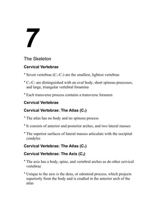
1. 7
The Skeleton
Cervical Vertebrae
* Seven vertebrae (C1-C7) are the smallest, lightest vertebrae
* C3-C7 are distinguished with an oval body, short spinous processes,
and large, triangular vertebral foramina
* Each transverse process contains a transverse foramen
Cervical Vertebrae
Cervical Vertebrae: The Atlas (C1)
* The atlas has no body and no spinous process
* It consists of anterior and posterior arches, and two lateral masses
* The superior surfaces of lateral masses articulate with the occipital
condyles
Cervical Vertebrae: The Atlas (C1)
Cervical Vertebrae: The Axis (C2)
* The axis has a body, spine, and vertebral arches as do other cervical
vertebrae
* Unique to the axis is the dens, or odontoid process, which projects
superiorly from the body and is cradled in the anterior arch of the
atlas
2. * The dens is a pivot for the rotation of the atlas
Cervical Vertebrae: The Axis (C2)
Cervical Vertebrae: The Atlas (C2)
Regional Characteristics of Vertebrae
Regional Characteristics of Vertebrae
Thoracic Vertebrae
* There are twelve vertebrae (T1-T12) all of which articulate with ribs
*Major markings include two facets and two demifacets on the heart-shaped
body, the circular vertebral foramen, transverse processes, and
a long spinous process
* The location of the articulate facets prevents flexion and extension,
but allows rotation of this area of the spine
Thoracic Vertebrae
Lumbar Vertebrae
* The five lumbar vertebrae (L1-L5) are located in the small of the back
and have an enhanced weight-bearing function
* They have short, thick pedicles and laminae, flat hatchet-shaped
spinous processes, and a triangular-shaped vertebral foramen
* Orientation of articular facets locks the lumbar vertebrae together to
provide stability
Lumbar Vertebrae
Sacrum
* Sacrum
* Consists of five fused vertebrae (S1-S5), which shape the posterior wall
of the pelvis
3. * It articulates with L5 superiorly, and with the auricular surfaces of the
hip bones
* Major markings include the sacral promontory, transverse lines, alae,
dorsal sacral foramina, sacral canal, and sacral hiatus
Coccyx
* Coccyx (Tailbone)
* The coccyx is made up of four (in some cases three to five) fused
vertebrae that articulate superiorly with the sacrum
Sacrum and Coccyx: Anterior View
Sacrum and Coccyx: Posterior View
Bony Thorax (Thoracic Cage)
* The thoracic cage is composed of the thoracic vertebrae dorsally, the
ribs laterally, and the sternum and costal cartilages anteriorly
Bony Thorax (Thoracic Cage)
* Functions
* Forms a protective cage around the heart, lungs, and great blood
vessels
* Supports the shoulder girdles and upper limbs
* Provides attachment for many neck, back, chest, and shoulder muscles
* Uses intercostal muscles to lift and depress the thorax during breathing
Bony Thorax (Thoracic Cage)
Bony Thorax (Thoracic Cage)
Sternum (Breastbone)
* A dagger-shaped, flat bone that lies in the anterior midline of the
thorax
4. * Results from the fusion of three bones – the superior manubrium, the
body, and the inferior xiphoid process
* Anatomical landmarks include the jugular (suprasternal) notch, the
sternal angle, and the xiphisternal joint
Ribs
* There are twelve pair of ribs forming the flaring sides of the thoracic
cage
* All ribs attach posteriorly to the thoracic vertebrae
* The superior 7 pair (true, or vertebrosternal ribs) attach directly to the
sternum via costal cartilages
* Ribs 8-10 (false, or vertebrocondral ribs) attach indirectly to the
sternum via costal cartilage
* Ribs 11-12 (floating, or vertebral ribs) have no anterior attachment
Ribs
Structure of a Typical True Rib
* Bowed, flat bone consisting of a head, neck, tubercle, and shaft
Structure of a Typical True Rib
* Bowed, flat bone consisting of a head, neck, tubercle, and shaft
Appendicular Skeleton
* The appendicular skeleton is made up of the bones of the limbs and
their girdles
* Pectoral girdles attach the upper limbs to the body trunk
* Pelvic girdle secures the lower limbs
Pectoral Girdles (Shoulder Girdles)
5. * The pectoral girdles consist of the anterior clavicles and the posterior
scapulae
* They attach the upper limbs to the axial skeleton in a manner that
allows for maximum movement
* They provide attachment points for muscles that move the upper
limbs
Pectoral Girdles (Shoulder Girdles)
Clavicles (Collarbones)
* Slender, doubly curved long bones lying across the superior thorax
* The acromial (lateral) end articulates with the scapula, and the sternal
(medial) end articulates with the sternum
* Provide attachment points for numerous muscles, and act as braces to
hold the scapulae and arms out laterally away from the body
Clavicles (Collarbones)
Scapulae (Shoulder Blades)
* Triangular, flat bones lying on the dorsal surface of the rib cage,
between the second and seventh ribs
* Scapulae have three borders and three angles
*Major markings include the suprascapular notch, the supraspinous and
infraspinous fossae, the spine, the acromion, and the coracoid process
Scapulae (Shoulder Blades)
Scapulae (Shoulder Blades)
Scapulae (Shoulder Blades)
6. * The pectoral girdles consist of the anterior clavicles and the posterior
scapulae
* They attach the upper limbs to the axial skeleton in a manner that
allows for maximum movement
* They provide attachment points for muscles that move the upper
limbs
Pectoral Girdles (Shoulder Girdles)
Clavicles (Collarbones)
* Slender, doubly curved long bones lying across the superior thorax
* The acromial (lateral) end articulates with the scapula, and the sternal
(medial) end articulates with the sternum
* Provide attachment points for numerous muscles, and act as braces to
hold the scapulae and arms out laterally away from the body
Clavicles (Collarbones)
Scapulae (Shoulder Blades)
* Triangular, flat bones lying on the dorsal surface of the rib cage,
between the second and seventh ribs
* Scapulae have three borders and three angles
*Major markings include the suprascapular notch, the supraspinous and
infraspinous fossae, the spine, the acromion, and the coracoid process
Scapulae (Shoulder Blades)
Scapulae (Shoulder Blades)
Scapulae (Shoulder Blades)