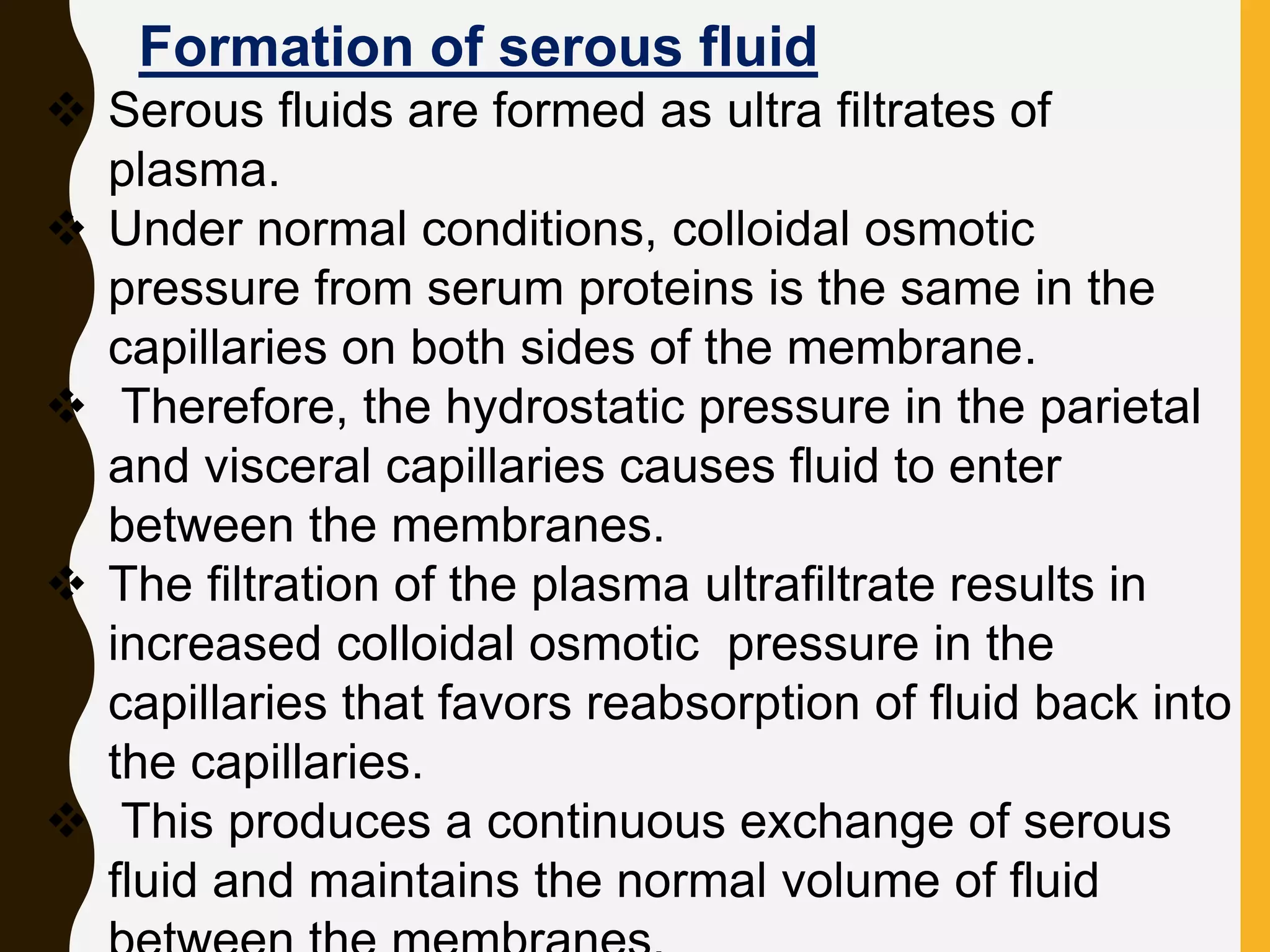
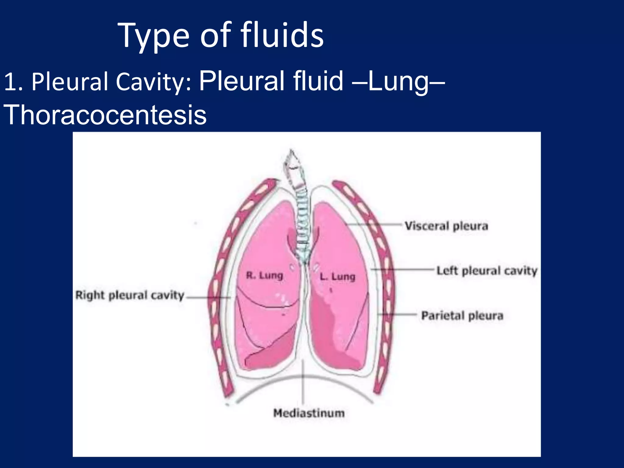
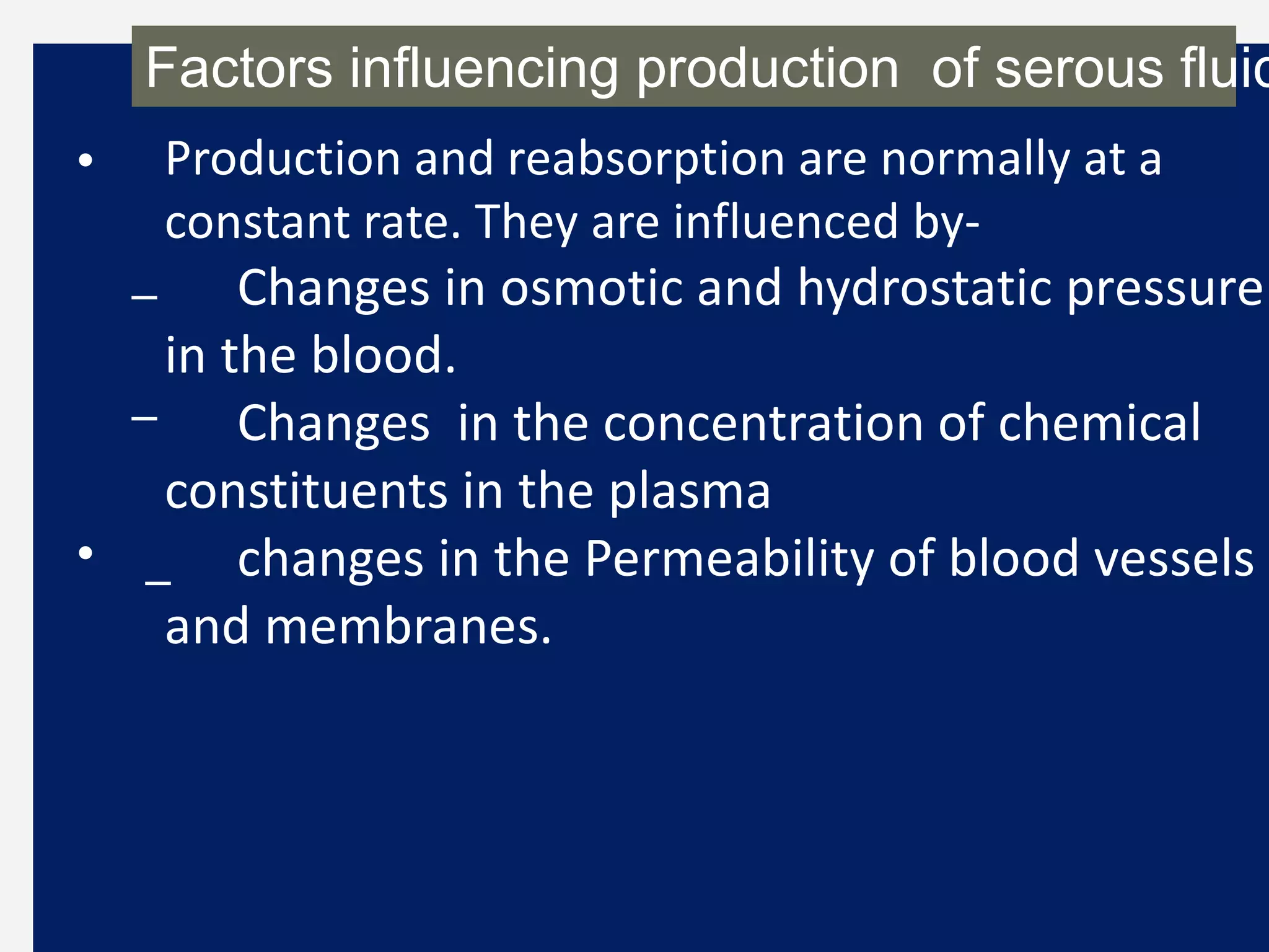
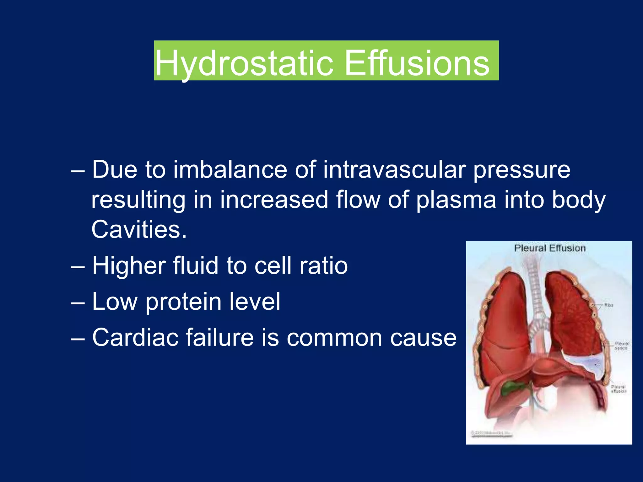
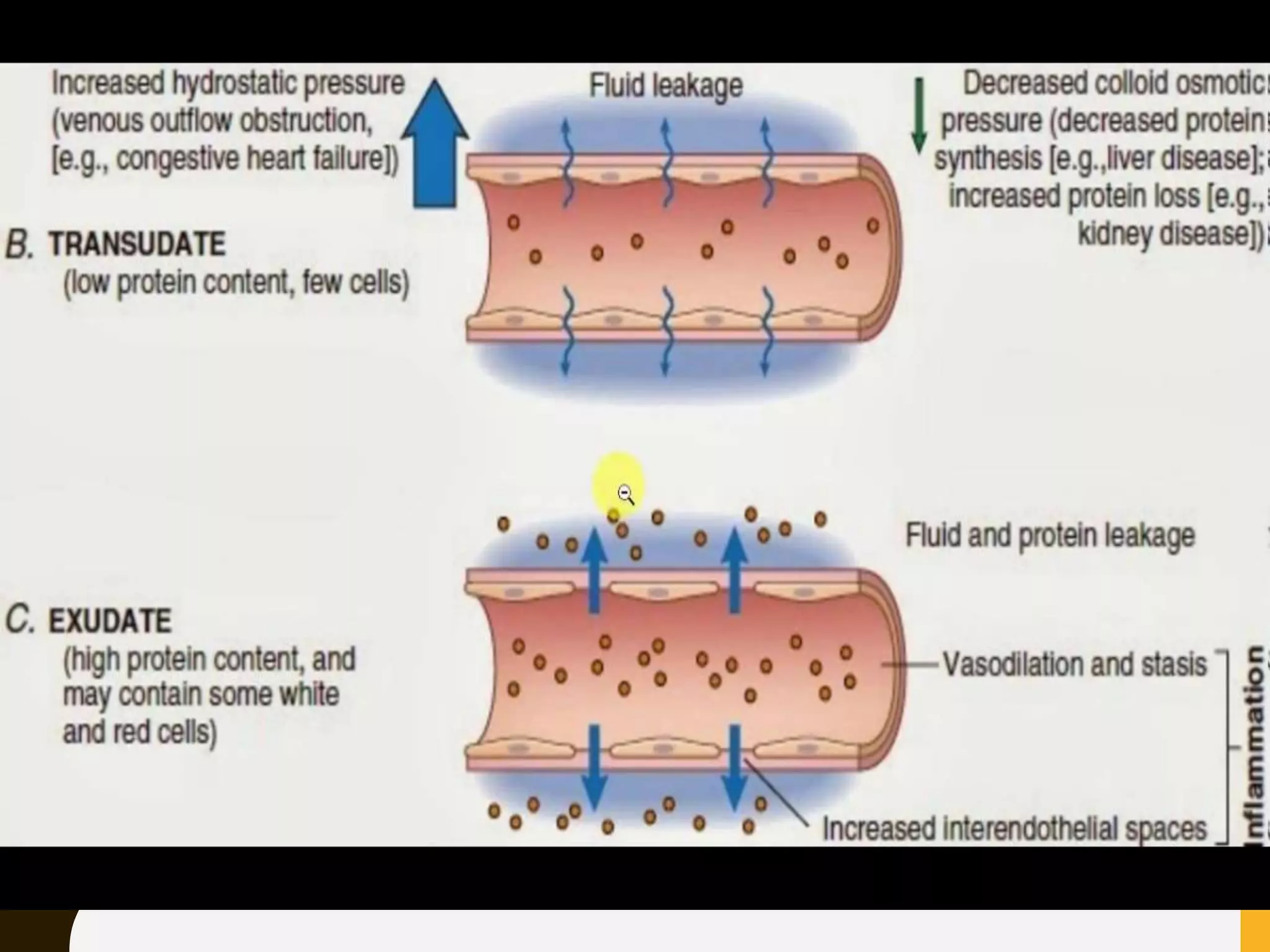
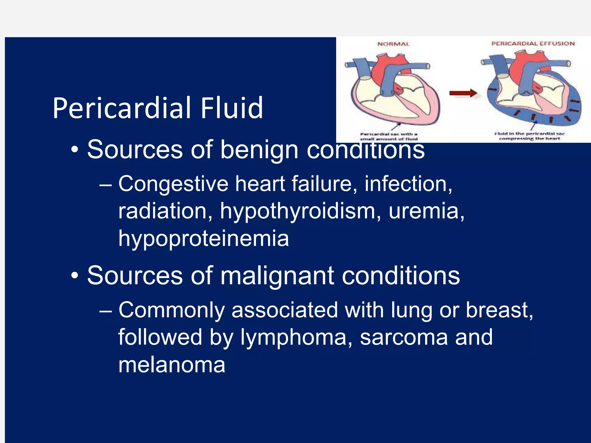
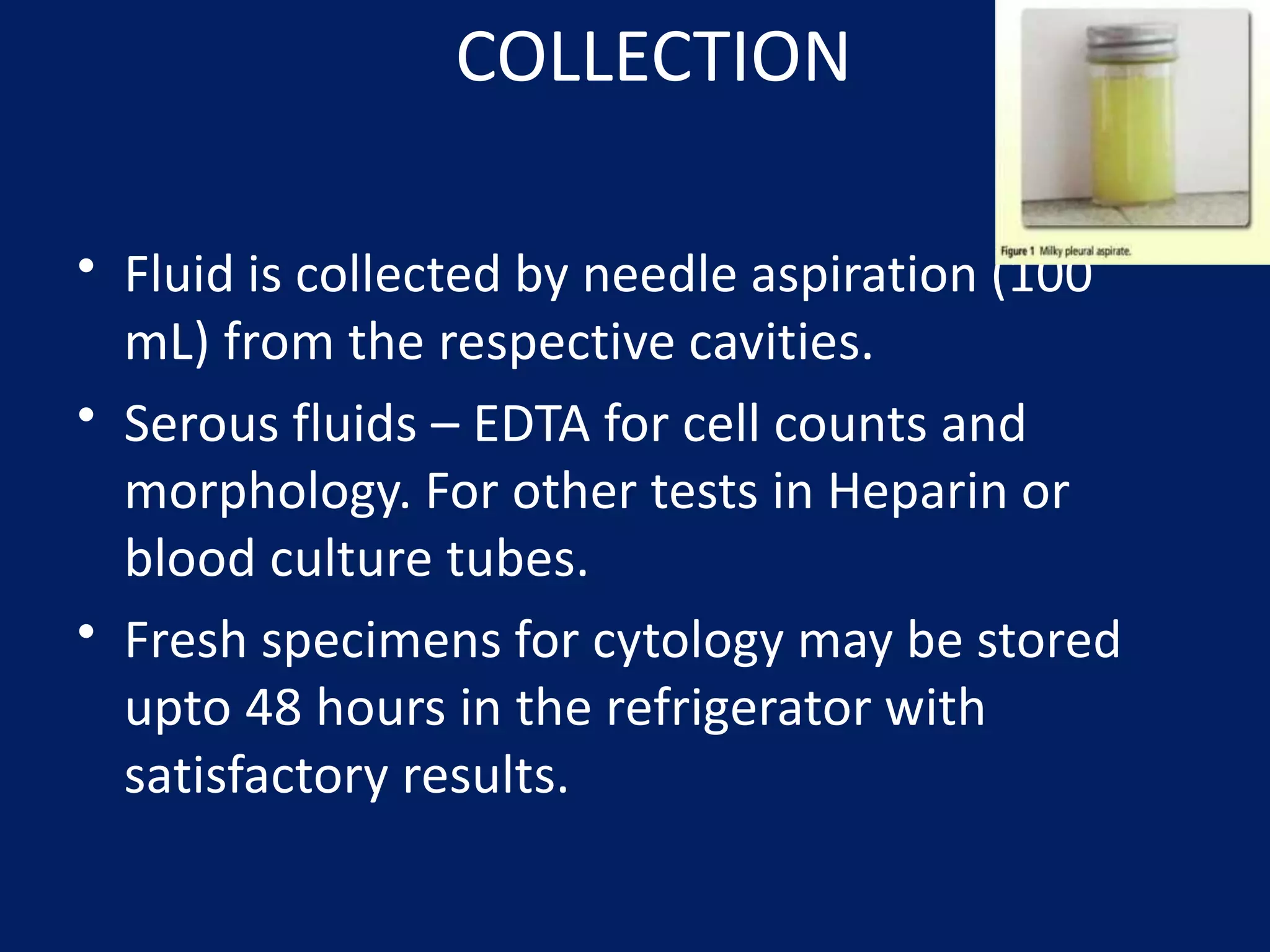
SEROUS BODY
FLUID
LECT-6
2
 The closed cavities of the body—namely, the pleural,
pericardial, and peritoneal cavities—are each lined
by two membranes referred to as the serous
membranes.
 One membrane lines the cavity wall (parietal
membrane), and the other covers the organs within
the cavity (visceral membrane).
 The fluid between the membranes is called serous
fluid, and it provides lubrication between the parietal
and visceral membranes.
 Lubrication is necessary to prevent the friction
between the two membranes that occurs as a result
Formation of serous fluid
 Serous fluids are formed as ultra filtrates of
plasma.
 Under normal conditions, colloidal osmotic
pressure from serum proteins is the same in the
capillaries on both sides of the membrane.
 Therefore, the hydrostatic pressure in the parietal
and visceral capillaries causes fluid to enter
between the membranes.
 The filtration of the plasma ultrafiltrate results in
increased colloidal osmotic pressure in the
capillaries that favors reabsorption of fluid back into
the capillaries.
 This produces a continuous exchange of serous
fluid and maintains the normal volume of fluid
Type of fluids
1. Pleural Cavity: Pleural fluid –Lung–
Thoracocentesis
Pericardial fluid – Heart
Pericardiocentesis.
2. Pericardial Cavity
Peritoneal fluid – Abdominal cavity –
Paracentesis/Peritoneal washing.
3. Peritoneal Cavity
• Production and reabsorption are normally at a
constant rate. They are influenced by-
Changes in osmotic and hydrostatic pressure
in the blood.
Changes in the concentration of chemical
constituents in the plasma
changes in the Permeability of blood vessels
and membranes.
–
–
•
Factors influencing production of serous fluid
–
WHAT IS SEROUS EFFUSION
• An accumulation of fluid, called an effusion.
• It results from an imbalance of fluid
production and reabsorption.
• This fluid accumulation in the pleural,
pericardial, and peritoneal cavities is known as
serous effusion.
Conditions related to abnormal
production of fluids
1. Increased Hydrostatic Pressure
• Congestive heart failure
2. Decreased Colloid Pressure
• Hypoproteinemia
• Increased capillary permeability (inflammation
and infection)
• Lymphatic obstruction (tumors)
Biologic nature of effusion
• Effusions are classified into four categories
– Hydrostatic
– Infectious
– Noninfectious inflammatory
– Malignant
• Each category can be of two types
– Transudate
– Exudate
Hydrostatic Effusions
– Due to imbalance of intravascular pressure
resulting in increased flow of plasma into body
Cavities.
– Higher fluid to cell ratio
– Low protein level
– Cardiac failure is common cause
Infectious Effusions
– Due to direct effects of organism or as by-
product of inflammation.
– Varying types of inflammatory cells can be
Found .
– Cultures and/or gram and acid fast stains may
be useful to identify an organism
Noninfectious Inflammatory Effusions
– Due to autoimmune conditions
– Autoimmune conditions include rheumatoid
arthritis and systemic lupus erythematosus
(SLE)
– Reactive conditions include tissue necrosis or
radiation therapy
Malignant Effusions
– Due to a primary tumor (such as
mesothelioma) or a metastatic tumor
– Patient history is extremely helpful in
rendering a diagnosis
– Knowledge of the appearance of the fluid is
also helpful (cloudy, bloody, partially clotted)
Sub-classification
of serous fluids
Transudates & Exudates
Transudative- low protein content,
increased hydrostatic pressure as in the
conditions of Congestive heart failure
•
• Decreased colloidal pressure- Hepatic cirrhosis ( no
protein synthesis)
• and Nephrotic Syndrome ( increased protein loss)
Exudative
• Bacterial pneumonia/peritonoitis.
• Tuberculosis, other granulomatous diseases
• Viral or mycoplasma infection
• Pancreatitis, bile peritonitis.
• Neoplasms: Bronchogenic carcinoma, metastatic
carcinoma, lymphoma, mesothelioma, pulmonary
infarct, hepatoma, ovarian cancer, prostate cancer.
• Non Infectious inflammatory disease: rheumatoid
disease, SLE
Pleural Fluid
• Sources of benign conditions
– Primary pulmonary infection or infarction
– Secondary due to abdominal disease, i.e.
cirrhosis
• Sources of malignant conditions
– Most common sources are lung, breast,
lymphoid neoplasms
CELL COUNT – PLEURAL FLUID
Total WBC count is almost never diagnostic
• >10,000/uL : Inflammation
• >50,000/uL : Parapneumonic effusions, usually
empyesma.
• <5,000/uL : Chronic exudates(malignancy, TB)
Pericardial Fluid
• Sources of benign conditions
– Congestive heart failure, infection,
radiation, hypothyroidism, uremia,
hypoproteinemia
• Sources of malignant conditions
– Commonly associated with lung or breast,
followed by lymphoma, sarcoma and
melanoma
Peritoneal Fluid
• Sources of benign conditions
– Portal venous hypertension,
hypoproteinemia and salt retention
• Sources of malignant conditions
– Commonly associated with ovary, breast,
GI, lymphoid neoplasms and mesothelioma
– In malignancies of unknown origin,
consider genital tract for women and GI
tract for men
SAAG (serum ascites Albumin
gradient)
Diagnosis of ascites due to portal hypertension
is established by measurement of serum
COLLECTION
• Fluid is collected by needle aspiration (100
mL) from the respective cavities.
• Serous fluids – EDTA for cell counts and
morphology. For other tests in Heparin or
blood culture tubes.
• Fresh specimens for cytology may be stored
upto 48 hours in the refrigerator with
satisfactory results.
THANK YOU !

Lect-6Serous body fluid.pptx

  • 1.
  • 2.
  • 3.
     The closedcavities of the body—namely, the pleural, pericardial, and peritoneal cavities—are each lined by two membranes referred to as the serous membranes.  One membrane lines the cavity wall (parietal membrane), and the other covers the organs within the cavity (visceral membrane).  The fluid between the membranes is called serous fluid, and it provides lubrication between the parietal and visceral membranes.  Lubrication is necessary to prevent the friction between the two membranes that occurs as a result
  • 5.
    Formation of serousfluid  Serous fluids are formed as ultra filtrates of plasma.  Under normal conditions, colloidal osmotic pressure from serum proteins is the same in the capillaries on both sides of the membrane.  Therefore, the hydrostatic pressure in the parietal and visceral capillaries causes fluid to enter between the membranes.  The filtration of the plasma ultrafiltrate results in increased colloidal osmotic pressure in the capillaries that favors reabsorption of fluid back into the capillaries.  This produces a continuous exchange of serous fluid and maintains the normal volume of fluid
  • 7.
    Type of fluids 1.Pleural Cavity: Pleural fluid –Lung– Thoracocentesis
  • 8.
    Pericardial fluid –Heart Pericardiocentesis. 2. Pericardial Cavity
  • 9.
    Peritoneal fluid –Abdominal cavity – Paracentesis/Peritoneal washing. 3. Peritoneal Cavity
  • 10.
    • Production andreabsorption are normally at a constant rate. They are influenced by- Changes in osmotic and hydrostatic pressure in the blood. Changes in the concentration of chemical constituents in the plasma changes in the Permeability of blood vessels and membranes. – – • Factors influencing production of serous fluid –
  • 11.
    WHAT IS SEROUSEFFUSION • An accumulation of fluid, called an effusion. • It results from an imbalance of fluid production and reabsorption. • This fluid accumulation in the pleural, pericardial, and peritoneal cavities is known as serous effusion.
  • 12.
    Conditions related toabnormal production of fluids 1. Increased Hydrostatic Pressure • Congestive heart failure 2. Decreased Colloid Pressure • Hypoproteinemia • Increased capillary permeability (inflammation and infection) • Lymphatic obstruction (tumors)
  • 13.
    Biologic nature ofeffusion • Effusions are classified into four categories – Hydrostatic – Infectious – Noninfectious inflammatory – Malignant • Each category can be of two types – Transudate – Exudate
  • 14.
    Hydrostatic Effusions – Dueto imbalance of intravascular pressure resulting in increased flow of plasma into body Cavities. – Higher fluid to cell ratio – Low protein level – Cardiac failure is common cause
  • 15.
    Infectious Effusions – Dueto direct effects of organism or as by- product of inflammation. – Varying types of inflammatory cells can be Found . – Cultures and/or gram and acid fast stains may be useful to identify an organism
  • 16.
    Noninfectious Inflammatory Effusions –Due to autoimmune conditions – Autoimmune conditions include rheumatoid arthritis and systemic lupus erythematosus (SLE) – Reactive conditions include tissue necrosis or radiation therapy
  • 17.
    Malignant Effusions – Dueto a primary tumor (such as mesothelioma) or a metastatic tumor – Patient history is extremely helpful in rendering a diagnosis – Knowledge of the appearance of the fluid is also helpful (cloudy, bloody, partially clotted)
  • 18.
    Sub-classification of serous fluids Transudates& Exudates Transudative- low protein content, increased hydrostatic pressure as in the conditions of Congestive heart failure • • Decreased colloidal pressure- Hepatic cirrhosis ( no protein synthesis) • and Nephrotic Syndrome ( increased protein loss)
  • 20.
    Exudative • Bacterial pneumonia/peritonoitis. •Tuberculosis, other granulomatous diseases • Viral or mycoplasma infection • Pancreatitis, bile peritonitis. • Neoplasms: Bronchogenic carcinoma, metastatic carcinoma, lymphoma, mesothelioma, pulmonary infarct, hepatoma, ovarian cancer, prostate cancer. • Non Infectious inflammatory disease: rheumatoid disease, SLE
  • 21.
    Pleural Fluid • Sourcesof benign conditions – Primary pulmonary infection or infarction – Secondary due to abdominal disease, i.e. cirrhosis • Sources of malignant conditions – Most common sources are lung, breast, lymphoid neoplasms
  • 22.
    CELL COUNT –PLEURAL FLUID Total WBC count is almost never diagnostic • >10,000/uL : Inflammation • >50,000/uL : Parapneumonic effusions, usually empyesma. • <5,000/uL : Chronic exudates(malignancy, TB)
  • 23.
    Pericardial Fluid • Sourcesof benign conditions – Congestive heart failure, infection, radiation, hypothyroidism, uremia, hypoproteinemia • Sources of malignant conditions – Commonly associated with lung or breast, followed by lymphoma, sarcoma and melanoma
  • 24.
    Peritoneal Fluid • Sourcesof benign conditions – Portal venous hypertension, hypoproteinemia and salt retention • Sources of malignant conditions – Commonly associated with ovary, breast, GI, lymphoid neoplasms and mesothelioma – In malignancies of unknown origin, consider genital tract for women and GI tract for men SAAG (serum ascites Albumin gradient) Diagnosis of ascites due to portal hypertension is established by measurement of serum
  • 25.
    COLLECTION • Fluid iscollected by needle aspiration (100 mL) from the respective cavities. • Serous fluids – EDTA for cell counts and morphology. For other tests in Heparin or blood culture tubes. • Fresh specimens for cytology may be stored upto 48 hours in the refrigerator with satisfactory results.
  • 26.