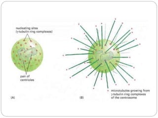
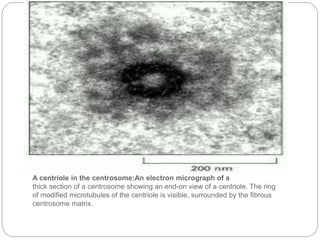
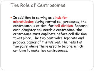
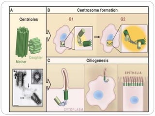
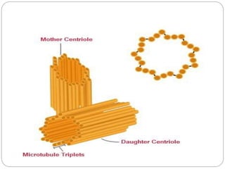
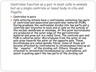
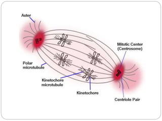
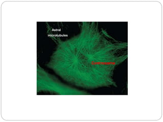
Centrioles are discs composed of microtubules arranged in a 9+3 pattern. They occur in pairs and function to organize microtubules and participate in cell division. As a pair, centrioles migrate to opposite poles during cell division and nucleate microtubules to form the mitotic spindle. As a single organelle, the centriole acts as a basal body at the base of cilia and flagella to organize their microtubules and direct movement. Centrioles replicate during cell division to ensure each daughter cell receives a centriole pair.
Cytoskeleton - microtubules ,microfilaments and intermediate filamentsBIOTECH SIMPLIFIED
The cytoskeleton is a structure that helps cells maintain their shape and internal organization, and it also provides mechanical support that enables cells to carry out essential functions like division and movement. There is no single cytoskeletal component. Rather, several different components work together to form the cytoskeleton.
The Indian Dental Academy is the Leader in continuing dental education , training dentists in all aspects of dentistry and
offering a wide range of dental certified courses in different formats.for more details please visit
www.indiandentalacademy.com
Motor molecules also carry vesicles or organelles to various destinations along “monorails’ provided by the cytoskeleton.
Interactions of motor proteins and the cytoskeleton circulates materials within a cell via streaming.
Recently, evidence is accumulating that the cytoskeleton may transmit mechanical signals that re-arrange the nucleoli and other structures.
Cell biology
Want to know the difference between Centrioles and Centrosome?
Check this ppt to know more about it!
-- Importance of these cellular organelles in a life.
-- Disorders related to these organelles.
-- Recent advancements in the field of Cancer and Cell biology.
By Mohammed Valikarimwala
SY BSc Biotechnology
Fergusson college, Pune.
STRUCTURE AND FUNCTIONS OF NUCLEUS OF A CELL.pptxRASHMI M G
The nucleus is the part of a cell that contains DNA organized into chromosomes and is located in the middle of the cell. It is surrounded by the nuclear envelope, which is a double membrane that separates the nucleus from the cytoplasm. The nuclear envelope contains nuclear pores, which are gateways that allow molecules to move into and out of the nucleus.
The Cell: The Histology Guide
Nucleus - The Cell: The Histology Guide - University of Leeds
The nucleus is found in the middle of the cells, and it contains DNA arranged in chromosomes. It is surrounded by the nuclear envelope, a double nuclear membrane (outer and inner), which separates the nucleus from the cytoplasm. The outer membrane is continuous with the rough endoplasmic reticulum.
open.baypath.edu
Nucleus – BIO109 Biology I Introduction to Biology
The boundary of the nucleus, called the nuclear envelope, is a double membrane that contains small openings called nuclear pores. These pores are gateways that allow molecules to move into and out of the nucleus, enabling it to communicate with the rest of the cell.
The nucleus has three main parts:
Nuclear membrane: A protective barrier of the nucleus
Nucleoplasm: The cytoplasm of the nucleus, which is a semifluid matrix that contains chromatin, the less condensed form of DNA that organizes into chromosomes during mitosis or cell division
Nucleolus: A spherical structure that produces and assembles the cell's ribosomes
The nucleus controls and regulates the activities of the cell, such as growth and metabolism.
What are the 4 types of nucleus?
What are the 3 parts of a nucleus?
How many nuclei are in a cell?
Ask a follow up
The Indian economy is classified into different sectors to simplify the analysis and understanding of economic activities. For Class 10, it's essential to grasp the sectors of the Indian economy, understand their characteristics, and recognize their importance. This guide will provide detailed notes on the Sectors of the Indian Economy Class 10, using specific long-tail keywords to enhance comprehension.
For more information, visit-www.vavaclasses.com
Unit 8 - Information and Communication Technology (Paper I).pdfThiyagu K
This slides describes the basic concepts of ICT, basics of Email, Emerging Technology and Digital Initiatives in Education. This presentations aligns with the UGC Paper I syllabus.
Welcome to TechSoup New Member Orientation and Q&A (May 2024).pdfTechSoup
In this webinar you will learn how your organization can access TechSoup's wide variety of product discount and donation programs. From hardware to software, we'll give you a tour of the tools available to help your nonprofit with productivity, collaboration, financial management, donor tracking, security, and more.
Model Attribute Check Company Auto PropertyCeline George
In Odoo, the multi-company feature allows you to manage multiple companies within a single Odoo database instance. Each company can have its own configurations while still sharing common resources such as products, customers, and suppliers.
This is a presentation by Dada Robert in a Your Skill Boost masterclass organised by the Excellence Foundation for South Sudan (EFSS) on Saturday, the 25th and Sunday, the 26th of May 2024.
He discussed the concept of quality improvement, emphasizing its applicability to various aspects of life, including personal, project, and program improvements. He defined quality as doing the right thing at the right time in the right way to achieve the best possible results and discussed the concept of the "gap" between what we know and what we do, and how this gap represents the areas we need to improve. He explained the scientific approach to quality improvement, which involves systematic performance analysis, testing and learning, and implementing change ideas. He also highlighted the importance of client focus and a team approach to quality improvement.
Palestine last event orientationfvgnh .pptxRaedMohamed3
An EFL lesson about the current events in Palestine. It is intended to be for intermediate students who wish to increase their listening skills through a short lesson in power point.
Instructions for Submissions thorugh G- Classroom.pptxJheel Barad
This presentation provides a briefing on how to upload submissions and documents in Google Classroom. It was prepared as part of an orientation for new Sainik School in-service teacher trainees. As a training officer, my goal is to ensure that you are comfortable and proficient with this essential tool for managing assignments and fostering student engagement.
The French Revolution, which began in 1789, was a period of radical social and political upheaval in France. It marked the decline of absolute monarchies, the rise of secular and democratic republics, and the eventual rise of Napoleon Bonaparte. This revolutionary period is crucial in understanding the transition from feudalism to modernity in Europe.
For more information, visit-www.vavaclasses.com
2. Edouard van Beneden and Theodor
Boveri made the first observation and
identification of centrioles in 1883 and 1888
respectively
while the pattern of centriole duplication was
first worked out independently by Etienne de
Harven and Joseph G. Gall c. 1950
3. Centrioles are discs built from microtubule
material, with nine triplet microtubules
arranged in a cylinder
When the cell is not dividing, two centrioles
are paired with each other at 90 degree
angles. Along with MTOC, the two centrioles
combine to make the centrosome.
4.
5. A centriole in the centrosome:An electron micrograph of a
thick section of a centrosome showing an end-on view of a centriole. The ring
of modified microtubules of the centriole is visible, surrounded by the fibrous
centrosome matrix.
6. The Role of Centrosomes
In addition to serving as a hub for
microtubules during normal cell processes, the
centrosome is critical for cell division. Because
each daughter cell needs a centrosome, the
centrosome must duplicate before cell division
takes place. The two centrioles separate and
produce copies of themselves. The result is
two pairs where there used to be one, which
combine to make two centrosomes.
7. Centriole duplication
Before DNA replication , cells contain two centrioles.
The older of the two centrioles is termed the mother
centriole , the other the daughter. During the cell
division cycle , a new centriole grows from the side of
each mother centriole. After duplication, the two
centriole pairs will remain attached to each other
orthogonally until mitosis . At that point the mother
and daughter centrioles separate dependently on an
enzyme called separase.
The two centrioles in the centrosome are tied to one
another. The mother centriole has radiating appendages
at the distal end of its long axis and is attached to its
daughter at the proximal end. Each daughter cell
formed after cell division will inherit one of these pairs.
Centrioles start duplicating when DNA replicates.
8.
9. Occurrence
Centrioles are present in the cells of most
eukaryotes, for example those of animals .
However, they are absent from conifers (
pinophyta ), flowering plants ( angiosperms ) and
most fungi , and are only present in the male
gametes of charophytes , bryophytes , seedless
vascular plants , cycads , and ginkgo .
Centrioles are present in (1) animal cells and (2)
the basal region of cilia and flagella in animals and
lower plants (e.g. chlamydomonas). In cilia and
flagella centrioles are called ‘basal bodies’
11. Found only in animal cells and some lower plants, a
centriole is composed of short lengths of
microtubules lying parallel to one another and
arranged around a central cavity to form a
cylinder.
composed mainly of a protein called tubulin that is
found in most eukaryotic cells.
An associated pair of centrioles, surrounded by a
shapeless mass of dense material, called the
pericentriolar material , or PCM, makes up a
compound structure called a centrosome .
12.
13. A centriole is composed of short lengths of
microtubules arranged in the form of an open-
ended cylinder about 500nm long and 200nm in
diameter. The microtubules forming the wall of the
cylinder are grouped into nine sets of bundles of
three microtubules each.(9+3)
In cilia and flagella where centrioles are at the
base of the structure, and are called basal bodies,
the wall and cavity architecture is slightly
different. In addition to cylinder walls composed
of nine sets of bundles of three microtubules,
there are walls of nine sets of two bundles. In both
types there is a central matrix from which spokes
radiate as in a cart wheel.
In animal cells centrioles usually reside in pairs
with the cylindrical centrioles at right angles to
each other.
14. The centrioles are made of groups of microtubules ,these microtubules are
arranged in a pattern of 9 + 3 . The pattern of the microtubules for a ring of 9
microtubules known as "triplets" and the microtubules are arranged at right
angles to one another
15. At the anaphase and telophase stages the
centrioles appear as two cylindrical structures .
They are open at both the ends and are located at
right angles to each other .
The length of the centriole is about 3 ,000 to 5
,000 A and it is about 1 ,500 to 1 ,800 Ã in
diameter.
The triplets are tilted , they form an angle of 40
degrees to the radius of the cylinder .
16. Each triplet fiber is composed of three sub -tubules or
sub - fibres . Each sub - tubule is of 250 Ã in diameter .
These sub - tubules are hollow structures and their
walls are made of monomeric units of proteins . The sub
- tubules are made up of protein tubulin .
The centriole internally shows a characteristic cart
wheel structure . The cart wheel structure has a
prominent central rod , and nine spokes radiating from
the central rod .
All the structures that surrounds the centriole
together constitute the centriole satellite . The
number of these satellites varies .
The centrosome structure is made of lipids and
proteins . However , it also contains carbohydrates and
nucleic acids too.
17. Centrioles in Plant Cell
Plant cells do not have centrioles . Hence , the
structure of the poles is different to that of
the cells that possess centrioles . The poles of
the cells of plants are broader when compared
to the cells of animals . This is probably
because of the absence of a cellular organelle
that is defined to act as a focal point . In some
of the plant spindle , the spindles are
delocalized in a such a way that the spindle
barely narrower than the rest of the spindle.
The poles of the plant cells are more diffuse
and have many fewer astral microtubules .
18. Cilia and flagella
The entire ciliary or flagellar projection is covered by a
membrane that is continuous with the plasma membrane
of the cell. The core of the cilium, called the axoneme,
contains an array of microtubules that runs longitudinally
through the entire organelle.
With rare exceptions, the axoneme of a motile cilium or
flagellum consists of nine peripheral doublet microtubules
surrounding a central pair of single microtubules.
All microtubules of the axoneme have the same polarity:
their plus ends are at the tip of the projection, and their
minus ends the base. Each peripheral doublet consists of
one complete microtubule, the A tubule, and one
incomplete microtubule, the B tubule, the latter containing
10 or 11 subunits rather than the usual 13.
19. The central tubules were seen to be enclosed by
the central sheath, which is connected to the A
tubules of the peripheral doublets by a set of
radial spokes.
The doublets are connected to one another by an
interdoublet bridge composed of an elastic
protein, nexin.
22. • Cell division
• Functions as a pair and as a
single centriole
• Cellular organisation
• Ciliogenesis
• Replication
23. Centrioles function as a pair in most cells in animals
but as a single centriole or basal body in cilia and
flagella
Centrioles in pairs
Cells entering mitosis have a centrosome containing two pairs
of centrioles and associated pericentriolar material (PCM).
During prophase the centrosome divides into two parts and a
centriole pair migrates to each end or pole on the outside of
the nuclear membrane or envelope. At this point microtubules
are produced at the outer edge of the pericentriolar
material and grow out in a radial form. The centriole pair and
PCM is called an aster. Microtubules from the aster at one
pole grow towards the aster at the opposite pole. These
microtubules are called spindle fibres. Some of these will
become attached by centromeres to chromosomes lined up on
the ‘equator’ of the dividing cell. Others, though not
attached to chromatids/chromosomes by centromeres, will
assist in pushing apart the two parts of the dividing cell
26. A single centriole or basal body.
At the base of each cilium or flagellum there is
a single centriole. This structure and
associated pericentriolar material, construct
microtubules in a linear direction. These
microtubules form most of the inside of cilia
and flagella and are largely responsible, using
protein motors, for the mechanical aspects of
their movement. The centriole at the base of
each one also appears to exert some degree of
direction and control over the movement of the
cilia and flagella.
27. The centrioles may produce flagella or cilia .
The fiber of the tail of sperms also arises
from the centriole .
The dysfunctioning of the centrosome is also
responsible for the development of certain
cancers .
28. Cellular organization:Centrioles are a very
important part of centrosomes , which are
involved in organizing microtubules in the
cytoplasm. The position of the centriole
determines the position of the nucleus and
plays a crucial role in the spatial arrangement
of the cell.
29. Ciliogenesis
In organisms with flagella and cilia , the
position of these organelles is determined by
the mother centriole, which becomes the basal
body . An inability of cells to use centrioles to
make functional cilia and flagella has been
linked to a number of genetic and
developmental diseases. In particular, the
inability of centrioles to properly migrate prior
to ciliary assembly has recently been linked to
Meckel-Gruber syndrome.
30. Replication
In cells where centrioles are present as a pair,
replication takes place during the whole of the cell
cycle. In phase G1 the two centriole cylinders move
very slightly apart from one another. During S
phase new cylinders of microtubules form near, and
at right angles to, the two ‘mother’ cylinders.
The two pairs of centrioles keep very close to one
another until the prophase stage of mitosis. At
this point they separate with both pairs of
centrioles moving over the outer surface of the
nuclear envelope to opposite ends or ‘poles’ of
the cell, to form the astral poles of the dividing
cell.
31. SUMMARY
Centrioles occur as paired cylindrical organelles together
with pericentriolar material (PCM) in the centrosome of an
animal cell.
Centrioles are found as single structures in cilia and flagella
in animal cells and some lower plant cells.
Centrioles are constructed of microtubules.
In animal cells centrioles organise the pericentriolar material
to produce microtubules including mitotic spindle fibres.
Centrioles present something of an enigma; they appear to
have an effect on the outcome of mitosis in animal cells.
When present there is a satisfactory division but without
them mitosis still takes place but sometimes with an
unsatisfactory outcome. Centrioles are absent from the cells
of higher plants but normal mitosis takes place and with
satisfactory results.