Download to read offline


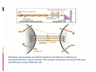


The document discusses cell-cell adhesion, specifically cadherins. Cadherins are calcium-dependent cell adhesion molecules that are critical for establishing cell connections and segregating cell types during development. The major classes of cadherins include E-cadherin, P-cadherin, N-cadherin, and protocadherins. Cadherins interact with catenins and the actin cytoskeleton to bind cells together. Different cadherin expression patterns play important roles in tissue formation and separation during embryogenesis.


