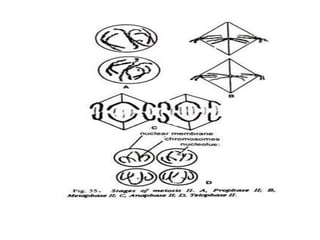
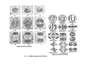
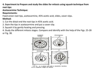
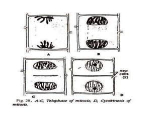
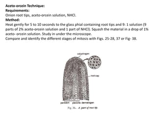
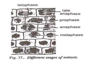
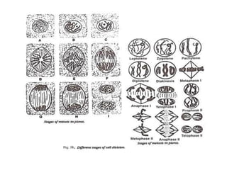
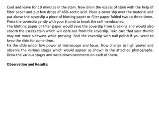
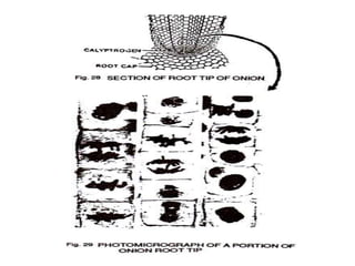
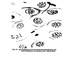
1. Experiments on Cell Biology
Dr. Naveen Gaurav
Associate Professor and Head
Department of Biotechnology
Shri Guru Ram Rai University
Dehradun
2. Experiments on Cell Biology
1. Experiment to study the effect of colchicine on the mitotic division of the root tip cells of
onion:
Requirements:
Rooted bulbs of onion, small beakers, water, 1% aqueous solution of colchicine, acetocarmine,
45% acetic acid, coverslips, slides, burner, and microscope.
Method:
1. Fill water in a small beaker and place on it a rooted bulb of onion so that the roots hang in
water.
2. Fill 1% aqueous solution of colchicine in another small beaker and place on it also a similar type
of rooted bulb of onion. Allow the roots to grow in both the beakers for some time.
3. Place the root tips from both the beakers on two separate slides and prepare the squash by the
“acetocarmine technique” mentioned above in Exercise No. 2. Observe under microscope.
Observations:
Normal stages of mitosis are observed in the dividing cells of water-treated root tips.
On the other hand, following abnormalities are observed in the dividing cells of the colchicine-
treated root tips:
(i) Separation of daughter chromosomes to their respective poles is checked in the metaphase
spindles due to colchicine.
(ii) Colchicine treatment shows doubling of chromosomes in the nucleus, and thus induces
polyploidy.
Results:
Colchicine, an alkaloid obtained from some species of Colchicum (e.g., C. autumnale), results in
doubling of chromosomes and thus induces polyploidy in plants.
3. 2. Experiment to study various stages of mitosis by preparing bone marrow squash of
mice:
Requirements: Chloroformed mice, razor, blade, sodium citrate (1.2%), specimen tube,
needle, Camoy’s fluid, centrifuge, distilled water, HCl, water bath, ice-cold water, Basic
Fuchsin stain, acetic acid, slide, cover-slips, microscope.
Method:
1. Remove the femur of a chloroformed mice, cut the epiphysis with the help of a razor or
blade, and collect the bone marrow in a specimen tube containing sodium citrate (1.2%).
2. With the help of a neelde mix the bone marrow thoroughly to prepare a fine suspension.
3. Fix the bone marrow suspension in Carnoy’s fluid for about 20 minutes, centrifuge it and
discard the supernatant of the centrifuge tube.
4. Repeat this process of centrifuging 2-3 times by adding fresh fixative each time. This will
remove the fat completely.
5. Rinse with distilled water and hydrolyze the bone marrow for 5 minutes with normal HCl
on a water bath at 60°C.
6. Stop hydrolysis by immediately transferring the tissue in ice-cold water.
7. Stain the bone marrow with Basic Fuchsin for about half an hour and keep it in acetic
acid (45%).
8. Prepare a squash on a slide by smearing, mount with a coverslip and observe under a
microscope.
Observations and Results:
Various stages of mitosis (prophase, metaphase, anaphase and telophase) are seen clearly
in the actively dividing cells of bone marrow of mice.
4. 3. Experiment to prepare and study slides for meiotic stages using plant material:
Requirements:
Plant material (male flowers of Bajra or Maize or some other plants), needle,
acetocarmine, slides, coverslips.
Method:
1. Take the male flower buds of the given material and arrange them in a gradual
sequence from smallest to largest in size.
2. Dissect out the anthers from the lowers and keep one of them in a drop of
acetocarmine. Put the coverslip and press it a little with the help of needle, and observe
whether any stage of meiosis is present or not. Try again and again so far you are unable
to get any meiotic stage.
3. Heat a little to the slide containing any stage and squash it again by pressing it with
your thumb and finger, and study the various available details.
Compare and identify the stage of your slide with the Figs.: 29-35 or Fig. 38.
5.
6.
7. 4. Experiment to Prepare and study the slides for mitosis using squash technique from
root tips:
Acetocarmine Technique:
Requirements:
Fixed onion root tips, acetocarmine, 45% acetic acid, slides, cover-slips.
Method:
1. Cut the distal end the root tips in 45% acetic acid.
2. Stain the tips in acetocarmine and put a cover-slip.
3. Squash it by gently heating and pressing.
4. Study the different mitosis stages. Compare and identify with the help of the Figs. 25-28
or Fig. 38.
8.
9. Aceto-orcein Technique:
Requirements:
Onion root tips, aceto-orcein solution, NHCl.
Method:
Heat gently for 5 to 10 seconds to the glass phial containing root tips and 9: 1 solution (9
parts of 2% aceto-orcein solution and 1 part of NHCl). Squash the material in a drop of 1%
aceto- orcein solution. Study in under the microscope.
Compare and identify the different stages of mitosis with Figs. 25-28, 37 or Fig- 38.
10.
11.
12. 5. Experiment on Mitotic Cell Division by squash method :
To prepare a slide of root tip of onion by squash method arid to identify, study and draw
the various mitotic stages.
Material required:
Onion root tips fixed in Carnoy fluid (6 parts absolute alc + 3 parts glacial acetic acid + 1
part Chloroform); Aceto carmine stain; slides; cover slip; blotting paper; spirit lamp; watch
glass; 1 -N HCI, 45% Acetic acid and filter paper.
Procedure:
Take large sized mature onions about a week before the experiment. Cut all the dried roots
from the stem at the base of the bulb. Now place these onions on the mouth of couplin jars
filled with water in such a way that the stem portion must be dipped continuously in water.
In about a weeks’ time new roots would develop and would start growing downward in the
water. Take the onions and cut the milky white portion of root tips viz., up to 5 mm length.
Cut each piece into smaller pieces and fix them in Carnoy fixative for half an hour.
Transfer the material to 90% alcohol and then to 70% alcohol keeping the material for 10
minutes in each. Now preserve the material in 70% alcohol. At the time of proceeding for
experiment in the laboratory take few pieces from 70% alcohol in a watch glass in few
drops of 1- N HCI and leave for 5 minutes. By this procedure the material would become
soft.
Now drain off the HCI and wash the pieces with a little distilled water at least twice or
thrice. Now put the root tips on a clear slide on right hand side and pour a few drops of 2%
Acetocarmine. Warm the slide gently on sprit lamp for few minutes at least 3-4 times but
never let it boil.
13. Cool and leave for 10 minutes in the stain. Now drain the excess of stain with the help of
filter paper and put few drops of 45% acetic acid. Place a cover slip over the material and
put above the coverslip a piece of blotting paper or filter paper folded two to three times.
Press the coverslip gently with your thumb to break the cell membranes.
The blotting paper or filter paper would save the coverslip from breaking and would also
absorb the excess stain which will ooze out from the coverslip. Take care that your thumb
may not move sideways while pressing. Seal the coverslip with nail polish if you want to
keep the slide for some time.
Fix the slide under low power of microscope and focus. Now change to high power and
observe the various stages which would appear as shown in the attached photographs.
Draw the various stages and write down comments on each of them.
Observation and Results:
14.
15. 6. Experiment on Meiotic Cell Division:
To prepare a slide of meiotic stages through squash method, buds of Tradescantia or
spikes of Bajra.
Material:Buds of Tradescantia or Flax or immature spikelet’s of Bajra or Testes of
Grasshopper or Chironomous larva or rat, carnoy fluid; Acetocarmine; needles; slide;
watch glass; 45% Acetic acid; coverslip; filter paper or blotting paper; spirit lamp;
normal saline; 1% sodium citrate; and 2.2% Trisodium citrate.
Procedure – 3 (For plant material):
Take young buds of Tradescantia or Flax or immature male spikelet’s of Bajra (Millet)
and fix them in freshly prepared Carnoy’s fluid for 10 hrs. After rehydrating in 90% and
70% alcohol store the material in 70% alcohol.
(1) Take a bud of Tradescantia or few smaller anthers from the male spikelet of Bajra in
70% alcohol in a watch glass.
(2) Dissect out the bud or dissect each anther taken from the spikelet with needles &
keep them in 45% acetic acid.
(3) Put few anthers on the slide and few drops of Acetocarmlne solution over them.
(4) Warm the slide gently a few times, but never let it boil.
(5) Drain off excess stain and put few drops of 45% acetic acid.
(6) Now put coverslip over the material and cover it with filter paper or blotting paper
folded 2 to 3 times.
(7) Press the coverslip with your thumb so as to rupture the anthers.
ADVERTISEMENTS:
(8) Observe under the microscope first in low power and then in high power.