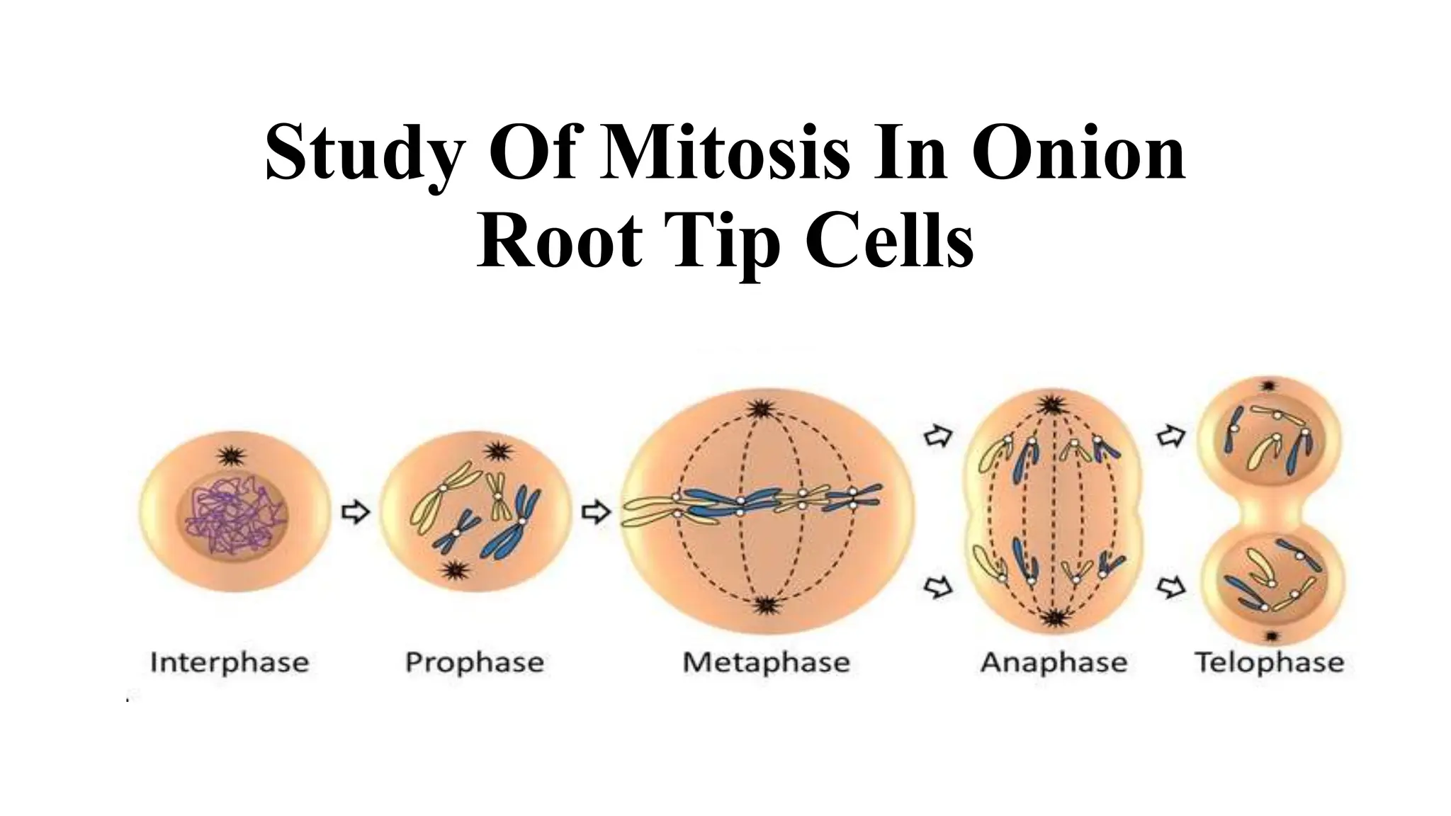
Study Of Mitosis In Onion
Root Tip Cells
Aim:To study and demonstrate mitosis by
preparing the mount of an onion root tip cells
Mitosis
In mitosis, the nucleus of the Eukaryotic cells divides into two,
subsequently resulting in the splitting of the parent cells into two
daughter cells. Hence, every cell division involves two chief stages:
Cytokinesis – Cytoplasm division
Karyokinesis – Nucleus division
Materials Required
• Compound microscope
• Acetocarmine stain
• Water
• Burner
• N/10 Hydrochloric acid
• Filter paper
• Coverslip
• Aceto alcohol (Glacial acetic acid and Ethanol in the ratio 1:3)
• Glass Slide
• Onion root peel
• Forceps
• Blade
• Watch glass
• Dropper
• Needle
Procedure Of The Experiment
• Place an onion on a tile
• With the help of a sharp blade, carefully snip the dry roots of the onion
• Place the bulbs in a beaker containing water to grow the root tips
• It may take around 4 to 6 days for the new roots to grow and appear
• Trim around 3 cm of the newly grown roots and place them in a watch
glass
• With the help of forceps, pick one root and set in on a new glass slide
• With the help of a dropper, allow one drop of 1N HCl to come in contact
with the tip of the root. Warm it lightly on the burner .
• In New watch glass take2-3 drops of Acetocarmine stain keep for15 min
After 15 min place section in 45% Glacial acetic acid.
• Take one clean slide, add 1 droplet of distilled water on it then put one
section
• With the help of a needle, a coverslip can be mounted on it
• Gently tap the coverslip with an unsharpened end of a needle in order
for the meristematic tissue of the root tip present under the coverslip to
be squashed properly and to be straightened out as a fine cell layer
• The onion root tip cells’ slide is now prepared and ready to be examined
for different stages of mitosis
• Observe and study mitosis by placing the slide under the compound
microscope. Focus as desired to obtain a distinct and clear image
Observations and Conclusion
• The slide containing the stained root tip cells is placed on the stage of
the compound microscope, changes taking place are noted and
sketched.
• The different phases of mitosis, such as prophase, metaphase,
anaphase and telophase can be observed
• Stages Of Mitosis
• The various stages of mitosis are:
1. Prophase
• The process of mitosis is initiated at this stage where in coiling and
thickening of the chromosomes occurs
• Shrinking and hence the disappearance of the nucleolus and nuclear
membrane takes place
• The stage reaches its final state when a cluster of fibres organizes to form
the spindle fibres
2. Metaphase
• Chromosomes turn thick in this phase. The two chromatids from each of
the chromosomes appear distinct
• Each of the chromosomes is fastened to the spindle fibres located on its
controller
• Chromosomes align at the centreline of the cell
3. Anaphase
• Each of the chromatid pair detaches from the centromere and
approaches the other end of the cell through the spindle fibre
• At this stage, compressing of the cell membrane at the centre takes
place
4. Telophase
• Chromatids have reached the other end of the cell
• The disappearance of the spindles
• Chromatin fibres are formed as a result of uncoiling of daughter
chromosomes
• The appearance of two daughter nuclei at the opposing ends due to
the reformation of the nucleolus and nuclear membrane
• At this phase, splitting of the cell or cytokinesis may also occur

mitosis practical of bio engineering field

  • 1.
    Study Of MitosisIn Onion Root Tip Cells
  • 2.
    Aim:To study anddemonstrate mitosis by preparing the mount of an onion root tip cells Mitosis In mitosis, the nucleus of the Eukaryotic cells divides into two, subsequently resulting in the splitting of the parent cells into two daughter cells. Hence, every cell division involves two chief stages: Cytokinesis – Cytoplasm division Karyokinesis – Nucleus division
  • 3.
    Materials Required • Compoundmicroscope • Acetocarmine stain • Water • Burner • N/10 Hydrochloric acid • Filter paper • Coverslip • Aceto alcohol (Glacial acetic acid and Ethanol in the ratio 1:3) • Glass Slide • Onion root peel • Forceps • Blade • Watch glass • Dropper • Needle
  • 4.
    Procedure Of TheExperiment • Place an onion on a tile • With the help of a sharp blade, carefully snip the dry roots of the onion • Place the bulbs in a beaker containing water to grow the root tips • It may take around 4 to 6 days for the new roots to grow and appear • Trim around 3 cm of the newly grown roots and place them in a watch glass • With the help of forceps, pick one root and set in on a new glass slide • With the help of a dropper, allow one drop of 1N HCl to come in contact with the tip of the root. Warm it lightly on the burner .
  • 5.
    • In Newwatch glass take2-3 drops of Acetocarmine stain keep for15 min After 15 min place section in 45% Glacial acetic acid. • Take one clean slide, add 1 droplet of distilled water on it then put one section • With the help of a needle, a coverslip can be mounted on it • Gently tap the coverslip with an unsharpened end of a needle in order for the meristematic tissue of the root tip present under the coverslip to be squashed properly and to be straightened out as a fine cell layer • The onion root tip cells’ slide is now prepared and ready to be examined for different stages of mitosis • Observe and study mitosis by placing the slide under the compound microscope. Focus as desired to obtain a distinct and clear image
  • 6.
    Observations and Conclusion •The slide containing the stained root tip cells is placed on the stage of the compound microscope, changes taking place are noted and sketched. • The different phases of mitosis, such as prophase, metaphase, anaphase and telophase can be observed
  • 7.
    • Stages OfMitosis • The various stages of mitosis are: 1. Prophase • The process of mitosis is initiated at this stage where in coiling and thickening of the chromosomes occurs • Shrinking and hence the disappearance of the nucleolus and nuclear membrane takes place • The stage reaches its final state when a cluster of fibres organizes to form the spindle fibres 2. Metaphase • Chromosomes turn thick in this phase. The two chromatids from each of the chromosomes appear distinct • Each of the chromosomes is fastened to the spindle fibres located on its controller • Chromosomes align at the centreline of the cell
  • 8.
    3. Anaphase • Eachof the chromatid pair detaches from the centromere and approaches the other end of the cell through the spindle fibre • At this stage, compressing of the cell membrane at the centre takes place 4. Telophase • Chromatids have reached the other end of the cell • The disappearance of the spindles • Chromatin fibres are formed as a result of uncoiling of daughter chromosomes • The appearance of two daughter nuclei at the opposing ends due to the reformation of the nucleolus and nuclear membrane • At this phase, splitting of the cell or cytokinesis may also occur