Download to read offline


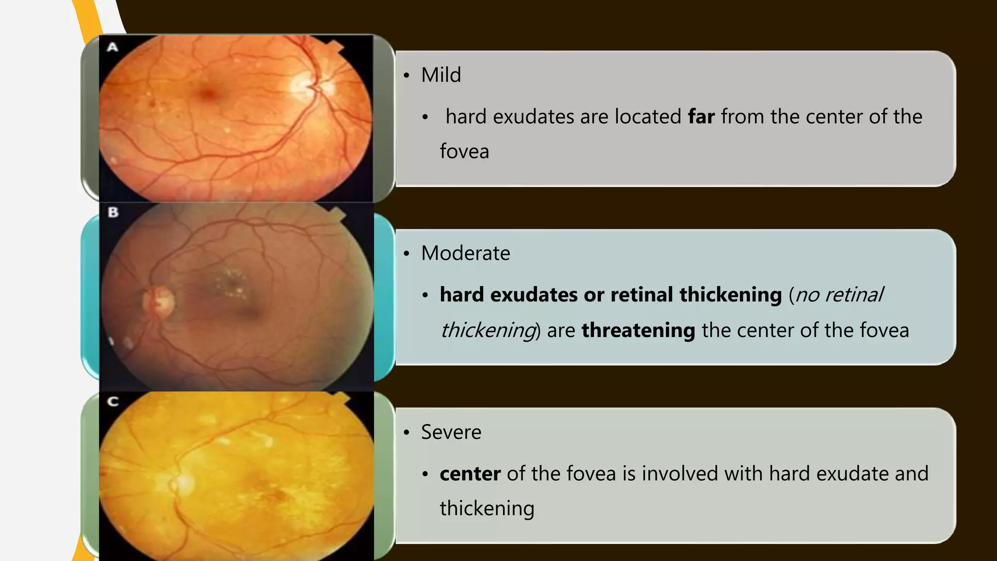


This document discusses diabetic retinopathy (DR), a complication of diabetes that can lead to blindness. It defines DR and outlines its prevalence, risk factors, signs and symptoms, pathogenesis, grading scales, screening recommendations, treatment approaches, and criteria for referral. DR affects the small blood vessels in the retina and ranges from mild non-proliferative DR to more severe proliferative DR. Screening is crucial and treatment depends on the stage, with options like laser therapy and intraocular injections.


