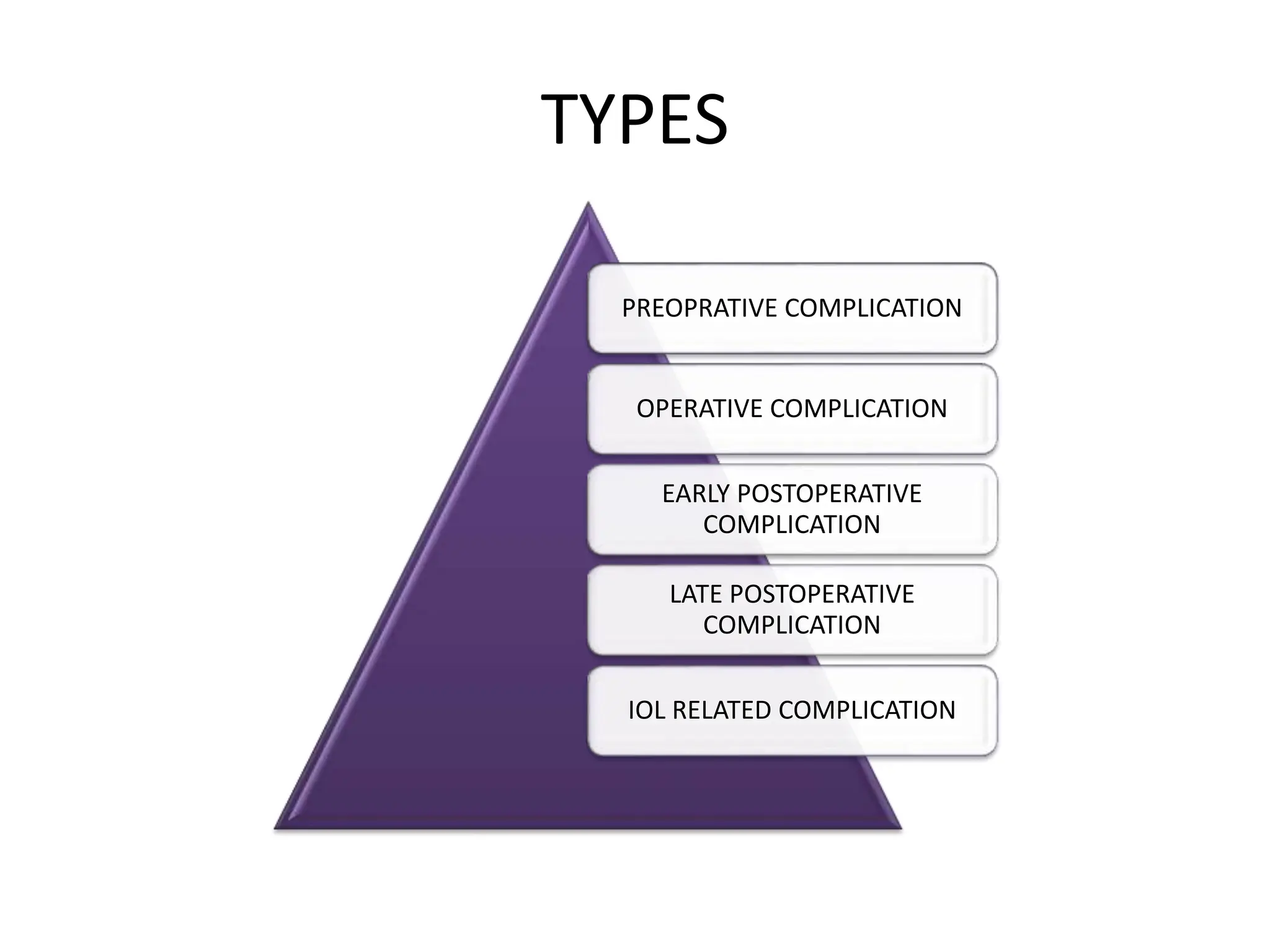
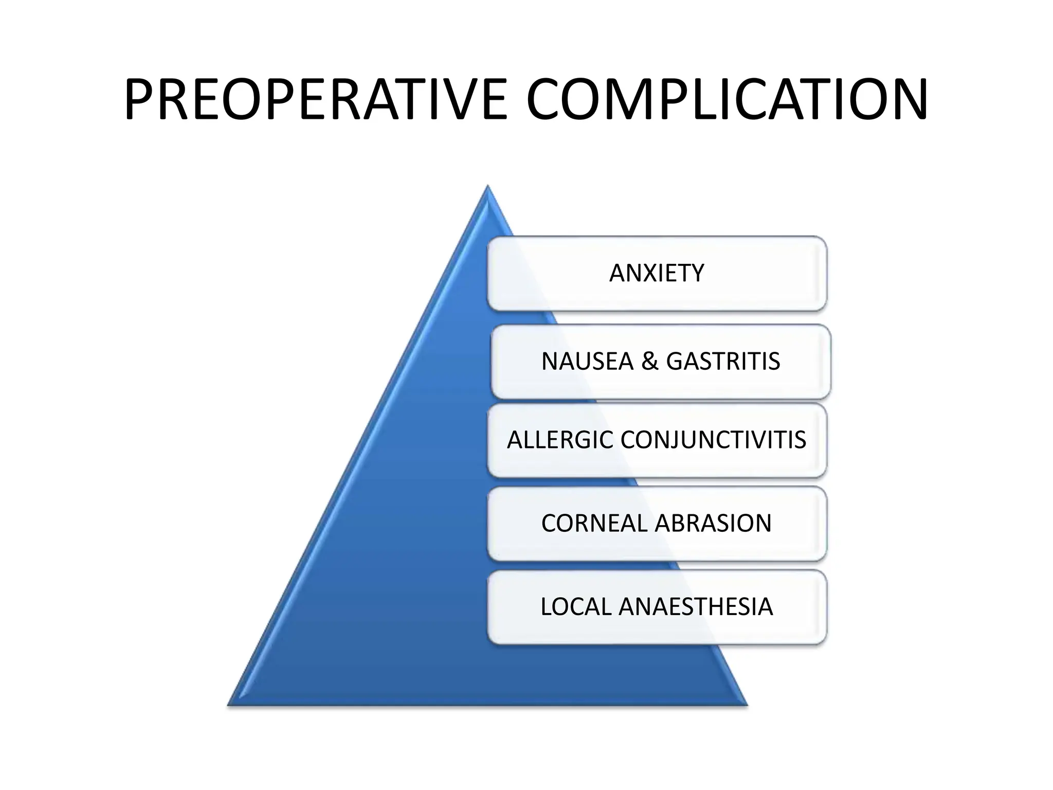
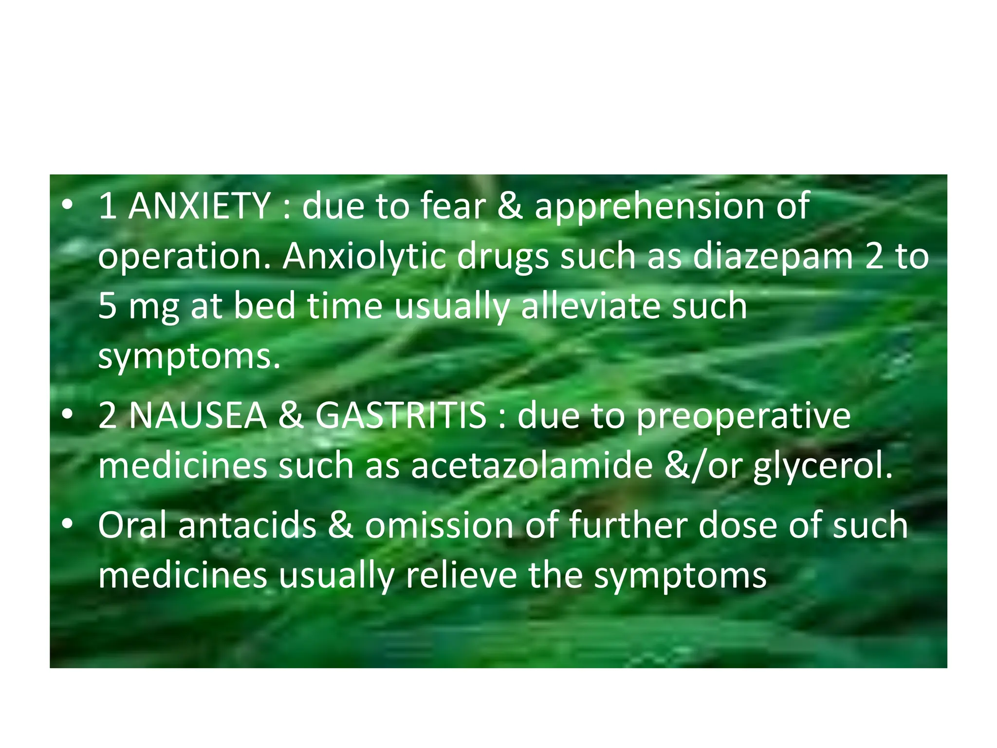
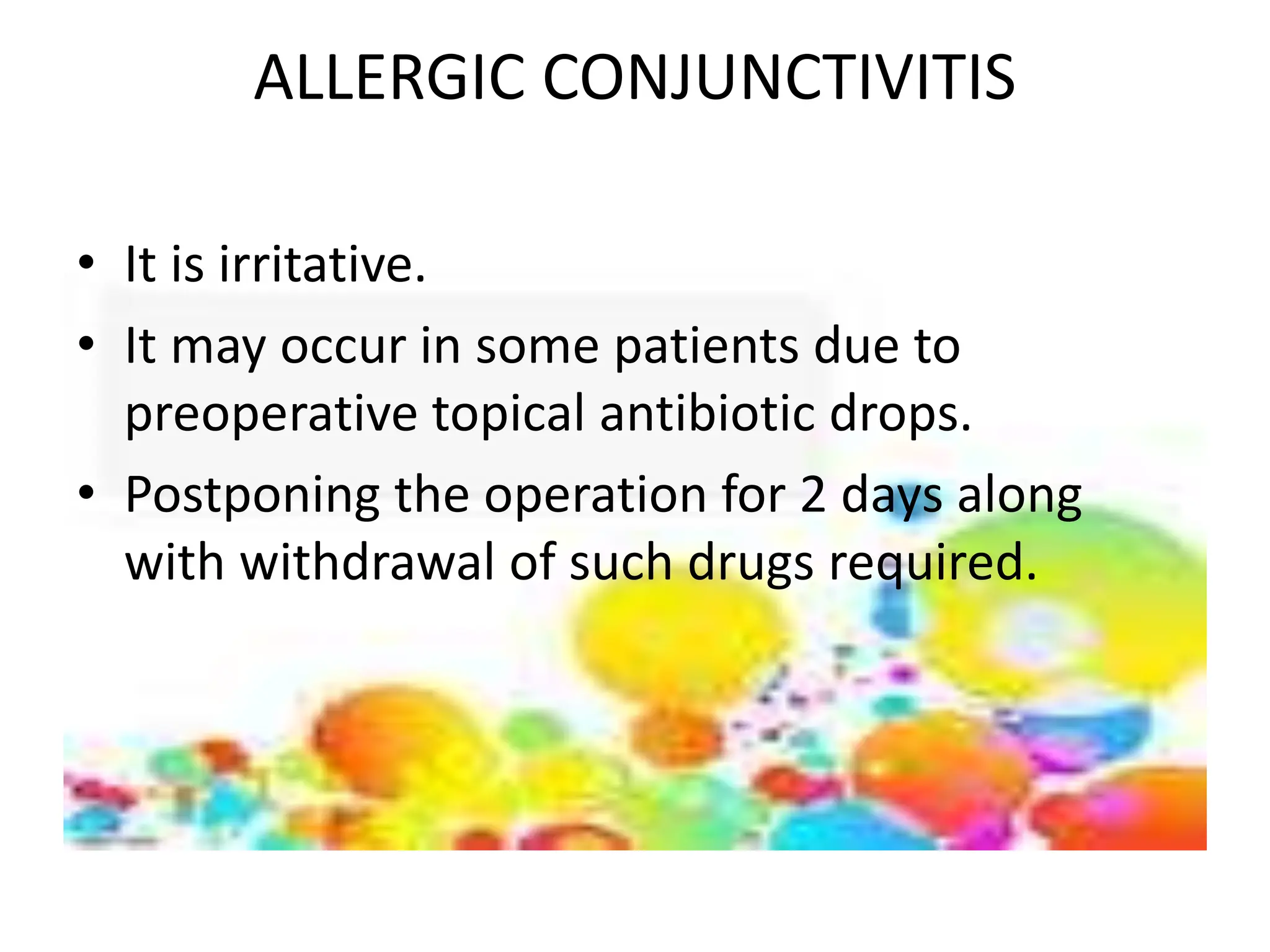
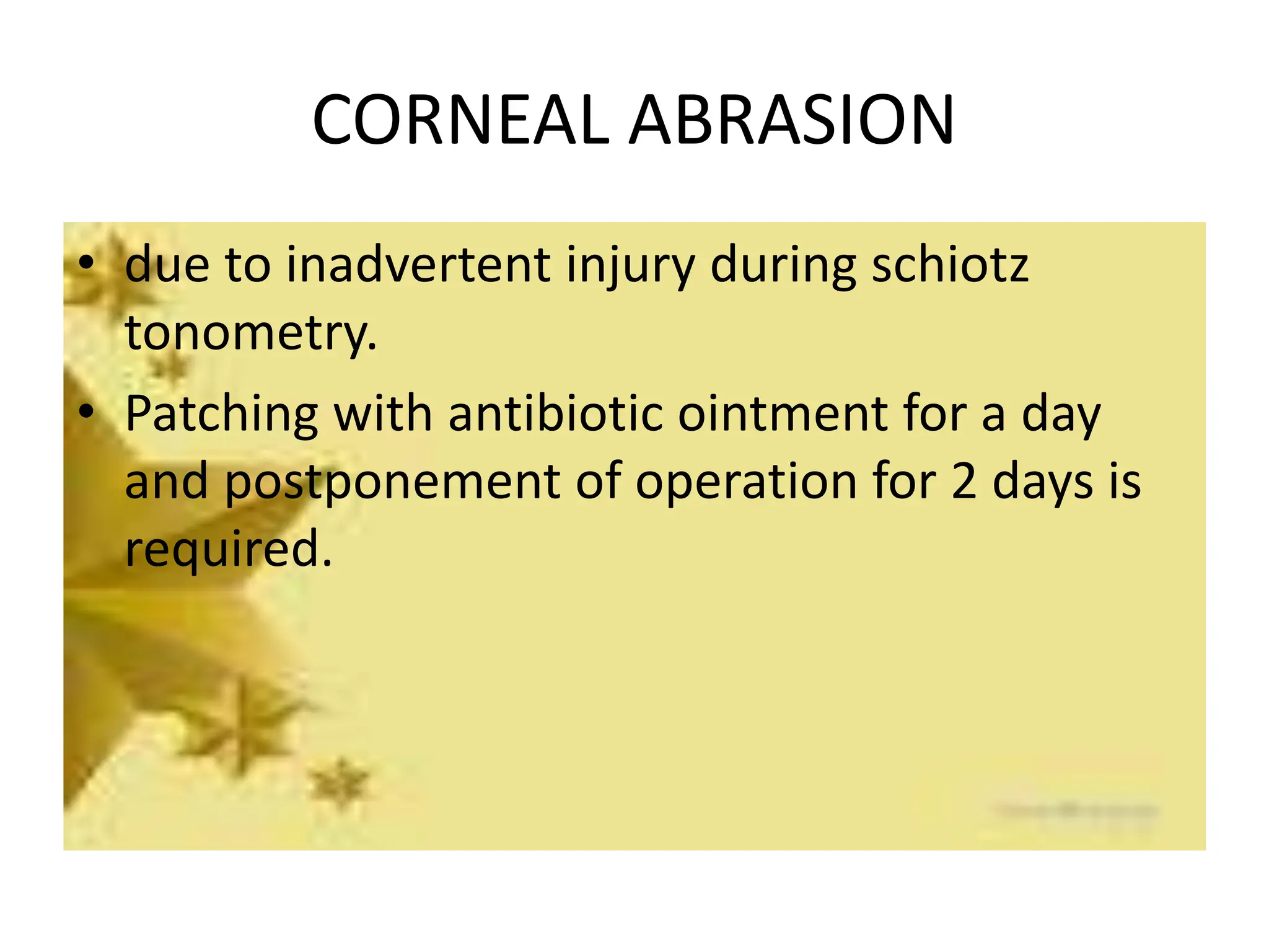
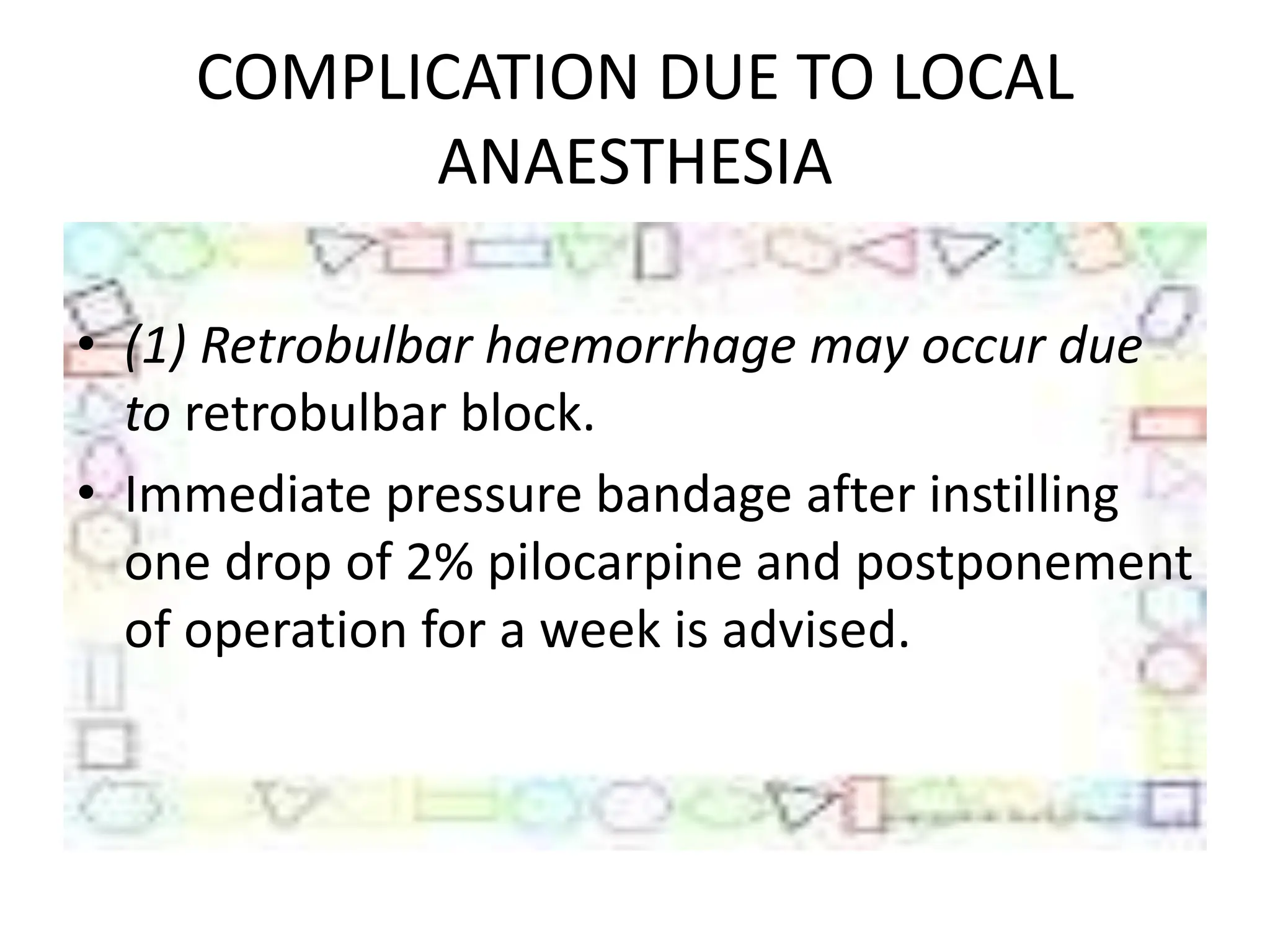
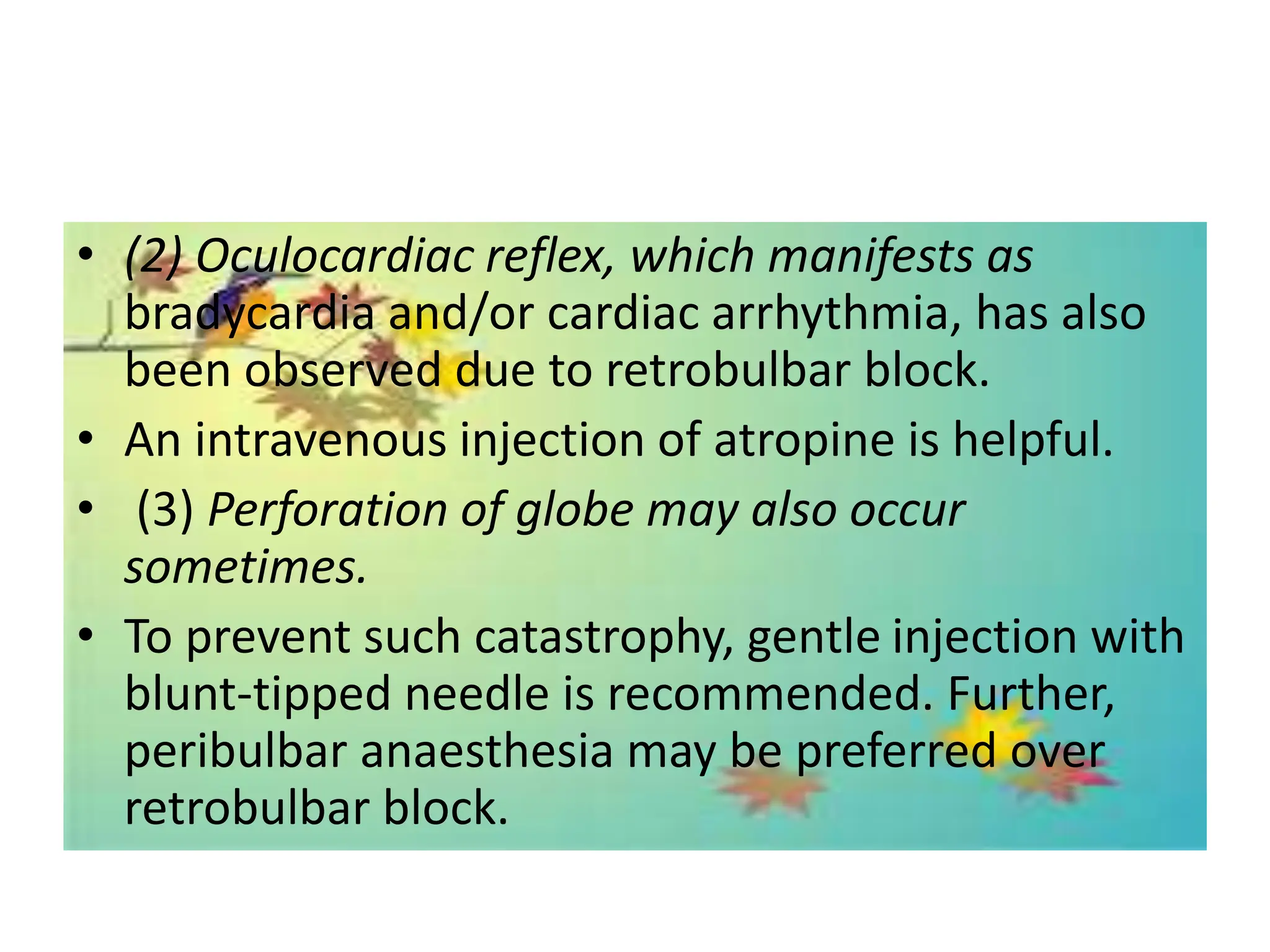
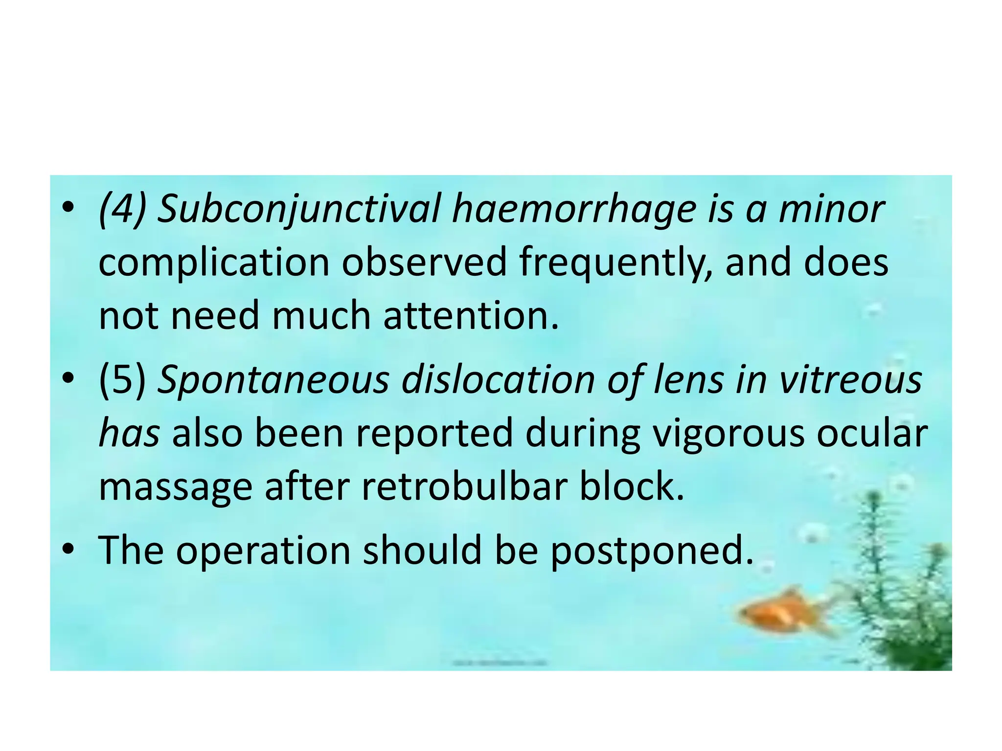
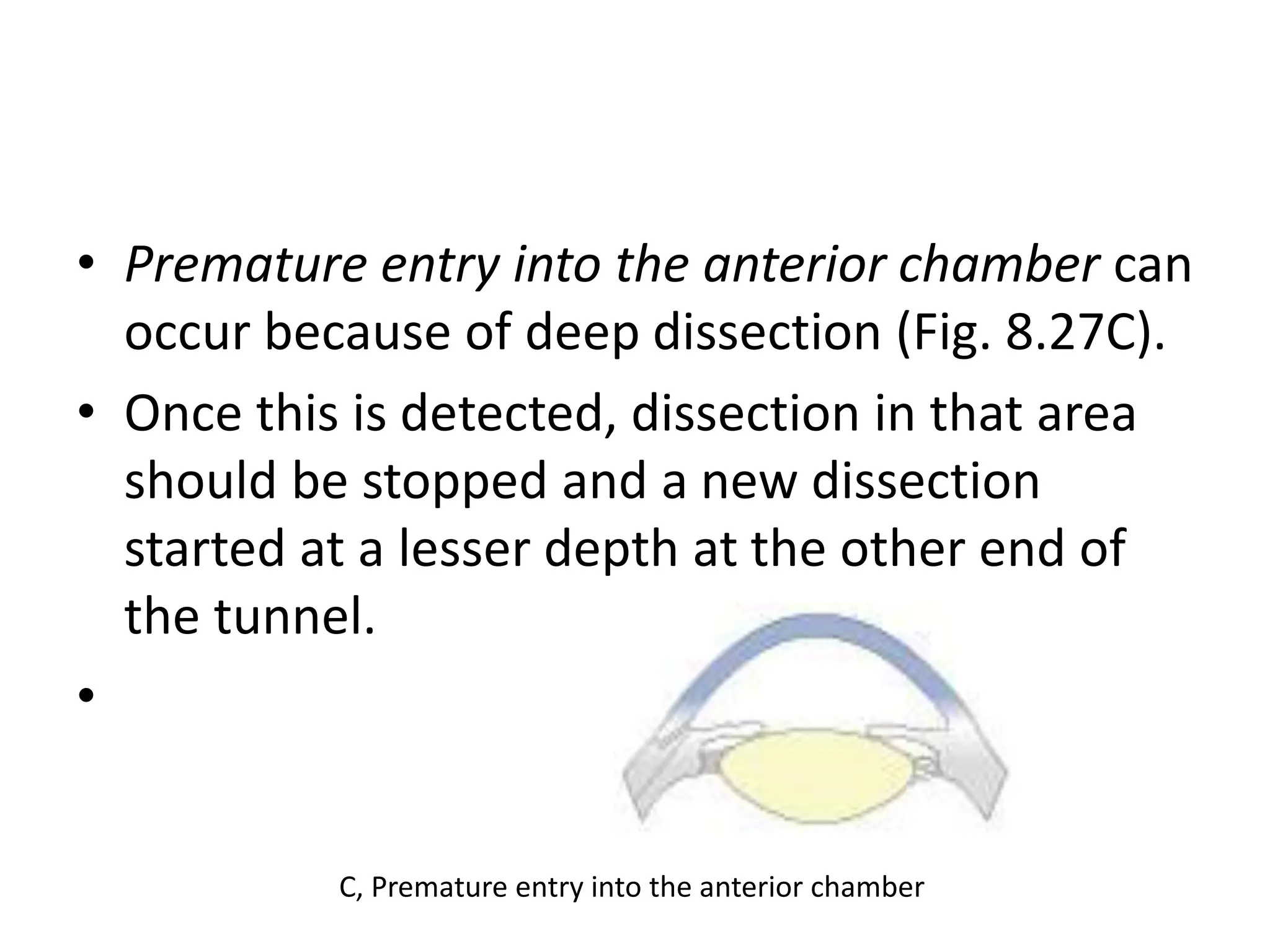
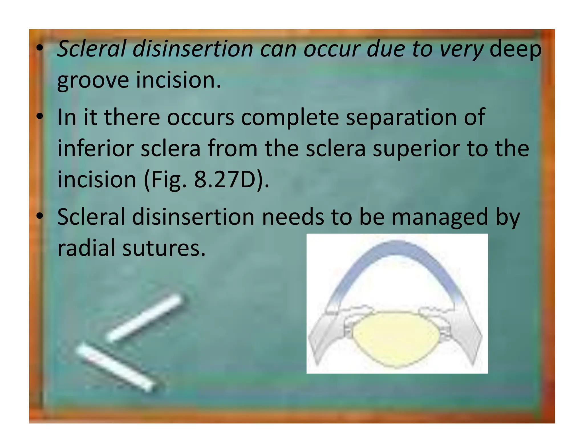
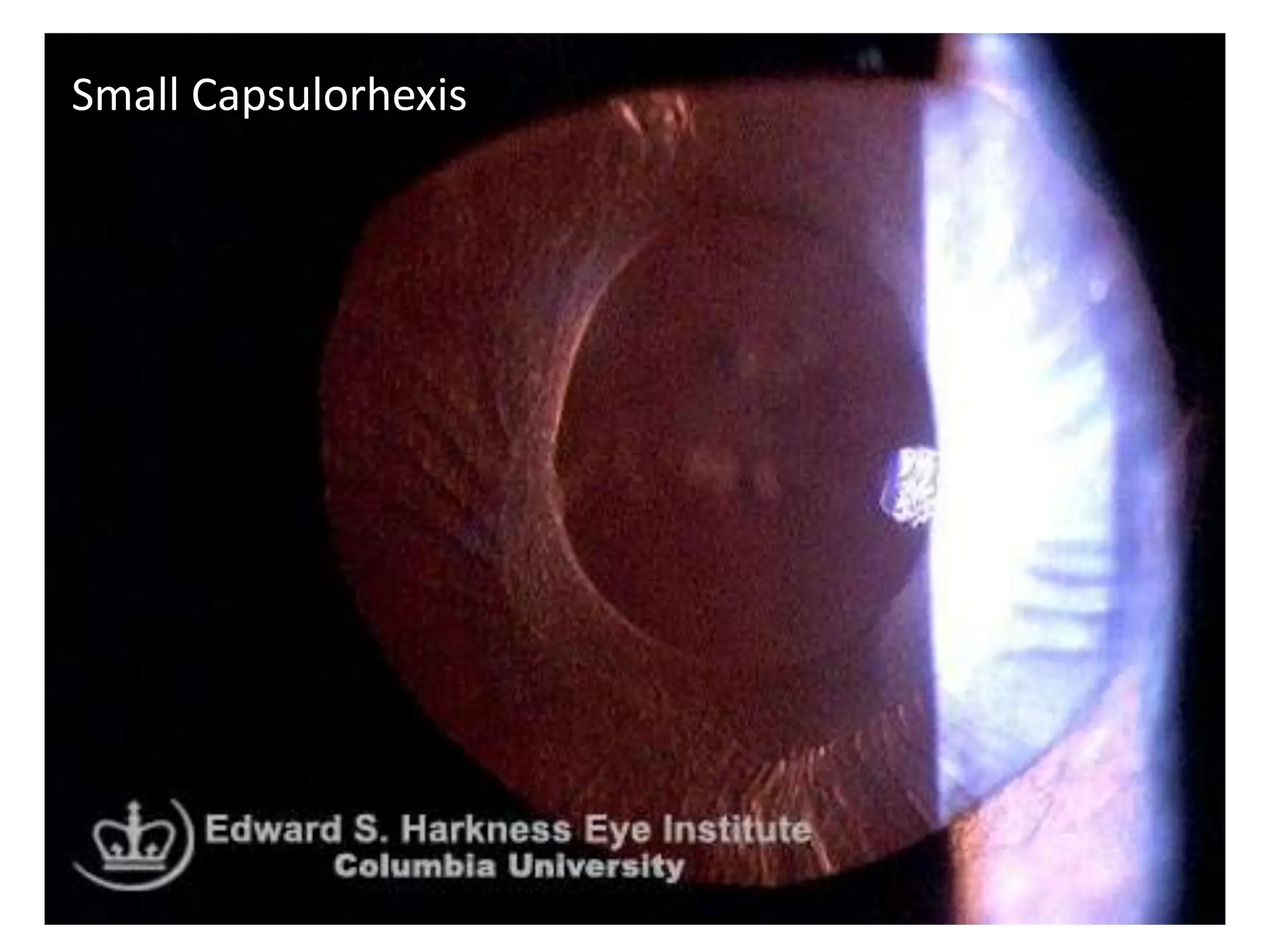
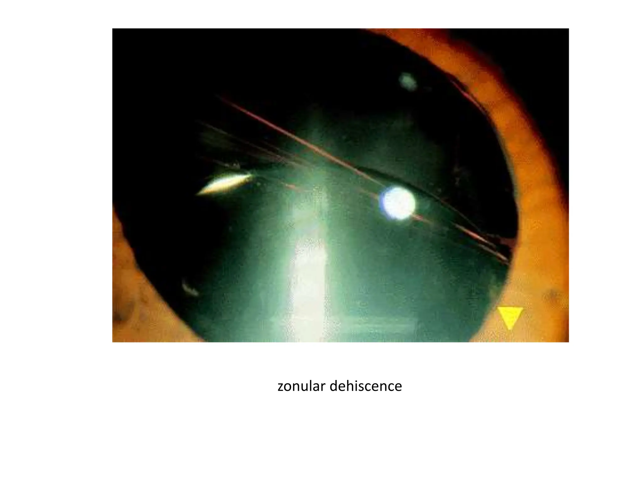
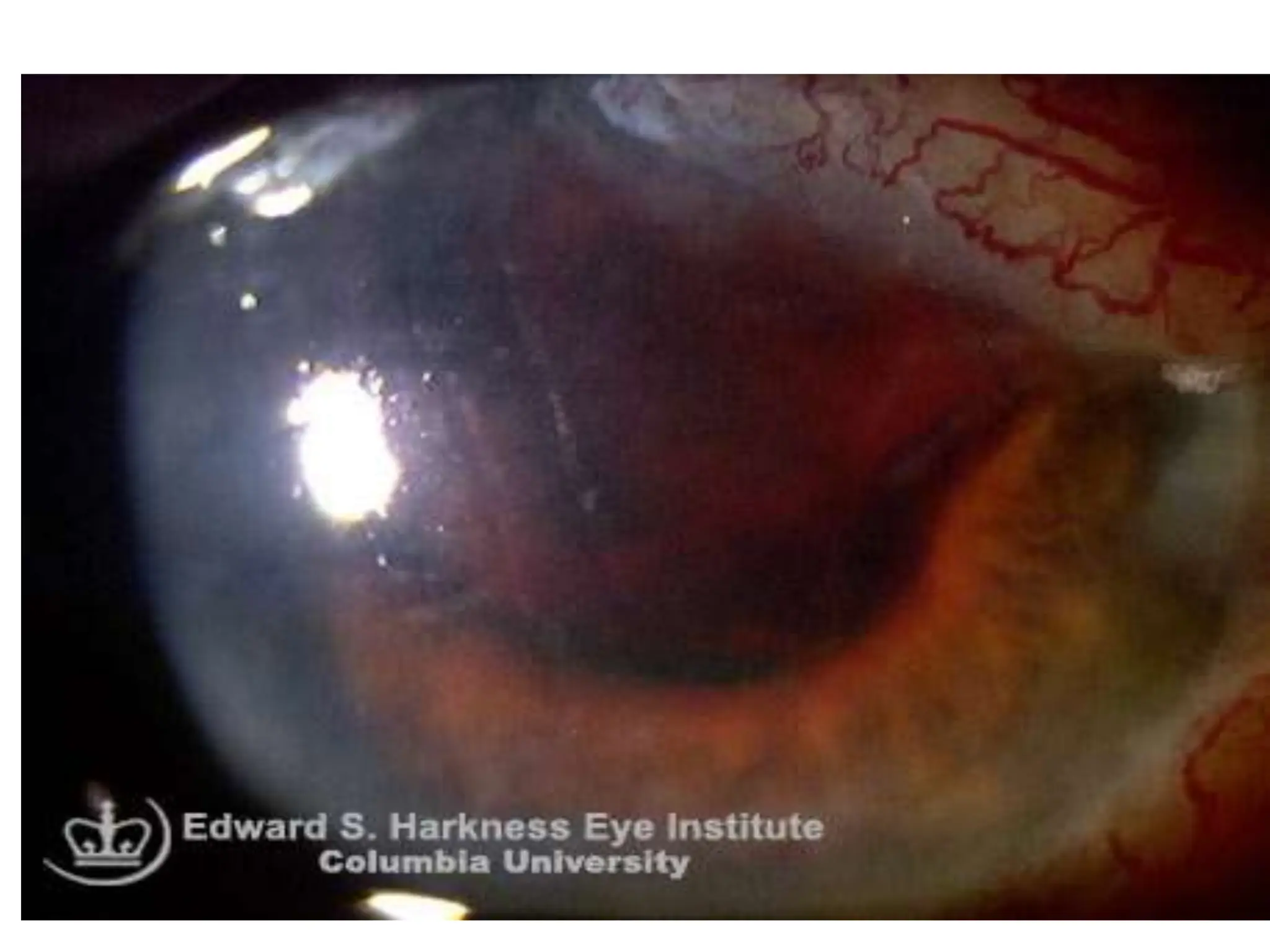
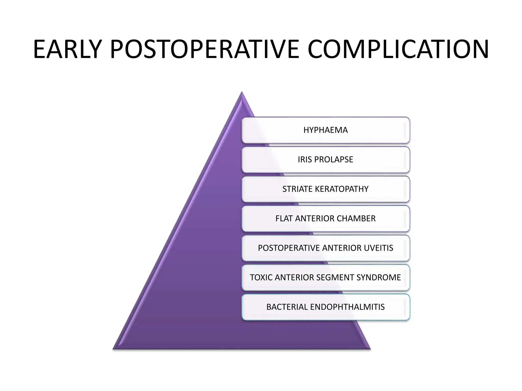
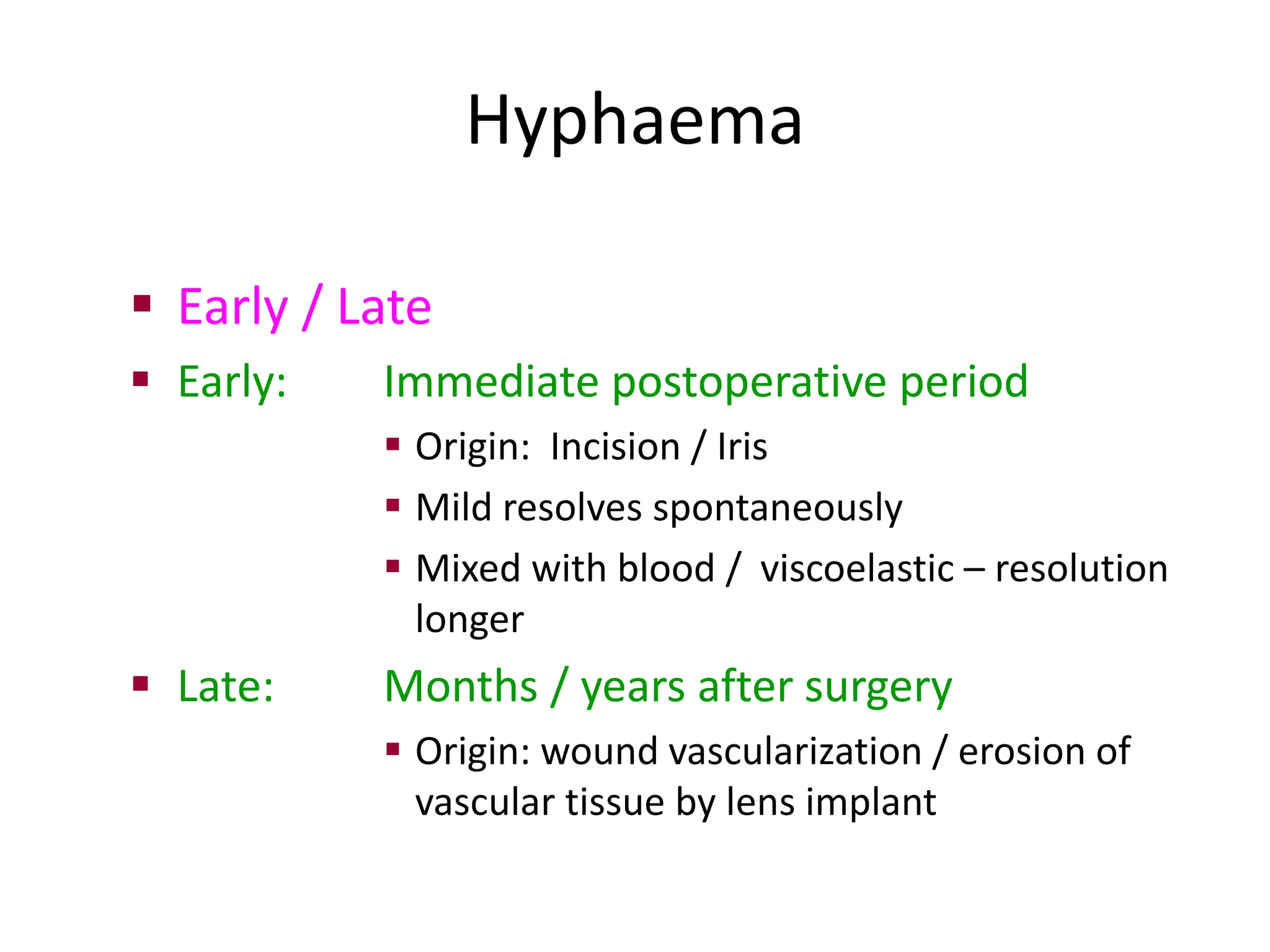
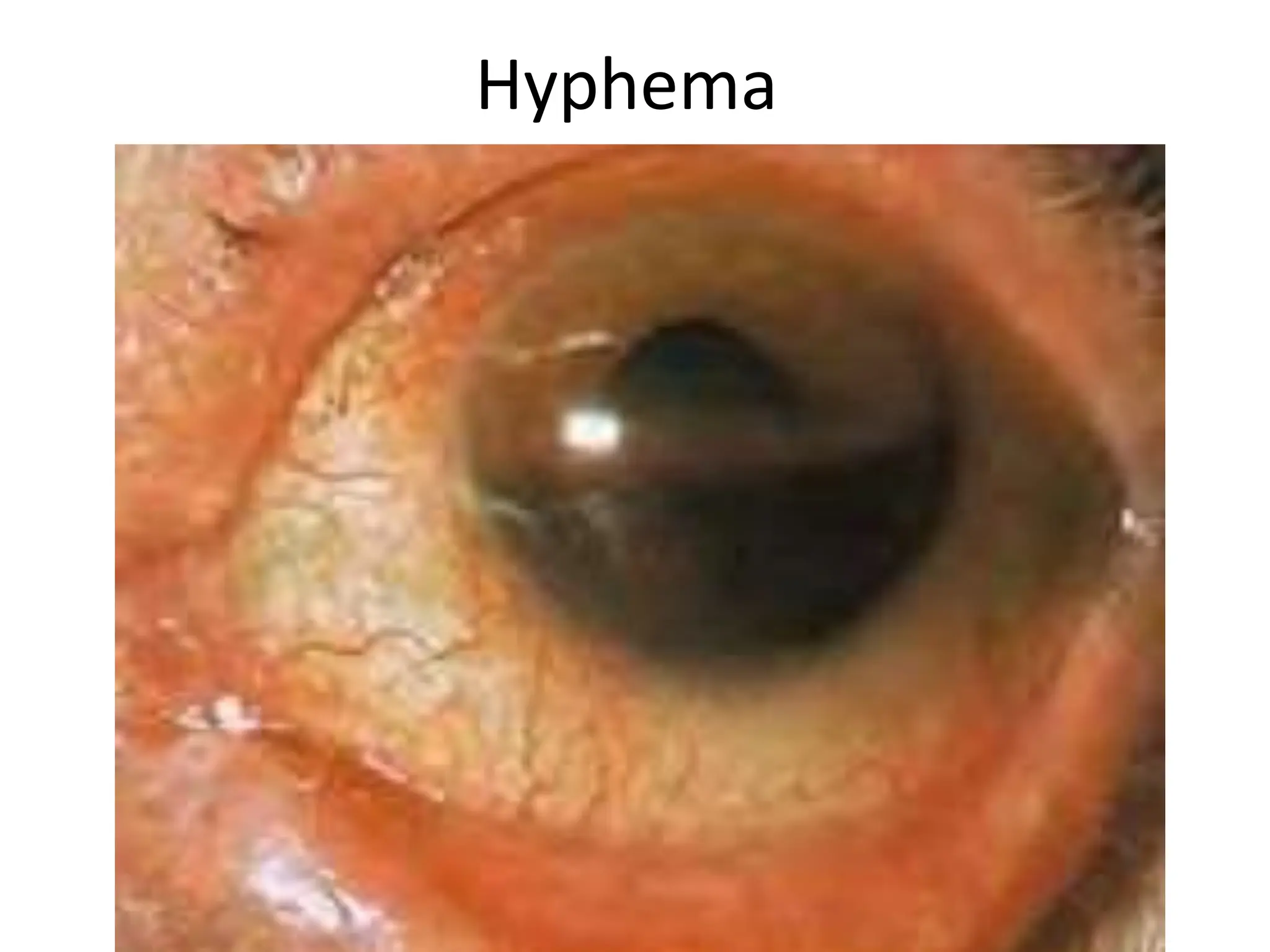
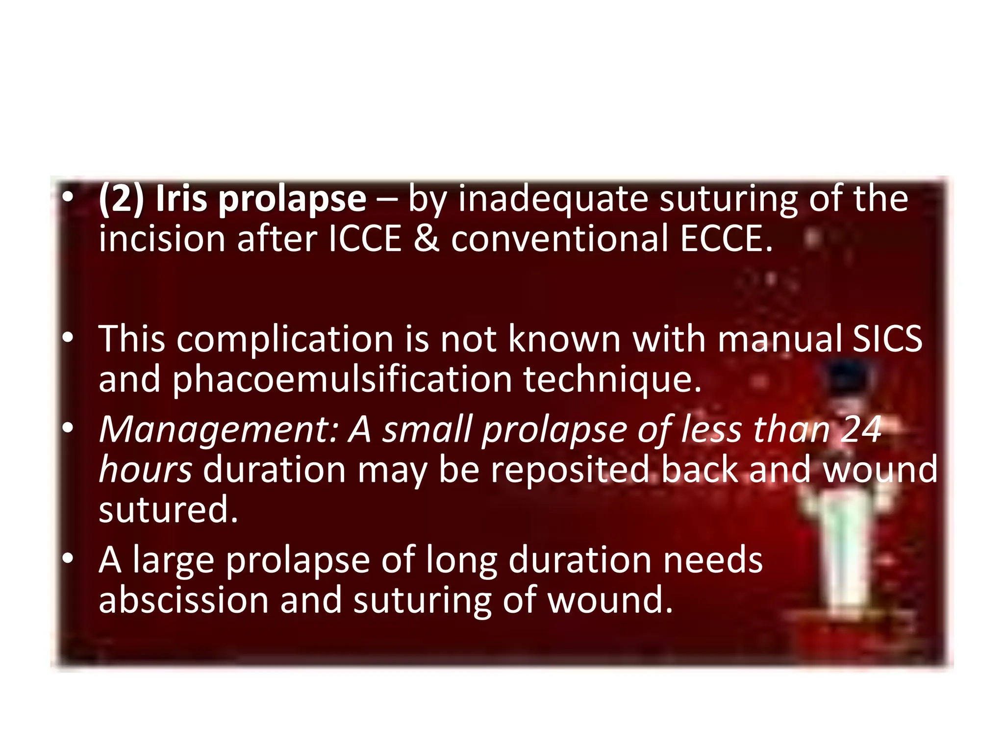
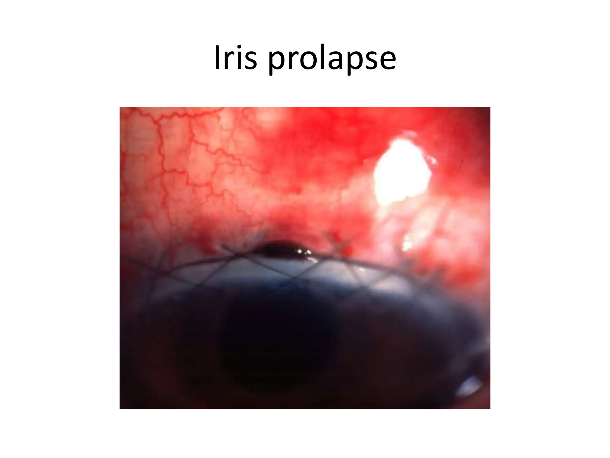
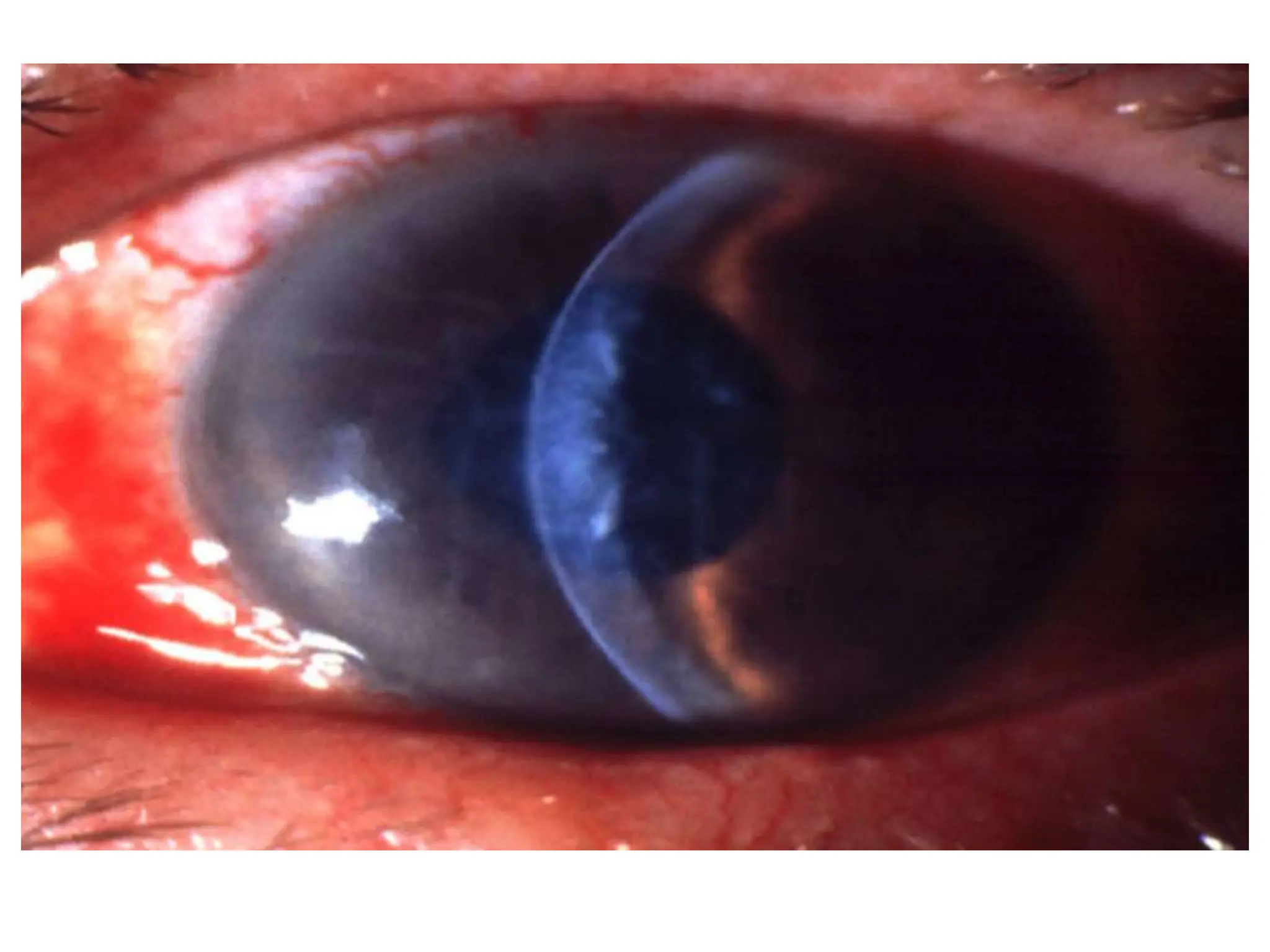
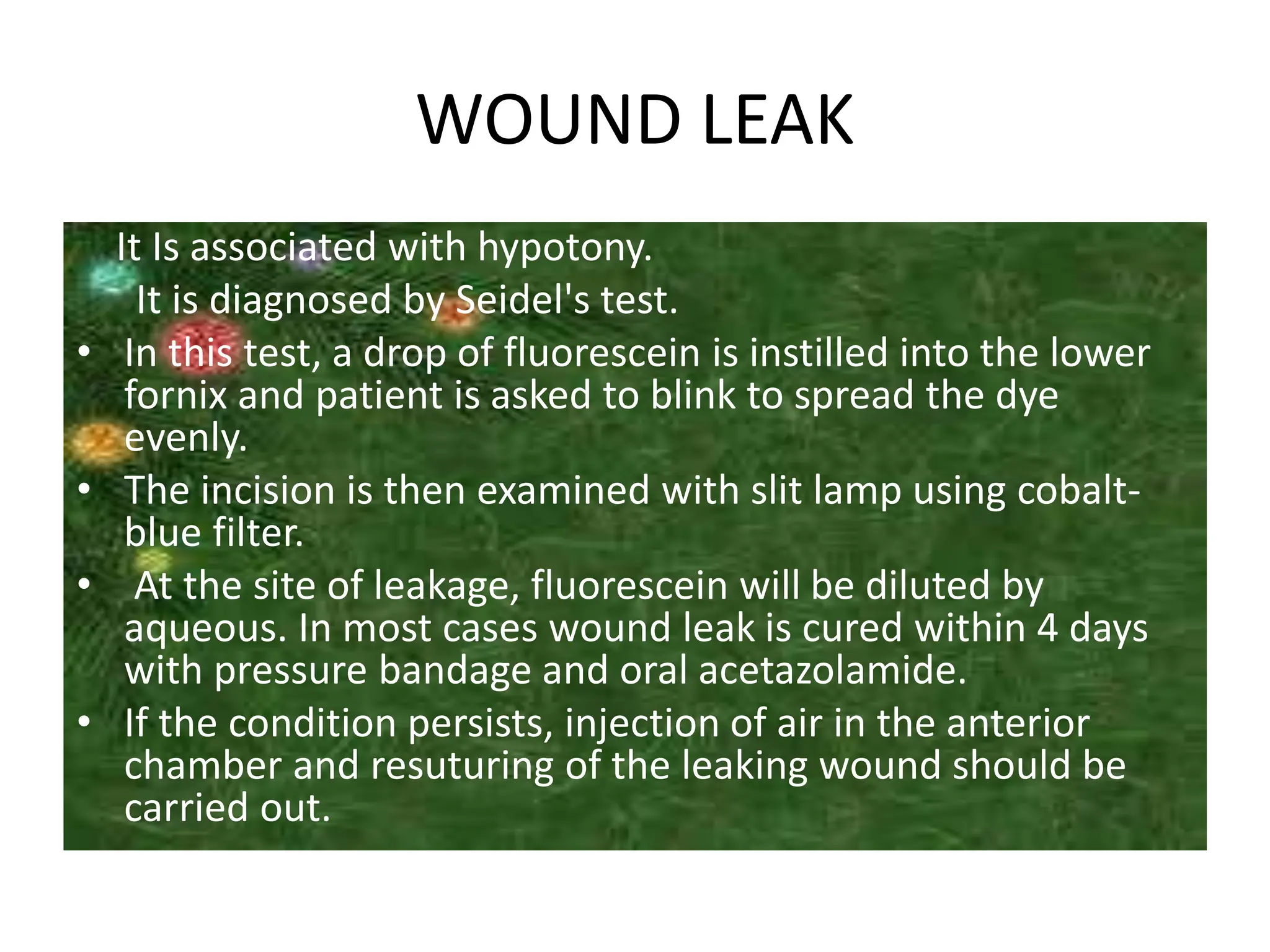
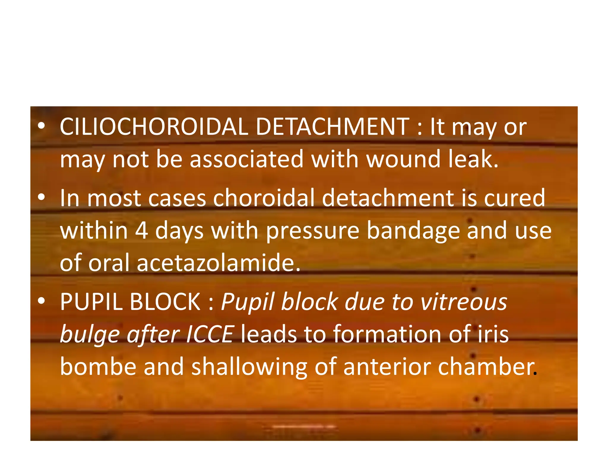
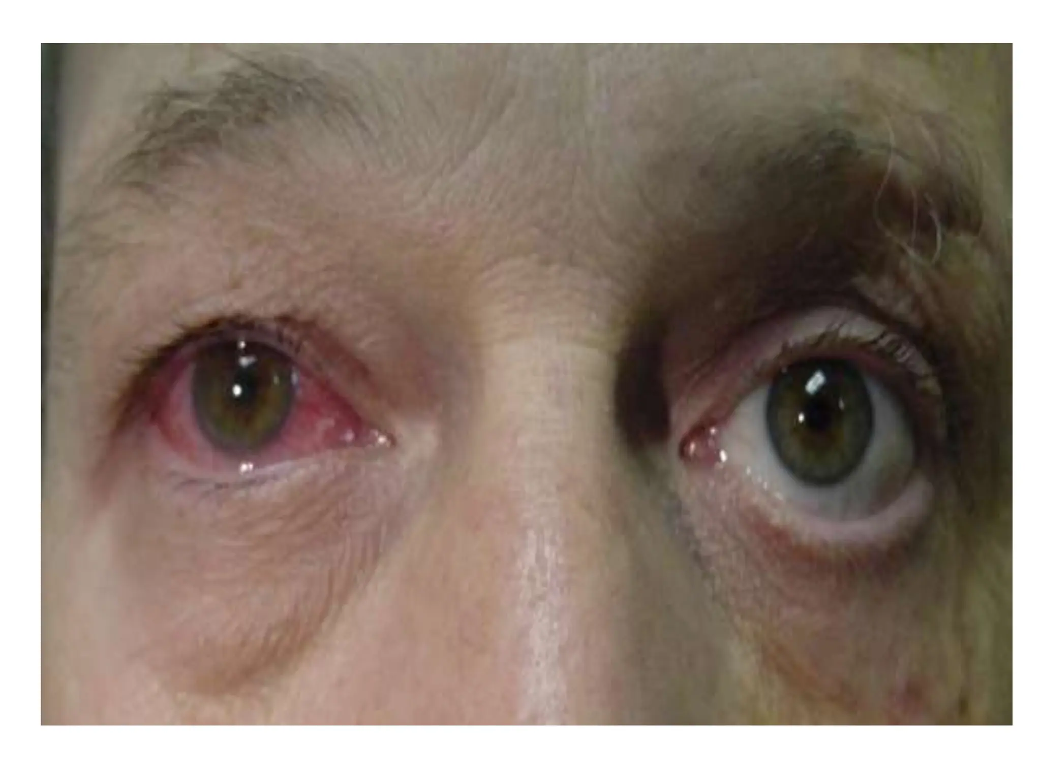
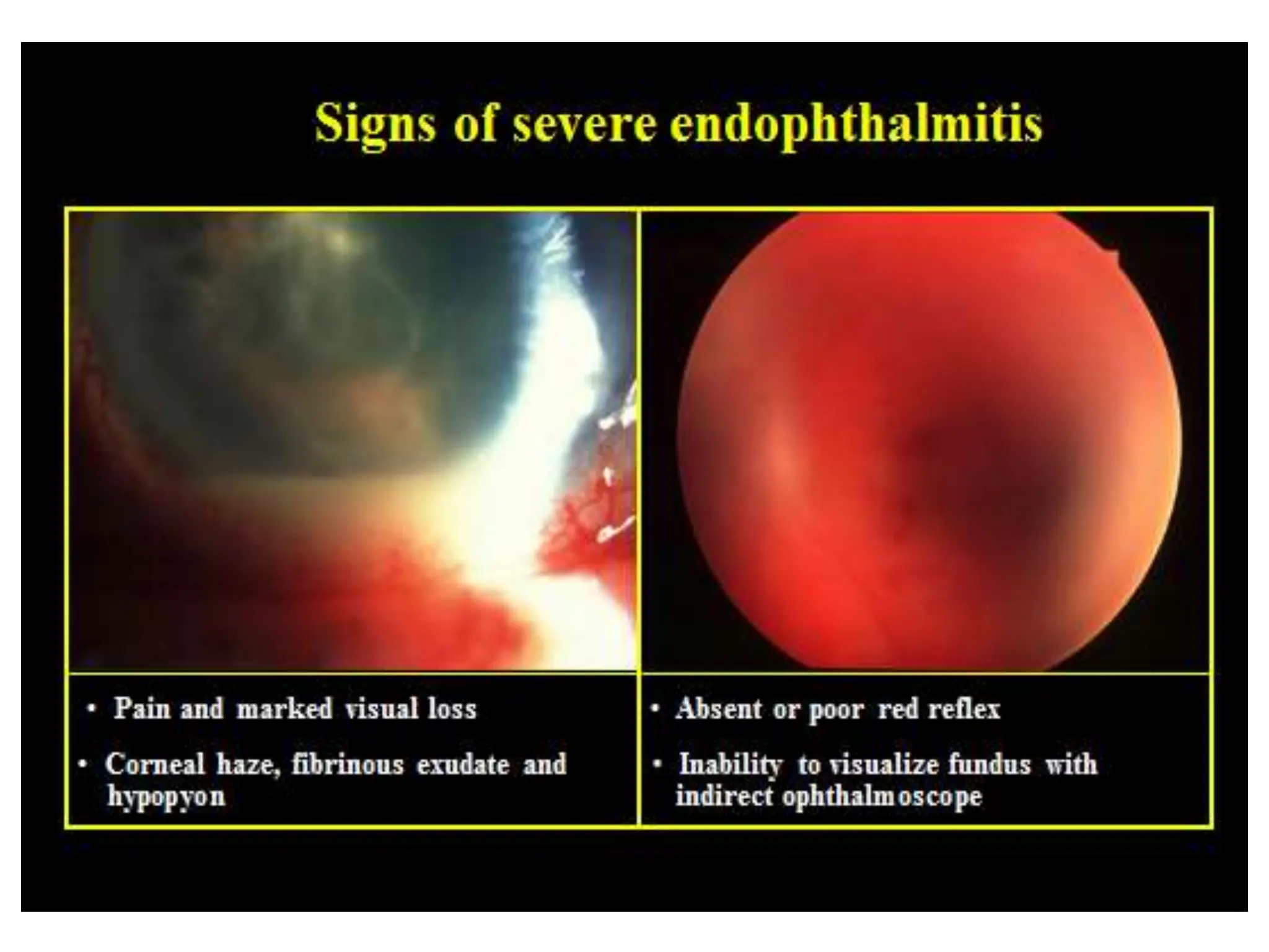
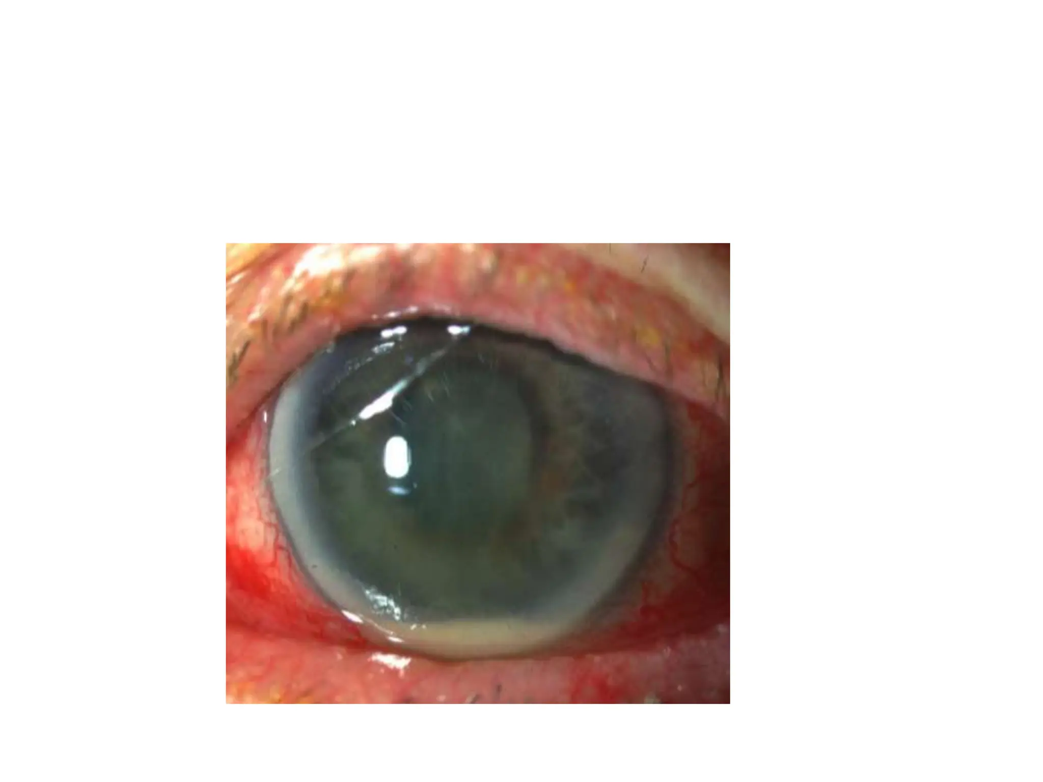
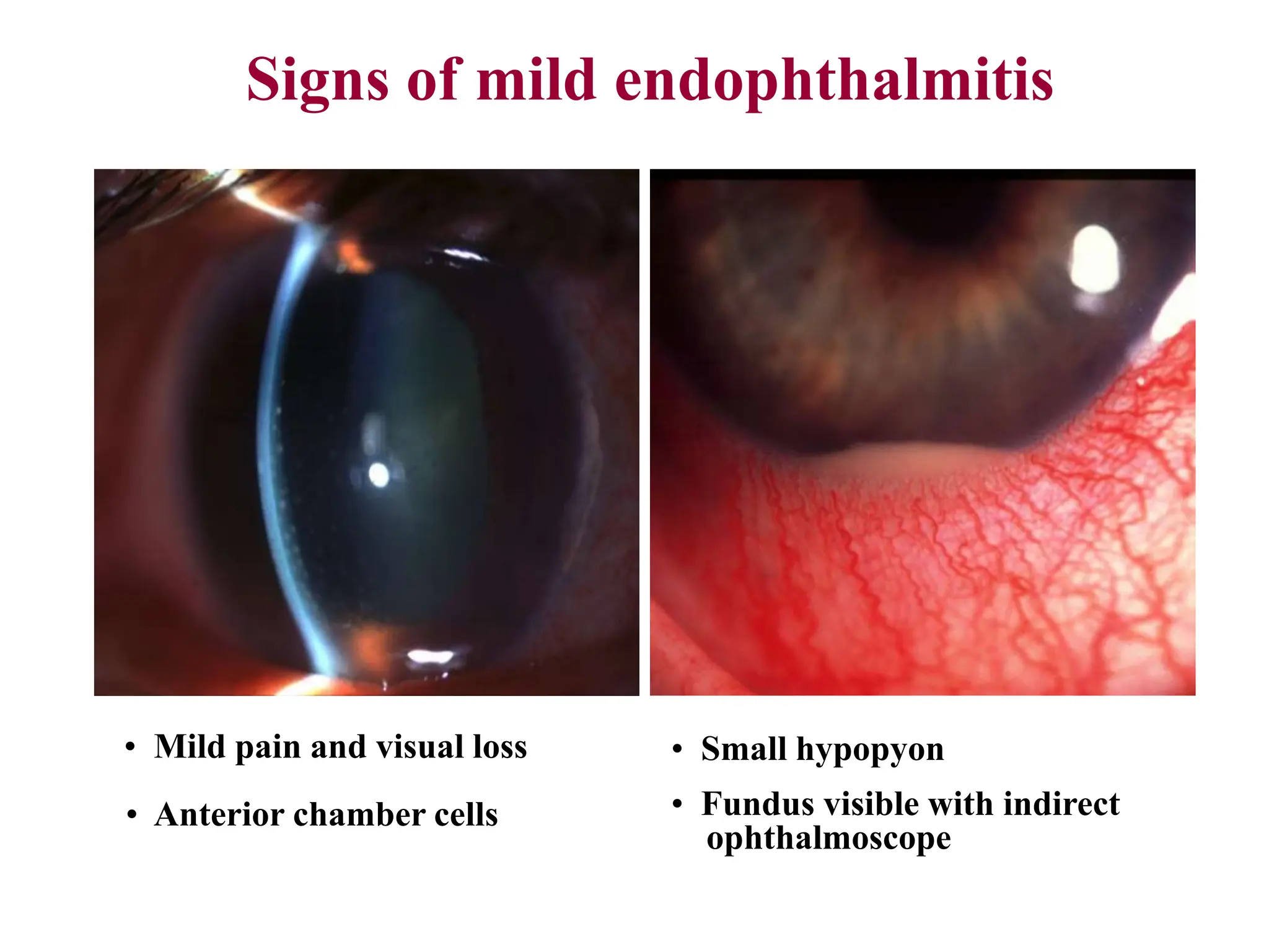
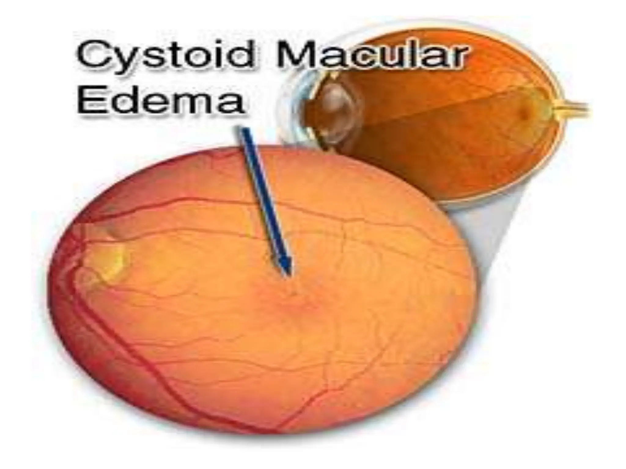
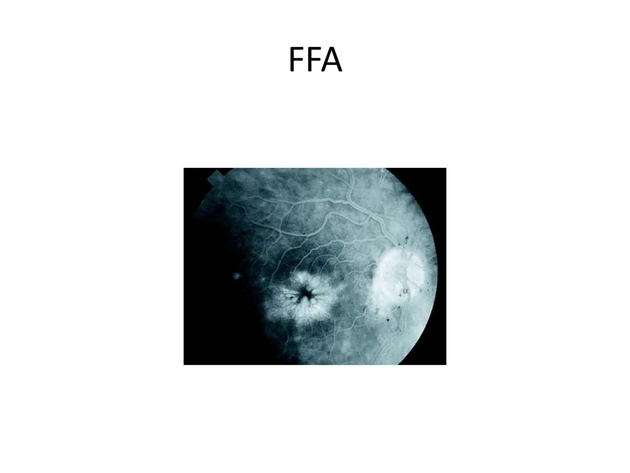
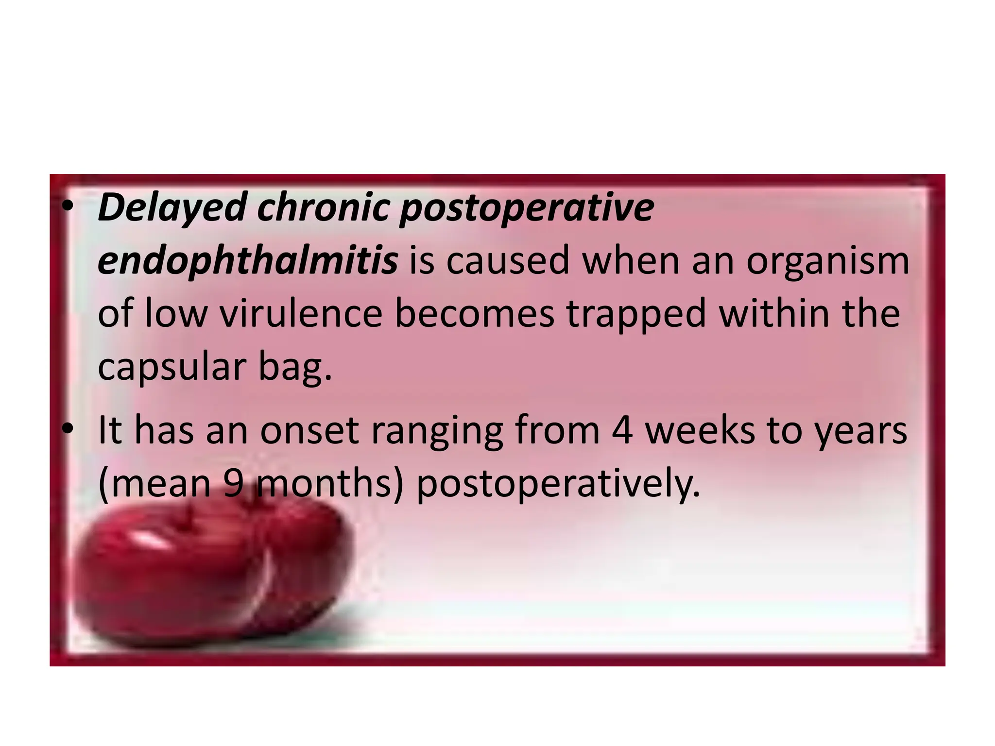
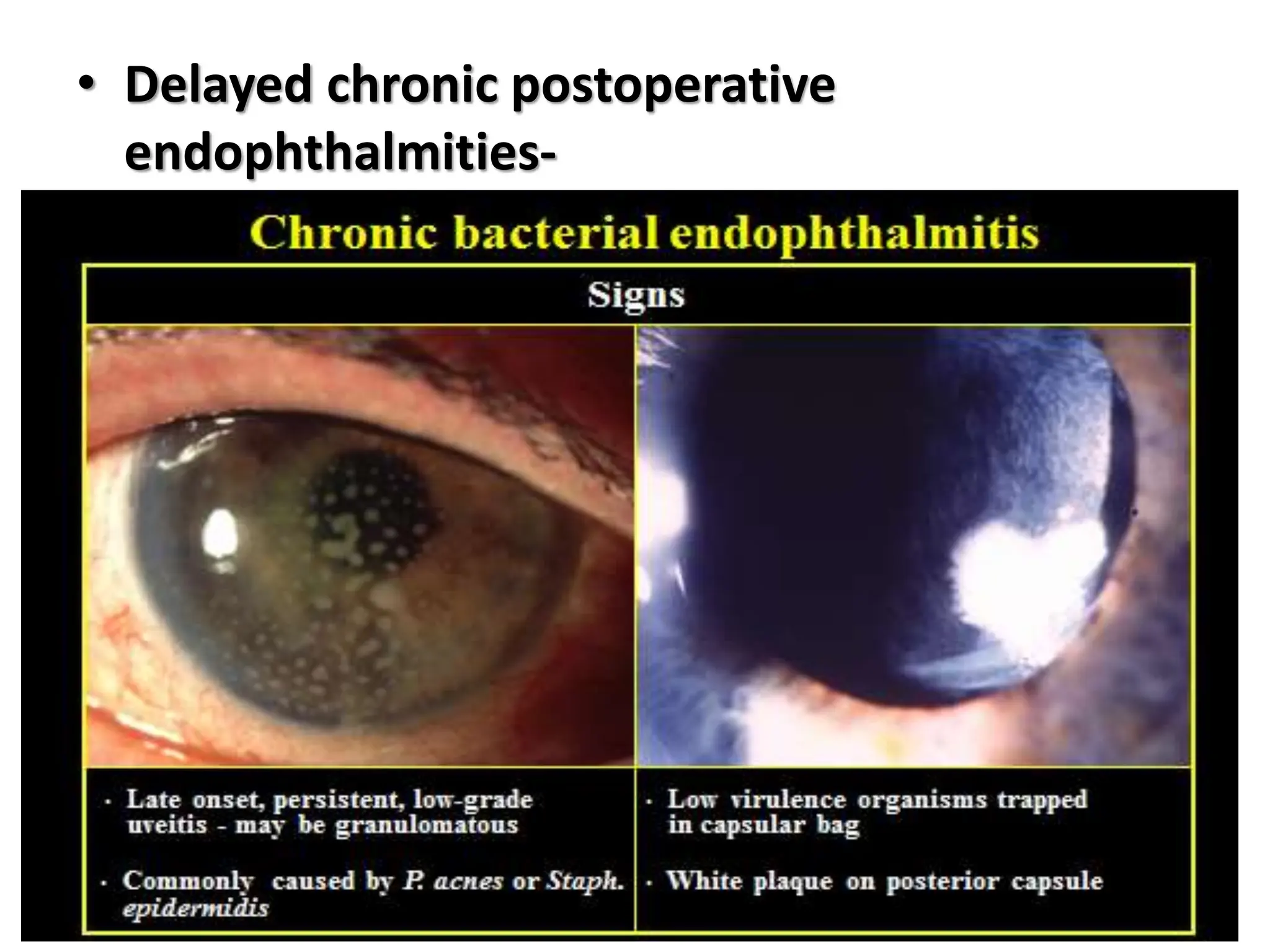
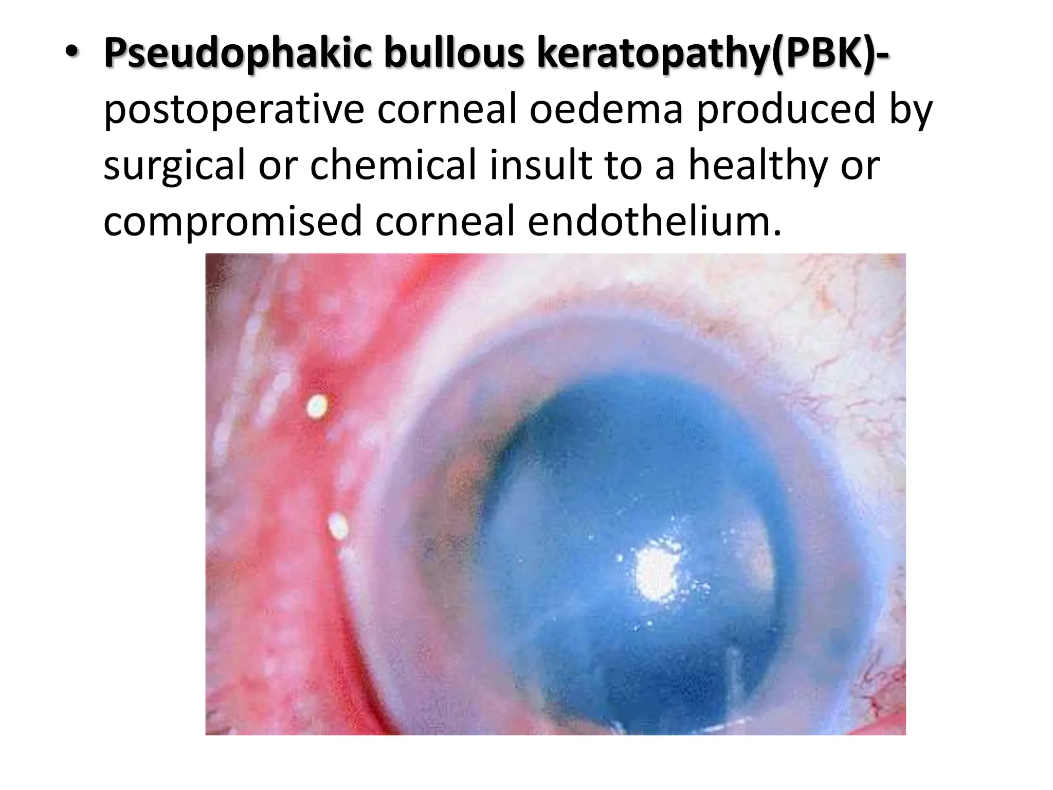
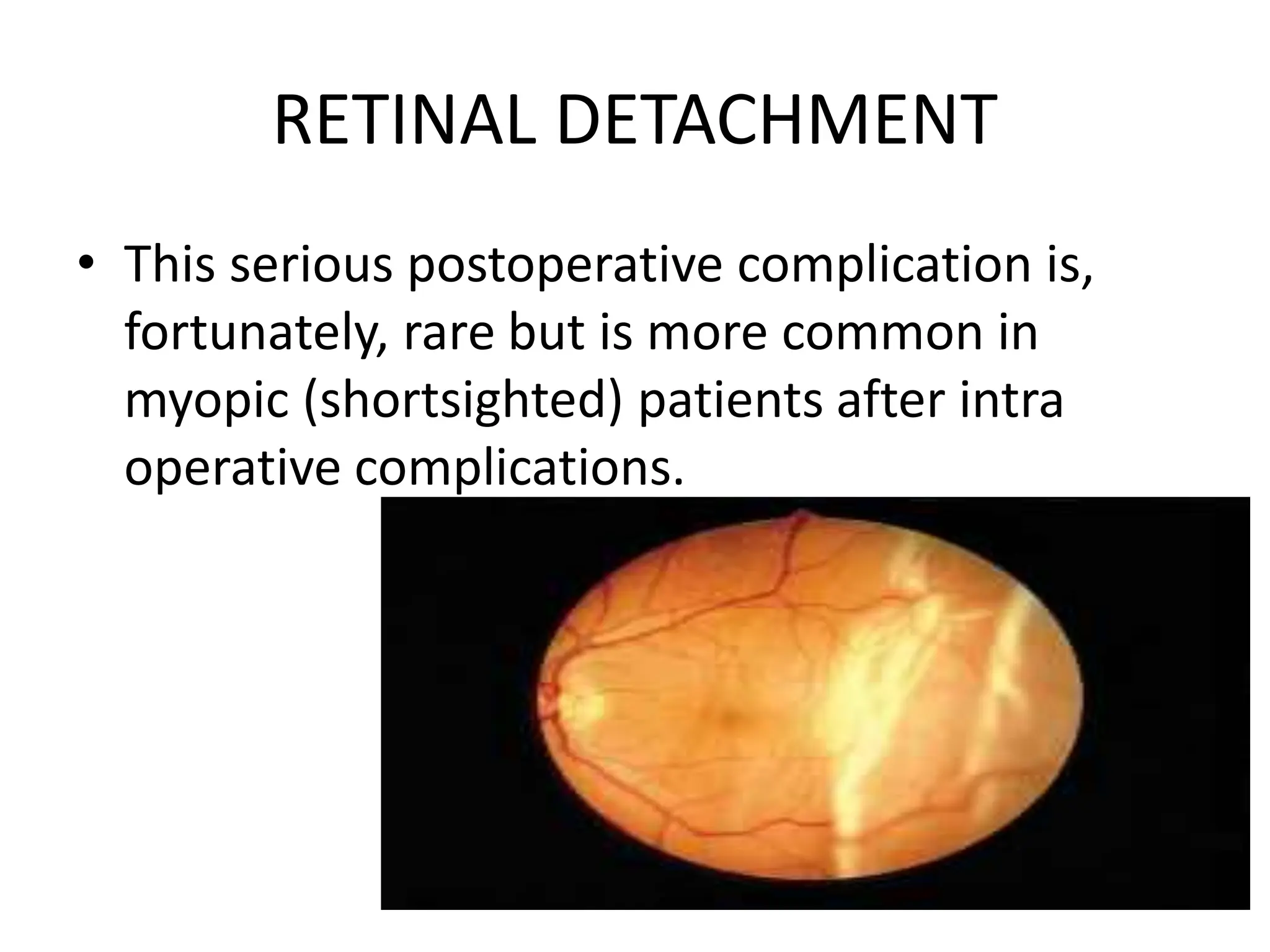
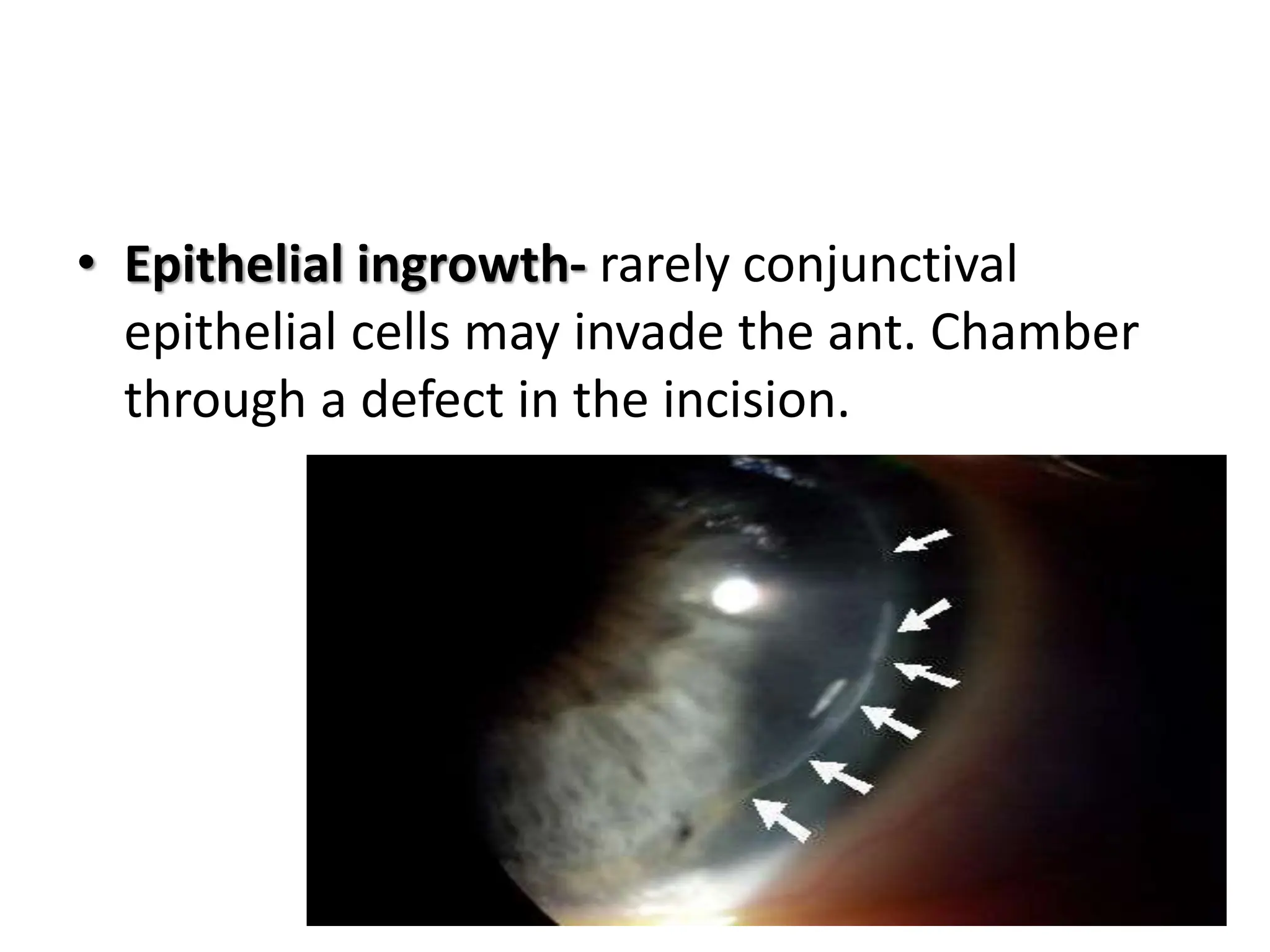
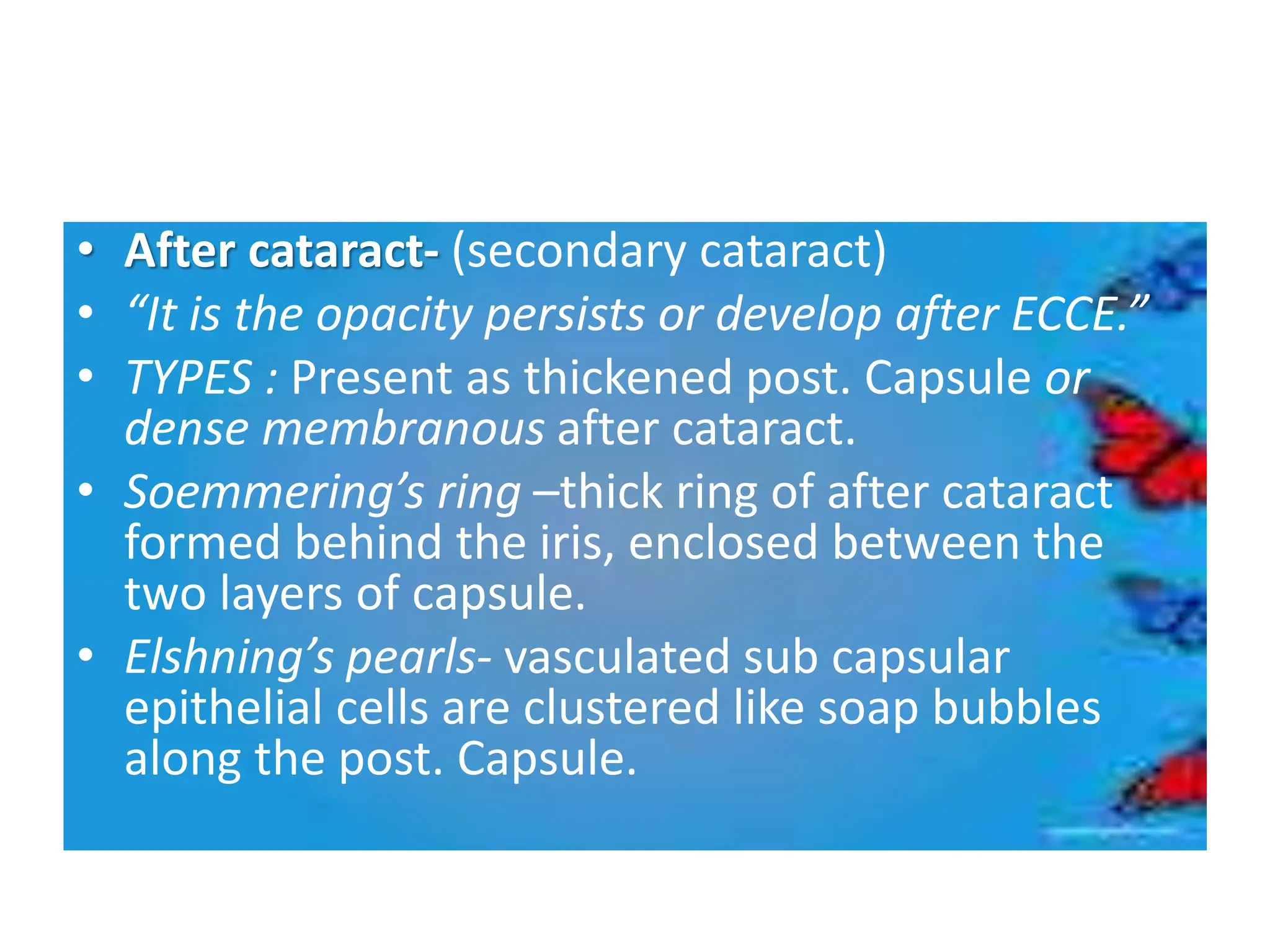
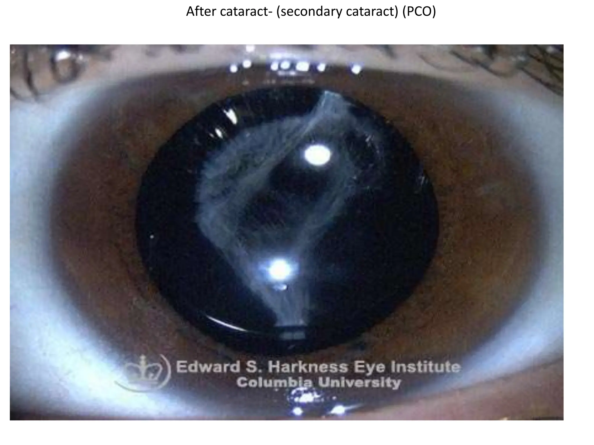
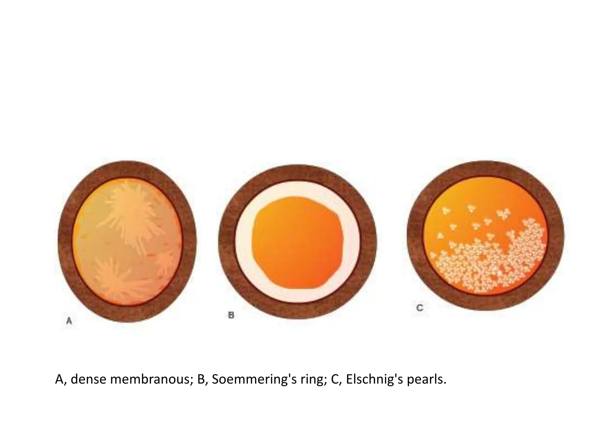
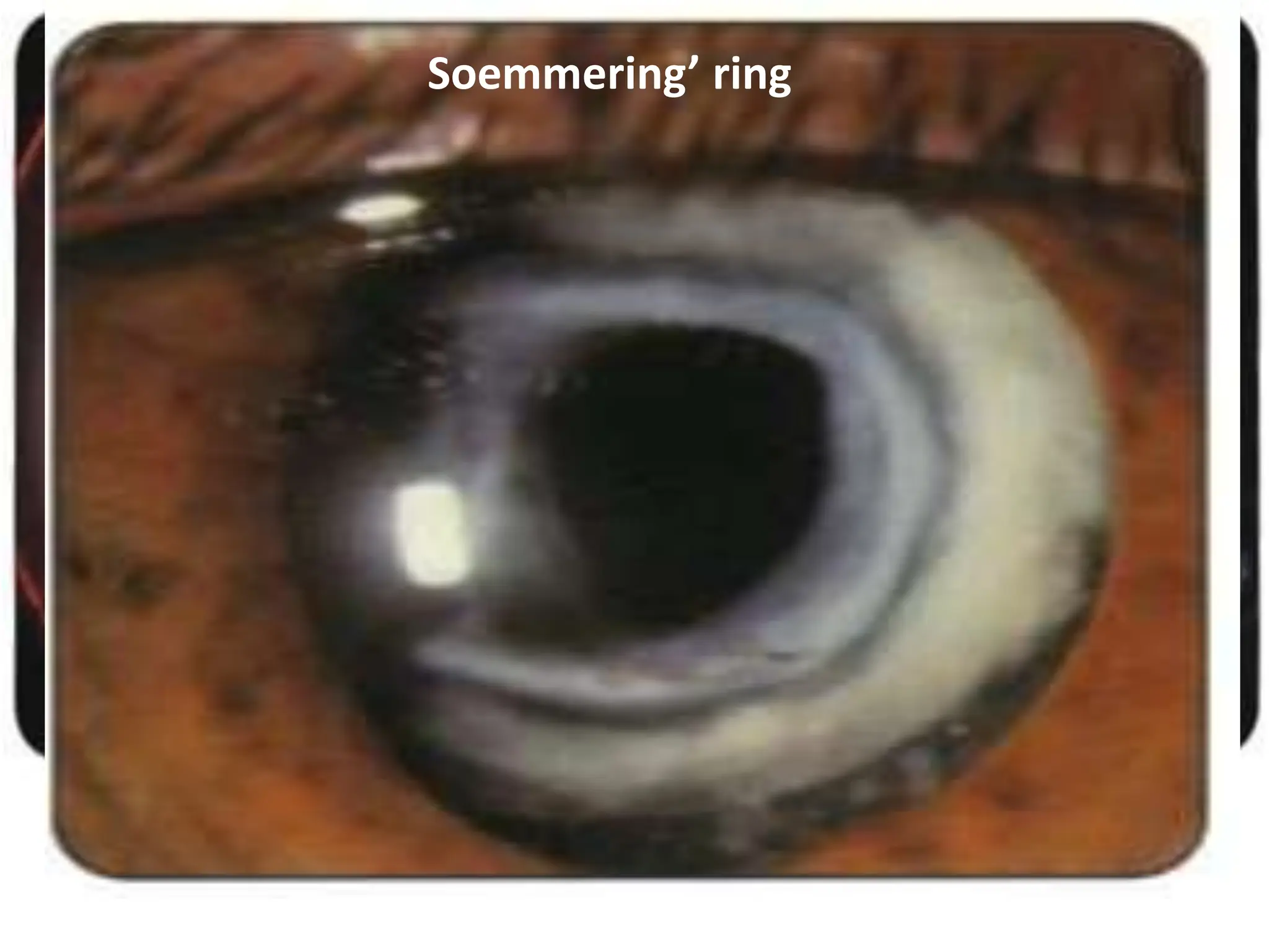
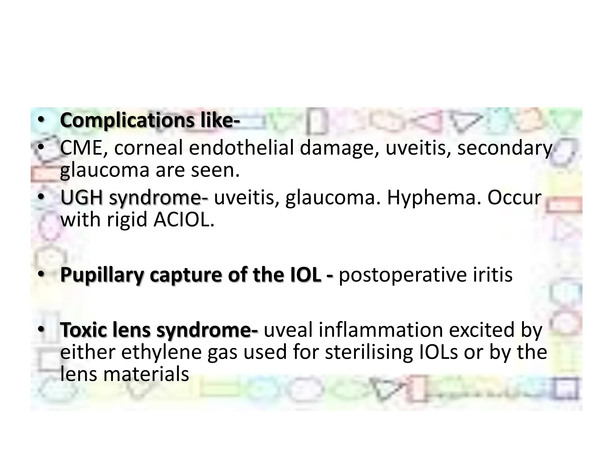
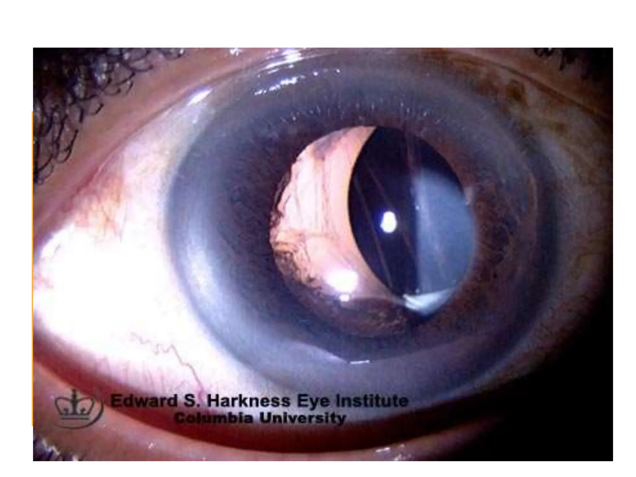
COMPLICATION OF
CATARACT SURGERY
MADE BY : SWATI PANARA
FROM : BHARTIMAIYA COLLEGE OF OPTOMETRY
2nd YEAR 4th SEMESTER
TYPES
PREOPRATIVE COMPLICATION
OPERATIVE COMPLICATION
EARLY POSTOPERATIVE
COMPLICATION
LATE POSTOPERATIVE
COMPLICATION
IOL RELATED COMPLICATION
PREOPERATIVE COMPLICATION
ANXIETY
NAUSEA & GASTRITIS
ALLERGIC CONJUNCTIVITIS
CORNEAL ABRASION
LOCAL ANAESTHESIA
• 1 ANXIETY : due to fear & apprehension of
operation. Anxiolytic drugs such as diazepam 2 to
5 mg at bed time usually alleviate such
symptoms.
• 2 NAUSEA & GASTRITIS : due to preoperative
medicines such as acetazolamide &/or glycerol.
• Oral antacids & omission of further dose of such
medicines usually relieve the symptoms
ALLERGIC CONJUNCTIVITIS
• It is irritative.
• It may occur in some patients due to
preoperative topical antibiotic drops.
• Postponing the operation for 2 days along
with withdrawal of such drugs required.
CORNEAL ABRASION
• due to inadvertent injury during schiotz
tonometry.
• Patching with antibiotic ointment for a day
and postponement of operation for 2 days is
required.
COMPLICATION DUE TO LOCAL
ANAESTHESIA
• (1) Retrobulbar haemorrhage may occur due
to retrobulbar block.
• Immediate pressure bandage after instilling
one drop of 2% pilocarpine and postponement
of operation for a week is advised.
• (2) Oculocardiac reflex, which manifests as
bradycardia and/or cardiac arrhythmia, has also
been observed due to retrobulbar block.
• An intravenous injection of atropine is helpful.
• (3) Perforation of globe may also occur
sometimes.
• To prevent such catastrophy, gentle injection with
blunt-tipped needle is recommended. Further,
peribulbar anaesthesia may be preferred over
retrobulbar block.
• (4) Subconjunctival haemorrhage is a minor
complication observed frequently, and does
not need much attention.
• (5) Spontaneous dislocation of lens in vitreous
has also been reported during vigorous ocular
massage after retrobulbar block.
• The operation should be postponed.
OPERATIVE COMPLICATION
• SUPERIOR RECTUS MUSCLE LACERATION
• EXCESSIVE BLEEDING
• INCISION RELATED COMPLICATION
• INJURY TO THE CORNEA , IRIS & LENS
• ANTERIOR CAPSULORHEXIS
• POSTERIOR CAPSULAR RUPTURE
• VITREOUS LOSS
• ZONULAR DEHISCENE
• NUCLEAR DROP IN TO THE VITREOUS CAVITY
• POSTERIOR LOSS OF LENS FRAGMENT
• EXPULSIVE CHOROIDAL HAEMORRHAGE
• SUPRACHOROIDAL HAEMORRHAGE
(1) Superior rectus muscle laceration and/or
haematoma, may occur while applying the bridle
suture.
• Usually no treatment is required.
(2). Excessive bleeding may be encountered during
the preparation of conjunctival flap or during
incision into the anterior chamber. Bleeding
vessels may be gently cauterized.
• 3. Incision related complications depend upon
the type of cataract surgery being performed.
• i. In conventional ECCE there may occur irregular
incision.
• In manual SICS and phacoemulsification following
complications may occur while making the self-
sealing tunnel incision.
• Button holing of anterior wall of tunnel can occur
because of superficial dissection of the scleral
flap (Fig. 8.27B).
A, correct incision;
B, Buttonholing of anterior wall of the tunnel;
• Premature entry into the anterior chamber can
occur because of deep dissection (Fig. 8.27C).
• Once this is detected, dissection in that area
should be stopped and a new dissection
started at a lesser depth at the other end of
the tunnel.
•
C, Premature entry into the anterior chamber
• Scleral disinsertion can occur due to very deep
groove incision.
• In it there occurs complete separation of
inferior sclera from the sclera superior to the
incision (Fig. 8.27D).
• Scleral disinsertion needs to be managed by
radial sutures.
• 4. Injury to the cornea (Descement's
detachment), iris and lens may occur when
anterior chamber is entered with a sharp-tipped
instrument such as keratome or a piece of razor
blade.
• A gentle handling with proper hypotony reduces
the incidence of such inadvertent injuries.
• 5. Iris injury and iridodialysis (tear of iris from
root) may occur inadvertently during intraocular
manipulation.
• 6. Complications related to anterior capsulorhexis. Continuous curvilinear
capsulorhexis (CCC) is the preferred technique for opening the anterior capsule for
SICS and phacoemulsification. Following complications may occur:
• Escaping capsulorhexis i.e., capsulorhexis moves peripherally and may extend to
the equator or posterior capsule.
• Small capsulorhexis. It predisposes to posterior capsular tear and nuclear drop
during hydrodissection.
• It also predisposes to occurrence of zonular deshiscence. Therefore, a small sized
capsulorhexis should always be enlarged by 2 or 3 relaxing incisions before
proceeding further.
• Very large capsulorhexis may cause problems for in the bag placement of IOL.
• Eccentric capsulorhexis can lead to IOL decentration at a later stage.
Small Capsulorhexis
• 7. Posterior capsular rupture (PCR).
• It is a dreaded complication during extra capsular
cataract extraction.
• In manual SICS and phacoemulsification PCR is even
more feared because it can lead to nuclear drop into
the vitreous.
• The PCR can occur in following situations:
• During forceful hydrodissection,
• By direct injury with some instrument such as
• Sinskey's hook, chopper or phacotip, and
• During cortex aspiration
• (8) Zonular dehiscence- during nucleus
prolapse into the anterior chamber in manual
SICS.
• (9) Vitreous loss.- occur following accidental
rupture of post. Capsule during any technique
of ECCE.
zonular dehiscence
• To decrease vitreous volume: Preoperative use of
hyperosmotic agents like 20 percent mannitol or oral
glycerol is suggested.
• To decrease aqueous volume: Preoperatively
acetazolamide 500 mg orally should be used and
adequate ocular massage should be carried out
digitally after injecting local anaesthesia.
• To decrease orbital volume adequate ocular massage
and orbital compression by use of superpinky, Honan's
ball, or 30 mm of Hg pressure by paediatric
sphygmomanometer should be carried out.
• Better ocular akinesia and anaesthesia decrease
the chances of pressure from eye muscle.
• Minimizing the external pressure on eyeball by
not using eye speculum, reducing pull on bridle
suture and overall gentle handling during surgery.
• Use of Flieringa ring to prevent collapse of sclera
especially in myopic patients decreases the
incidence of vitreous loss.
• When IOP is high in spite of all above
measures and operation cannot be
postponed, in that situation a planned
posterior-sclerotomy with drainage of vitreous
from pars plana will prevent rupture of the
anterior hyaloid face and vitreous loss.
• (10) Nuclear drop into the vitreous cavity. – it
occurs phacoemulsification , less frequently
with manual SICS.
• It is a dreadful complication which occur due
to sudden & large PCR.
Management.- ant. Vitrectomy & cortical
clean up.
• (11) Post. Loss of lens fragments- into the
vitreous cavity may occur after PCR or zonular
dehiscence during phaco.
- Result in glaucoma, chronic, uveitis, chronic
CME, RD.
- Management.- pars plana Vitrectomy &
removal of nuclear fragment.
• (12) Expulsive choroidal haemorrhage.-
• It is one of the most dramatic and serious complications of
cataract surgery.
• It usually occurs in hypertensive and patients with
arteriosclerotic changes.
• It may occur during operation or during immediate
postoperative period.
• Its incidence was high in ICCE and conventional ECCE but
has decreased markedly with valvular incision of manual
SICS and phaco emulsification technique.
- Characterized by spontaneous gaping of the wound followed
by expulsion of the lens, vitreous, retina, uvea, & finally a gush
of bright red blood.
EARLY POSTOPERATIVE COMPLICATION
HYPHAEMA
IRIS PROLAPSE
STRIATE KERATOPATHY
FLAT ANTERIOR CHAMBER
POSTOPERATIVE ANTERIOR UVEITIS
TOXIC ANTERIOR SEGMENT SYNDROME
BACTERIAL ENDOPHTHALMITIS
• (1) Hyphaema – collection of blood in ant.
Chamber may occur from conjunctival or
scleral vessels due to minor ocular trauma.
• Treatment. Most Hyphaema absorb
spontaneously and thus need no treatment.
Sometimes hyphaema may be large and
associated with rise in IOP.
Hyphaema
 Early / Late
 Early: Immediate postoperative period
 Origin: Incision / Iris
 Mild resolves spontaneously
 Mixed with blood / viscoelastic – resolution
longer
 Late: Months / years after surgery
 Origin: wound vascularization / erosion of
vascular tissue by lens implant
Hyphema
• (2) Iris prolapse – by inadequate suturing of the
incision after ICCE & conventional ECCE.
• This complication is not known with manual SICS
and phacoemulsification technique.
• Management: A small prolapse of less than 24
hours duration may be reposited back and wound
sutured.
• A large prolapse of long duration needs
abscission and suturing of wound.
Iris prolapse
• (3) Striate keratopathy.- by mild corneal oedema
with descement's fold is a common complication
observed during immediate postoperative period.
- Due to endothelial damage during surgery.
• Management : Mild striate keratopathy usually
disappears spontaneously within a week.
Moderate to severe keratopathy may be treated
by instillation of hypertonic saline drops (5%
sodium chloride) along with steroids.
FLAT ANTERIOR CHAMBER
 Shallow or flat anterior chamber
 Wound leak
 Choroidal detachment or hemorrhage
 Pupillary block
 Ciliary block
WOUND LEAK
It Is associated with hypotony.
It is diagnosed by Seidel's test.
• In this test, a drop of fluorescein is instilled into the lower
fornix and patient is asked to blink to spread the dye
evenly.
• The incision is then examined with slit lamp using cobalt-
blue filter.
• At the site of leakage, fluorescein will be diluted by
aqueous. In most cases wound leak is cured within 4 days
with pressure bandage and oral acetazolamide.
• If the condition persists, injection of air in the anterior
chamber and resuturing of the leaking wound should be
carried out.
• CILIOCHOROIDAL DETACHMENT : It may or
may not be associated with wound leak.
• In most cases choroidal detachment is cured
within 4 days with pressure bandage and use
of oral acetazolamide.
• PUPIL BLOCK : Pupil block due to vitreous
bulge after ICCE leads to formation of iris
bombe and shallowing of anterior chamber.
• (5) Postoperative anterior uveitis can be
induced by instrumental trauma, undue
handling of uveal tissue, reaction to residual
cortex or chemical reaction induced by
viscoelastics, pilocarpine etc.
• Management includes more aggressive use of
topical steroids, cycloplegics.
• Bacterial endophthalmitis- dreadful
complication with an incidence 0.2 to 0.5%.
- Sorce of infection- contaminated solution,
instruments, surgeon’s hands, pt’s own flora
from conjunctiva, eyelids, & air-borne
bacteria.
- Sign & symptom : ocular pain , diminished of
vision , corneal oedema
Signs of mild endophthalmitis
• Mild pain and visual loss
• Anterior chamber cells
• Small hypopyon
• Fundus visible with indirect
ophthalmoscope
LATE POSTOPERATIVE COMPLICATION
• CYSTOID MACULAR OEDEMA
• DELAYED CHRONIC POSTOPERATIVE
ENDOPHTHALMITIS
• PSEUDOPHAKIC BULLOUS KERATOPATHY
• RETINAL DETACHMENT
• EPITHELIAL INGROWTH
• FIBROUS DOWNGROWTH
• AFTER CATARACT
• GLAUCOMA
• CME- collection of fluid in the form of cystic
loculi in the henle’s layer of macula is a
frequent complication of cataract Surgery.
- On fundoscopy it gives a honeycomb
appearance.
- On FA- typical flower petal patterns due to
leakage of dye from perifoveal capillaries.
FFA
• Delayed chronic postoperative
endophthalmitis is caused when an organism
of low virulence becomes trapped within the
capsular bag.
• It has an onset ranging from 4 weeks to years
(mean 9 months) postoperatively.
• Delayed chronic postoperative
endophthalmities-
• Pseudophakic bullous keratopathy(PBK)-
postoperative corneal oedema produced by
surgical or chemical insult to a healthy or
compromised corneal endothelium.
RETINAL DETACHMENT
• This serious postoperative complication is,
fortunately, rare but is more common in
myopic (shortsighted) patients after intra
operative complications.
• Epithelial ingrowth- rarely conjunctival
epithelial cells may invade the ant. Chamber
through a defect in the incision.
• Fibrous down growth- into the ant. Chamber
may occur very rarely when the cataract
wound apposition is not perfect.
- May cause secondary glaucoma, phthisis
bulbi.
• After cataract- (secondary cataract)
• “It is the opacity persists or develop after ECCE.”
• TYPES : Present as thickened post. Capsule or
dense membranous after cataract.
• Soemmering’s ring –thick ring of after cataract
formed behind the iris, enclosed between the
two layers of capsule.
• Elshning’s pearls- vasculated sub capsular
epithelial cells are clustered like soap bubbles
along the post. Capsule.
After cataract- (secondary cataract) (PCO)
A, dense membranous; B, Soemmering's ring; C, Elschnig's pearls.
Capsule or dense membranous Elshning’s pearls-
Soemmering’ ring
ILO RELATED COMPLICATION
MALPOSITION OF IOL
PUPILLARY CAPTURE
OF THE IOL
TOXIC ANTERIOR
SEGMENT SYNDROME
• Complications like-
• CME, corneal endothelial damage, uveitis, secondary
glaucoma are seen.
• UGH syndrome- uveitis, glaucoma. Hyphema. Occur
with rigid ACIOL.
• Pupillary capture of the IOL - postoperative iritis
• Toxic lens syndrome- uveal inflammation excited by
either ethylene gas used for sterilising IOLs or by the
lens materials
• Malposotion of IOL – decentration,
subluxation, & dislocation.
• Sun-set syndrome- infer. Subluxation of IOL
• Sun- rise syndrome-sup. Subluxation of IOL
• Lost lens syndrome- IOL into the vitreous
cavity
• Windshield wiper syndrome- IOL is places
vertically in the sulcus.
complicationofcataractsurgery-140604025945-phpapp02.pdf

complicationofcataractsurgery-140604025945-phpapp02.pdf

  • 1.
    COMPLICATION OF CATARACT SURGERY MADEBY : SWATI PANARA FROM : BHARTIMAIYA COLLEGE OF OPTOMETRY 2nd YEAR 4th SEMESTER
  • 2.
    TYPES PREOPRATIVE COMPLICATION OPERATIVE COMPLICATION EARLYPOSTOPERATIVE COMPLICATION LATE POSTOPERATIVE COMPLICATION IOL RELATED COMPLICATION
  • 3.
    PREOPERATIVE COMPLICATION ANXIETY NAUSEA &GASTRITIS ALLERGIC CONJUNCTIVITIS CORNEAL ABRASION LOCAL ANAESTHESIA
  • 4.
    • 1 ANXIETY: due to fear & apprehension of operation. Anxiolytic drugs such as diazepam 2 to 5 mg at bed time usually alleviate such symptoms. • 2 NAUSEA & GASTRITIS : due to preoperative medicines such as acetazolamide &/or glycerol. • Oral antacids & omission of further dose of such medicines usually relieve the symptoms
  • 5.
    ALLERGIC CONJUNCTIVITIS • Itis irritative. • It may occur in some patients due to preoperative topical antibiotic drops. • Postponing the operation for 2 days along with withdrawal of such drugs required.
  • 6.
    CORNEAL ABRASION • dueto inadvertent injury during schiotz tonometry. • Patching with antibiotic ointment for a day and postponement of operation for 2 days is required.
  • 7.
    COMPLICATION DUE TOLOCAL ANAESTHESIA • (1) Retrobulbar haemorrhage may occur due to retrobulbar block. • Immediate pressure bandage after instilling one drop of 2% pilocarpine and postponement of operation for a week is advised.
  • 8.
    • (2) Oculocardiacreflex, which manifests as bradycardia and/or cardiac arrhythmia, has also been observed due to retrobulbar block. • An intravenous injection of atropine is helpful. • (3) Perforation of globe may also occur sometimes. • To prevent such catastrophy, gentle injection with blunt-tipped needle is recommended. Further, peribulbar anaesthesia may be preferred over retrobulbar block.
  • 9.
    • (4) Subconjunctivalhaemorrhage is a minor complication observed frequently, and does not need much attention. • (5) Spontaneous dislocation of lens in vitreous has also been reported during vigorous ocular massage after retrobulbar block. • The operation should be postponed.
  • 10.
    OPERATIVE COMPLICATION • SUPERIORRECTUS MUSCLE LACERATION • EXCESSIVE BLEEDING • INCISION RELATED COMPLICATION • INJURY TO THE CORNEA , IRIS & LENS • ANTERIOR CAPSULORHEXIS • POSTERIOR CAPSULAR RUPTURE • VITREOUS LOSS • ZONULAR DEHISCENE • NUCLEAR DROP IN TO THE VITREOUS CAVITY • POSTERIOR LOSS OF LENS FRAGMENT • EXPULSIVE CHOROIDAL HAEMORRHAGE • SUPRACHOROIDAL HAEMORRHAGE
  • 11.
    (1) Superior rectusmuscle laceration and/or haematoma, may occur while applying the bridle suture. • Usually no treatment is required. (2). Excessive bleeding may be encountered during the preparation of conjunctival flap or during incision into the anterior chamber. Bleeding vessels may be gently cauterized.
  • 12.
    • 3. Incisionrelated complications depend upon the type of cataract surgery being performed. • i. In conventional ECCE there may occur irregular incision. • In manual SICS and phacoemulsification following complications may occur while making the self- sealing tunnel incision. • Button holing of anterior wall of tunnel can occur because of superficial dissection of the scleral flap (Fig. 8.27B).
  • 13.
    A, correct incision; B,Buttonholing of anterior wall of the tunnel;
  • 14.
    • Premature entryinto the anterior chamber can occur because of deep dissection (Fig. 8.27C). • Once this is detected, dissection in that area should be stopped and a new dissection started at a lesser depth at the other end of the tunnel. • C, Premature entry into the anterior chamber
  • 15.
    • Scleral disinsertioncan occur due to very deep groove incision. • In it there occurs complete separation of inferior sclera from the sclera superior to the incision (Fig. 8.27D). • Scleral disinsertion needs to be managed by radial sutures.
  • 16.
    • 4. Injuryto the cornea (Descement's detachment), iris and lens may occur when anterior chamber is entered with a sharp-tipped instrument such as keratome or a piece of razor blade. • A gentle handling with proper hypotony reduces the incidence of such inadvertent injuries. • 5. Iris injury and iridodialysis (tear of iris from root) may occur inadvertently during intraocular manipulation.
  • 17.
    • 6. Complicationsrelated to anterior capsulorhexis. Continuous curvilinear capsulorhexis (CCC) is the preferred technique for opening the anterior capsule for SICS and phacoemulsification. Following complications may occur: • Escaping capsulorhexis i.e., capsulorhexis moves peripherally and may extend to the equator or posterior capsule. • Small capsulorhexis. It predisposes to posterior capsular tear and nuclear drop during hydrodissection. • It also predisposes to occurrence of zonular deshiscence. Therefore, a small sized capsulorhexis should always be enlarged by 2 or 3 relaxing incisions before proceeding further. • Very large capsulorhexis may cause problems for in the bag placement of IOL. • Eccentric capsulorhexis can lead to IOL decentration at a later stage.
  • 18.
  • 19.
    • 7. Posteriorcapsular rupture (PCR). • It is a dreaded complication during extra capsular cataract extraction. • In manual SICS and phacoemulsification PCR is even more feared because it can lead to nuclear drop into the vitreous. • The PCR can occur in following situations: • During forceful hydrodissection, • By direct injury with some instrument such as • Sinskey's hook, chopper or phacotip, and • During cortex aspiration
  • 20.
    • (8) Zonulardehiscence- during nucleus prolapse into the anterior chamber in manual SICS. • (9) Vitreous loss.- occur following accidental rupture of post. Capsule during any technique of ECCE.
  • 21.
  • 22.
    • To decreasevitreous volume: Preoperative use of hyperosmotic agents like 20 percent mannitol or oral glycerol is suggested. • To decrease aqueous volume: Preoperatively acetazolamide 500 mg orally should be used and adequate ocular massage should be carried out digitally after injecting local anaesthesia. • To decrease orbital volume adequate ocular massage and orbital compression by use of superpinky, Honan's ball, or 30 mm of Hg pressure by paediatric sphygmomanometer should be carried out.
  • 23.
    • Better ocularakinesia and anaesthesia decrease the chances of pressure from eye muscle. • Minimizing the external pressure on eyeball by not using eye speculum, reducing pull on bridle suture and overall gentle handling during surgery. • Use of Flieringa ring to prevent collapse of sclera especially in myopic patients decreases the incidence of vitreous loss.
  • 24.
    • When IOPis high in spite of all above measures and operation cannot be postponed, in that situation a planned posterior-sclerotomy with drainage of vitreous from pars plana will prevent rupture of the anterior hyaloid face and vitreous loss.
  • 25.
    • (10) Nucleardrop into the vitreous cavity. – it occurs phacoemulsification , less frequently with manual SICS. • It is a dreadful complication which occur due to sudden & large PCR. Management.- ant. Vitrectomy & cortical clean up.
  • 26.
    • (11) Post.Loss of lens fragments- into the vitreous cavity may occur after PCR or zonular dehiscence during phaco. - Result in glaucoma, chronic, uveitis, chronic CME, RD. - Management.- pars plana Vitrectomy & removal of nuclear fragment.
  • 27.
    • (12) Expulsivechoroidal haemorrhage.- • It is one of the most dramatic and serious complications of cataract surgery. • It usually occurs in hypertensive and patients with arteriosclerotic changes. • It may occur during operation or during immediate postoperative period. • Its incidence was high in ICCE and conventional ECCE but has decreased markedly with valvular incision of manual SICS and phaco emulsification technique. - Characterized by spontaneous gaping of the wound followed by expulsion of the lens, vitreous, retina, uvea, & finally a gush of bright red blood.
  • 28.
    EARLY POSTOPERATIVE COMPLICATION HYPHAEMA IRISPROLAPSE STRIATE KERATOPATHY FLAT ANTERIOR CHAMBER POSTOPERATIVE ANTERIOR UVEITIS TOXIC ANTERIOR SEGMENT SYNDROME BACTERIAL ENDOPHTHALMITIS
  • 29.
    • (1) Hyphaema– collection of blood in ant. Chamber may occur from conjunctival or scleral vessels due to minor ocular trauma. • Treatment. Most Hyphaema absorb spontaneously and thus need no treatment. Sometimes hyphaema may be large and associated with rise in IOP.
  • 30.
    Hyphaema  Early /Late  Early: Immediate postoperative period  Origin: Incision / Iris  Mild resolves spontaneously  Mixed with blood / viscoelastic – resolution longer  Late: Months / years after surgery  Origin: wound vascularization / erosion of vascular tissue by lens implant
  • 31.
  • 32.
    • (2) Irisprolapse – by inadequate suturing of the incision after ICCE & conventional ECCE. • This complication is not known with manual SICS and phacoemulsification technique. • Management: A small prolapse of less than 24 hours duration may be reposited back and wound sutured. • A large prolapse of long duration needs abscission and suturing of wound.
  • 33.
  • 34.
    • (3) Striatekeratopathy.- by mild corneal oedema with descement's fold is a common complication observed during immediate postoperative period. - Due to endothelial damage during surgery. • Management : Mild striate keratopathy usually disappears spontaneously within a week. Moderate to severe keratopathy may be treated by instillation of hypertonic saline drops (5% sodium chloride) along with steroids.
  • 35.
    FLAT ANTERIOR CHAMBER Shallow or flat anterior chamber  Wound leak  Choroidal detachment or hemorrhage  Pupillary block  Ciliary block
  • 36.
    WOUND LEAK It Isassociated with hypotony. It is diagnosed by Seidel's test. • In this test, a drop of fluorescein is instilled into the lower fornix and patient is asked to blink to spread the dye evenly. • The incision is then examined with slit lamp using cobalt- blue filter. • At the site of leakage, fluorescein will be diluted by aqueous. In most cases wound leak is cured within 4 days with pressure bandage and oral acetazolamide. • If the condition persists, injection of air in the anterior chamber and resuturing of the leaking wound should be carried out.
  • 37.
    • CILIOCHOROIDAL DETACHMENT: It may or may not be associated with wound leak. • In most cases choroidal detachment is cured within 4 days with pressure bandage and use of oral acetazolamide. • PUPIL BLOCK : Pupil block due to vitreous bulge after ICCE leads to formation of iris bombe and shallowing of anterior chamber.
  • 38.
    • (5) Postoperativeanterior uveitis can be induced by instrumental trauma, undue handling of uveal tissue, reaction to residual cortex or chemical reaction induced by viscoelastics, pilocarpine etc. • Management includes more aggressive use of topical steroids, cycloplegics.
  • 39.
    • Bacterial endophthalmitis-dreadful complication with an incidence 0.2 to 0.5%. - Sorce of infection- contaminated solution, instruments, surgeon’s hands, pt’s own flora from conjunctiva, eyelids, & air-borne bacteria. - Sign & symptom : ocular pain , diminished of vision , corneal oedema
  • 41.
    Signs of mildendophthalmitis • Mild pain and visual loss • Anterior chamber cells • Small hypopyon • Fundus visible with indirect ophthalmoscope
  • 42.
    LATE POSTOPERATIVE COMPLICATION •CYSTOID MACULAR OEDEMA • DELAYED CHRONIC POSTOPERATIVE ENDOPHTHALMITIS • PSEUDOPHAKIC BULLOUS KERATOPATHY • RETINAL DETACHMENT • EPITHELIAL INGROWTH • FIBROUS DOWNGROWTH • AFTER CATARACT • GLAUCOMA
  • 43.
    • CME- collectionof fluid in the form of cystic loculi in the henle’s layer of macula is a frequent complication of cataract Surgery. - On fundoscopy it gives a honeycomb appearance. - On FA- typical flower petal patterns due to leakage of dye from perifoveal capillaries.
  • 44.
  • 45.
    • Delayed chronicpostoperative endophthalmitis is caused when an organism of low virulence becomes trapped within the capsular bag. • It has an onset ranging from 4 weeks to years (mean 9 months) postoperatively.
  • 46.
    • Delayed chronicpostoperative endophthalmities-
  • 47.
    • Pseudophakic bullouskeratopathy(PBK)- postoperative corneal oedema produced by surgical or chemical insult to a healthy or compromised corneal endothelium.
  • 48.
    RETINAL DETACHMENT • Thisserious postoperative complication is, fortunately, rare but is more common in myopic (shortsighted) patients after intra operative complications.
  • 49.
    • Epithelial ingrowth-rarely conjunctival epithelial cells may invade the ant. Chamber through a defect in the incision.
  • 50.
    • Fibrous downgrowth- into the ant. Chamber may occur very rarely when the cataract wound apposition is not perfect. - May cause secondary glaucoma, phthisis bulbi.
  • 51.
    • After cataract-(secondary cataract) • “It is the opacity persists or develop after ECCE.” • TYPES : Present as thickened post. Capsule or dense membranous after cataract. • Soemmering’s ring –thick ring of after cataract formed behind the iris, enclosed between the two layers of capsule. • Elshning’s pearls- vasculated sub capsular epithelial cells are clustered like soap bubbles along the post. Capsule.
  • 52.
  • 53.
    A, dense membranous;B, Soemmering's ring; C, Elschnig's pearls.
  • 54.
    Capsule or densemembranous Elshning’s pearls- Soemmering’ ring
  • 55.
    ILO RELATED COMPLICATION MALPOSITIONOF IOL PUPILLARY CAPTURE OF THE IOL TOXIC ANTERIOR SEGMENT SYNDROME
  • 56.
    • Complications like- •CME, corneal endothelial damage, uveitis, secondary glaucoma are seen. • UGH syndrome- uveitis, glaucoma. Hyphema. Occur with rigid ACIOL. • Pupillary capture of the IOL - postoperative iritis • Toxic lens syndrome- uveal inflammation excited by either ethylene gas used for sterilising IOLs or by the lens materials
  • 57.
    • Malposotion ofIOL – decentration, subluxation, & dislocation. • Sun-set syndrome- infer. Subluxation of IOL • Sun- rise syndrome-sup. Subluxation of IOL • Lost lens syndrome- IOL into the vitreous cavity • Windshield wiper syndrome- IOL is places vertically in the sulcus.