Downloaded 87 times
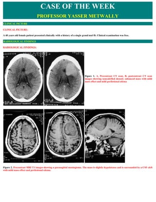
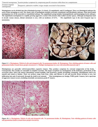
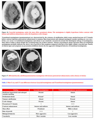
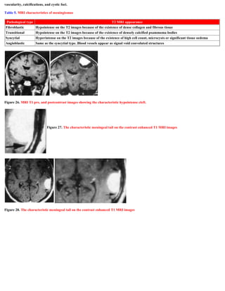
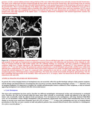
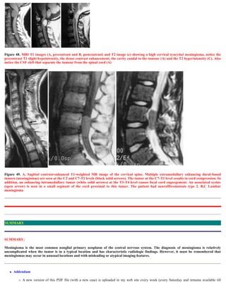
![Figure 22. Dural tail tissue adjacent to meningioma. Lower portion of the photomicrograph (original magnification, x250; hematoxylin-eosin
[H-E] stain) shows normal dura mater that is largely collagen. The upper region shows reactive changes characterized by vascular congestion
and loosening of the connective tissue. Slow flow within these vessels and accumulation of edema in the dura mater allow enhancement to be
visualized on gadolinium-enhanced T1-weighted MR images.
Grossly meningiomas are characterized, by the existence of a vascular rim that surrounds the meningioma and appears signal void on both
T1,T2 MRI images, this finding is consistent with the overall blood supply of meningiomas (the peripheries of meningiomas are supplied by
branches from the anterior or middle cerebral arteries that encircle the tumour and form the characteristic vascular rim). Meningiomas
encase, narrow and parasitize pial and meningeal vessels. Vascular rim is common in syncytial and angioblastic types and much less
commonly seen in transitional meningiomas.
Heterogeneous appearance of meningiomas in T2-weighted pulse sequence can be due to tumor vascularity, calcifications, and cystic foci.
MR imaging has also been found to be superior to CT in evaluating meningiomas for venous sinus invasion or internal carotid artery
encasement. Brain edema is observed in about 50% of meningiomas, with severe edema occurring with syncytial and angioblastic types. 5
Elster et al 5 reported a strong correlation between tumor histology and tumor intensity on T2-weighted images compared with those of the
cortex. Meningiomas are classified into four basic subtypes: fibroblastic, transitional, syncytial, and angioblastic. 4,6 Elster et al 5 have stated
that meningiomas significantly hyperintense to cortex tend to be primarily of syncytial or angioblastic type, whereas meningiomas
hypointense to cortex tend to be primarily of fibrous or transitional type.
Table 4. MRI appearance of the various types of meningiomas
Type Comment
Fibroblastic
meningiomas
Fibroblastic meningiomas are composed of large, narrow spindle cells. The distinct feature is the presence of
abundant reticulum and collagen fibers between individual cells. On MR imaging, fibroblastic meningiomas with
cells embedded in a dense collagenous matrix appear as low signal intensity in Tl-weighted and T2-weighted pulse
sequences.
Transitional
meningiomas
Transitional meningiomas are characterized by whorl formations in which the cells are wrapped together
resembling onion skins. The whorls may degenerate and calcify, becoming psammoma bodies. Marked calcifications
can be seen in this histologic type. MR imaging of transitional meningiomas thus also demonstrates low signal
intensity on Tl- weighted and T2-weighted images, with the calcifications contributing to the low signal intensity.
Syncytial
meningiomas
Syncytial (meningothelial, endotheliomatous) meningiomas contain polygonal cells, poorly defined and arranged in
lobules. Syncytial meningiomas composed of sheets of contiguous cells with sparse interstitium might account for
higher signal intensity in T2-weighted images. Microcystic changes and nuclear vesicles can also contribute to
increased signal intensity.
Angioblastic
meningiomas
Angioblastic meningiomas are highly cellular and vascular tumors with a spongy appearance. Increased signal in
T2-weighted pulse sequence of these tumors is due to high cellularity with increase in water content of tumor.](https://image.slidesharecdn.com/casemenin-1231854575757561-2/85/Case-record-Parasagittal-meningioma-12-320.jpg)
![Friday.)
To download the current version follow the link "http://pdf.yassermetwally.com/case.pdf".
You can also download the current version from my web site at "http://yassermetwally.com".
To download the software version of the publication (crow.exe) follow the link: http://neurology.yassermetwally.com/crow.zip
The case is also presented as a short case in PDF format, to download the short case follow the link:
http://pdf.yassermetwally.com/short.pdf
At the end of each year, all the publications are compiled on a single CD-ROM, please contact the author to know more details.
Screen resolution is better set at 1024*768 pixel screen area for optimum display.
For an archive of the previously reported cases go to www.yassermetwally.net, then under pages in the right panel, scroll down and click
on the text entry "downloadable case records in PDF format"
Also to view a list of the previously published case records follow the following link (http://wordpress.com/tag/case-record/) or click on it
if it appears as a link in your PDF reader
References
1. Burger PC, Scheithauer BW, Vogel FS: Surgical Pathology of the Nervous System and Its Coverings, ed 3. New York, Churchill-
Livingstone, 1991
2. Kepes Jj: Meningiomas: Biology, Pathology and Differential Diagnosis. New York, Masson Publishing, 1982
3. Shapiro JR, Coons SW: Genetics of adult malignant gliomas. BNI Quarterly 14:27- 38, 1998
4. Courville CB: Pathology of the Central Nervous System, ed 3. Mountain View, CA, Pacific, 1950, p 383
5. Elster AD, Challa VR, Gilbert TH, et al: Meningiomas: MR and histopathologic features. Radiology 170:857, 1989
6. Russell DS, Rubinstein Lj: Pathology of Tumors of the Nervous System, ed 4. Baltimore, Williams & Wilkins, 1977, p 48
7. Russel D, Rubenstein L (ed): Pathology of Tumors of the Nervous System. Baltimore, Williams and Wilkins, 1989
8. Atlas SW: Adult supratentorial tumors. Semin Roentgenol 25:130-154,1990
9. Osborne A (ed): Diagnostic Neuroradiology St. Louis, Mosby-Year Book, 1994
10. Zulch K (ed): Brain Tumors: Their Biology and Pathology, ed 3. New York, Spxinger-Verlag, 1986
11. Smith F, Slavik M, McDonald L: Association of breast cancer with meningioma. Cancer 42:1992-1994, 1978
12. Roelvink N, Kamphorst W, Alphen HV: Pregnancy related primary brain and spinal tumors. Arch Neurol 44:209-215,1987
13. Som P, Sacher M, Strenger S, et al: 'Benign" metastasizing meningiomas. AJNR Am j Neuroradiol 8:127-130, 1987
14. Claveria L, Sutton D, Tress B: The radiological diagnosis of meningiomas: The impact of EMI scanning. Br j Radiol 50:15-22, 1977
15. Louis DN, Scheithauer BW, Budka H, et al: Meningiomas. In Kleihues P, Cavenee WC (eds): Pathology and Genetics-Tumours of the
Nervous System. Lyon, World Health Organization and International Agency for Research on Cancer, 2000, p 176
16. Masaryk Tj: Neoplastic diseases of the spine. Radiol Clin North Am 29:829,1991
17. Chamberlain MC, Sandy AD, Press GA: Spinal cord tumors: gadolinium-DTPA-enhanced MR imaging. Neuroradiology 1991; 33(6):
469-74.
18. Derenda M, Bayassi S: [Thoracic spine meningioma mimicking intramedullary tumor]. Neurol Neurochir Pol 2000 Mar-Apr; 34(2): 357-
65.
19. Dillon WP, Norman D, Newton TH, et al: Intradural spinal cord lesions: Gd-DTPA-enhanced MR imaging. Radiology 1989 Jan; 170(1 Pt
1): 229-37.
20. Doita M, Harada T, Nishida K, et al: Recurrent calcified spinal meningioma detected by plain radiograph. Spine 2001 Jun 1; 26(11):
E249-52.
21. Egelhoff JC, Bates DJ, Ross JS, et al: Spinal MR findings in neurofibromatosis types 1 and 2. AJNR Am J Neuroradiol 1992 Jul-Aug; 13
(4): 1071-7.
REFERENCES](https://image.slidesharecdn.com/casemenin-1231854575757561-2/85/Case-record-Parasagittal-meningioma-27-320.jpg)
![22. Gamache FW Jr, Wang JC, Deck M, Heise C: Unusual appearance of an en plaque meningioma of the cervical spinal canal. A case report
and literature review. Spine 2001 Mar 1; 26(5): E87-9.
23. Levy WJ Jr, Bay J, Dohn D: Spinal cord meningioma. J Neurosurg 1982 Dec; 57(6): 804-12.
24. Li MH, Holtas S, Larsson EM: MR imaging of intradural extramedullary tumors. Acta Radiol 1992 May; 33(3): 207-12.
25. Masaryk TJ: Neoplastic disease of the spine. Radiol Clin North Am 1991 Jul; 29(4): 829-45.
26. Matsumoto S, Hasuo K, Uchino A, et al: MRI of intradural-extramedullary spinal neurinomas and meningiomas. Clin Imaging 1993 Jan-
Mar; 17(1): 46-52.
27. McCormick PC, Post KD, Stein BM: Intradural extramedullary tumors in adults. Neurosurg Clin N Am 1990 Jul; 1(3): 591-608.
28. Onofrio BM: Intradural extramedullary spinal cord tumors. Clin Neurosurg 1978; 25: 540-55.
29. Schroth G, Thron A, Guhl L, et al: Magnetic resonance imaging of spinal meningiomas and neurinomas. Improvement of imaging by
paramagnetic contrast enhancement. J Neurosurg 1987 May; 66(5): 695-700.
30. Solero CL, Fornari M, Giombini S, et al: Spinal meningiomas: review of 174 operated cases. Neurosurgery 1989 Aug; 25(2): 153-60.
31. Souweidane MM, Benjamin V: Spinal cord meningiomas. Neurosurg Clin N Am 1994 Apr; 5(2): 283-91.
32. Weck M, Pause M, Pinzer T: [Spinal meningioma as differential diagnosis of diabetic polyneuropathy]. Dtsch Med Wochenschr 2001
May 18; 126(20): 590-2.
33. Weil SM, Gewirtz RJ, Tew JM Jr: Concurrent intradural and extradural meningiomas of the cervical spine. Neurosurgery 1990 Oct; 27
(4): 629-31.
34. Zee CS, Henderson R, Huprich J: Imaging and relevant anatomy. In: Banzel E, Stillerman C, eds. The Thoracic Spine. Quality Medical
Publishing; 1999: 80-122.
35 . Russell DS, Rubinstein U. Pathology of tumors of the nervous system. 5th ed. Baltimore: Williams & Wilkins, 1989; 449-483.
36. Wood MW, White R, KernohanJ. One hundred meningiomas found incidentally at necropsy. J Neunopathol Exp Neurol 1957; 16:337-
340.
37. Russell EJ, George AE, Knicheffll, Budzilovich G. Atypical computed tomographic features of intracranial meningioma: radiological-
pathological correlation in a series of 131 consecutive cases. Radiology 1980; 135:673- 682. Rohninger M, Sutherland GR, Louw DF, Sima
AAF. Incidence and clinicopathological features of meningioma. J Neurosurg 1989; 71: 665-672.
38. Bradac GB, Ferszt R, Kendall BE. Cranial meningiomas. Berlin: Springer-Verlag, 1990; 1-128.
39. Zimmerman RD, Fleming CA, Saint-Louis LA, Lee BCP, ManningJJ, Deck MDF. Magnetic resonance imaging of meningiomas. AJNR
1985; 6: 149-157.
40. New PFJ, Anonow 5, HesselinkJR. National Cancer Institute study: evaluation of computed tomography in the diagnosis of intra- cranial
neoplasms. IV. Meningiomas. Radiology 1980; 136:665-675.
41. Kendall B, Symon L. Investigation of patients presenting with cerebellopontine angle syndromes. Neuroradiology 1977; 13:65-84.
42. Mikhael MA, Ciric IS, WoIifAP. Differentiation of cerebellopontine angle neuromas and meningiomas with MR imaging. J Comput
Assist Tomog. 1985; 9:852-856.
43. Wu E, Tang Y, Zhang Y, Bai R. CT in diagnosis of acoustic neuromas. AJNR 1986; 7:645- 650.
44 . Lusin JO, Nakagawa H. Multiple meningiomas evaluated by computed tomography. Neurosurgery 198i; 9:137-141.
45. Bydder GM, Kingsley DPE, BrownJ, Niendorf HP, Young IR. MR imaging of meningiomas including studies with and without
gadolinium- DTPA. J Comput Assist Tomogr 1985; 9:690-697.
46. Mani RL, Hedgcock MW, Mass SI, Gilmor RI, Enzmann DR, Eisenberg RI. Radiographic diagnosis of meningiomas of the lateral
ventriculo: review of 22 cases. J Neurosurg 1978; 49:249-255.
47. JelinekJ, Smirniotopoulos JG, Panisi JE, Kanzen M. Lateral ventricular neoplasms of the brain: differential diagnosis based on clinical
CT and MR findings. AJNR 1990; 11:567-574.
48. Geoffray A, Lee YY, Jing BS, Wallace S. Extracranial meningiomas of the head and neck. AJNR 1984; 5:599-604.](https://image.slidesharecdn.com/casemenin-1231854575757561-2/85/Case-record-Parasagittal-meningioma-28-320.jpg)

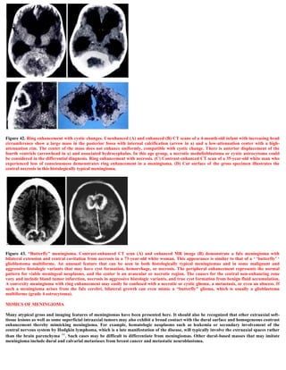
A 40-year-old female presented with a single grand mal seizure. MRI images showed a parasagittal meningioma that was hypointense on T1 images, surrounded by CSF, and causing mild mass effect and edema. Based on the hyperintensity on T2 images and lack of calcification, the meningioma was diagnosed as a syncytial subtype. Meningiomas are common, often located in the parasagittal region, and can cause symptoms by compressing brain structures. Histological subtypes include fibroblastic, transitional, and syncytial, which were determined based on imaging and pathology in this case.


