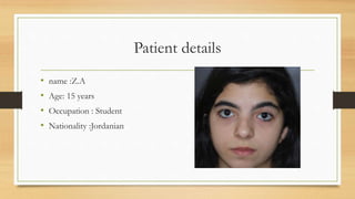
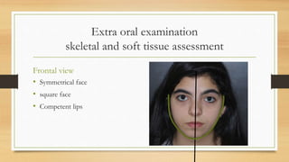
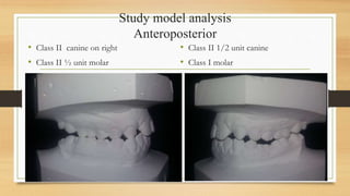
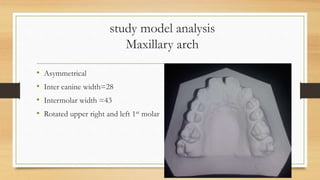
Case presentation
Done by : Dr.Raed Al-Abbadi
3rd year orthodontic resident
Supervised by :
Dr.Ahmad Al-Tarawneh
Dr.Raed Al-Rbata
Dr.Nancy Sarayrah
Patient details
• name :Z.A
• Age: 15 years
• Occupation : Student
• Nationality :Jordanian
Chief complaint
• My teeth in upper jaw are spaced
• Something wrong with bite on right side of my mouth
• Medical history : denied any medical history
• Dental history : routine dental visits
• Trauma or habits : denied any .
• TMJ findings : nothing abnormal
• Displacement : yes
Extra oral examination
skeletal and soft tissue assessment
Frontal view
• Symmetrical face
• square face
• Competent lips
Extra oral examination
skeletal and soft tissue assessment
Frontal smile
• Thin lips
• Normal level occlusal plane (no canting).
• upper midline to facial mildline : diastema ??
Middle of it >>Coincident.
• lower midline to upper midline:
Lower midline shifted to right
• Increased buccal corridor
• Increased incisal show
Extra oral examination
skeletal and soft tissue assessment
Profile view
• Slightly convex profile
• normal nasolabial angle
• Average frontonasal angle
• Obtuse labiomental angle
• Average throat length
• Average throat angle
• Average lower facial height
• Average FMPA
Intra oral examination
Frontal
• Fair oral hygiene
• Class I incisal classification
• Reduced overjet
• Missing upper right and left lateral incisors
• Midline diastema
• Fluorosis on lower right lateral and canine
• Cross bite on right side ( seems on both but
lower tilited lingually )
• Reduced overbite
• Lower midline shift to right by 1-2 mm
• Normal oral mucosa.
Right side
• Class II ½ unit molar relationship
• Canine relationship full unit class II
• Caries on upper right central
• Cross bite on right side ( 3,4,5,6)
• Amalgam filling on lower right 1st
molar
Left side
• Class I molar relationship
• Canine relationship class II ¼ unit
• Caries on upper left central
• Retained lower left primary 2nd
molar
• Calculus on upper left 1st molar
Upper arch
• U shaped arch
• Spacing
• Missing laterals on both sides
• Rotated 1st molars
• Teeth present 1,3,4,5,6,7
Lower arch
• U shaped arch
• Normal tongue size
• Carious lower right 2nd molar
• Rotated lower right 2nd premolar
• Retained lower right primary 2nd molar
• Lower right 1st molars filled with
amalgam
• Teeth present 1-7
Study model analysis
Anteroposterior
• Class II canine on right
• Class II ½ unit molar
• Class II 1/2 unit canine
• Class I molar
OJ = 1mm on upper right central , 2mm on
upper left
Vertical
• Reduced overbite
Transverse
• Cross bite on right side.
• Lower midline shifted to right by
2mm .
Curve of spee
Left sideRight side
flat
4 mm depth
study model analysis
Maxillary arch
• Asymmetrical
• Inter canine width=28
• Intermolar width =43
• Rotated upper right and left 1st molar
study model analysis
mandibular arch
• Asymmetrical
• Inter canine width=30
• Intermolar width =43
• Rotated lower right 2nd premolar
• Retained lower left primary 2nd molar
Teeth widths
5 4 3 2 1 1 2 3 4 5
6.5 7 7.5 **6.9 9.5 9.5 **6.9 7.5 6.5 6.5
7 7 6.5 6 6 6 6 6.5 6.5 8
5 4 3 2 1 1 2 3 4 E
If open space applied to replace missing laterals
Mesio distal needed according to anterior Bolton ratio >>>
37/0.772 = 2x+34 >>>2x =47.9-34=13.9
X=13.9/2=6.95mm
Space analysis
• Upper: Available space =70 ,,, Required =74.5 ( need 4.5 mm )
• Lower : Available space =67.5,,, Required = 65.5 ( there is extra 2mm )
Royal college of London analysis
Lower arch Upper arch
Crowding / spacing +1( estimated after extraction of
lower left E )
+1 de-rototaion of lower right 5
-4.5
Leveling curve of spee -1.5(4mm) on left side 0
Arch width change 0 +5mm ( .5mm for every 1mm
increase in width )
10 mm expansion>>+5
Insicior A/p change 0 0
Angulation change 0 0
Inclination change 0 0
Result +0.5 +0.5
IOTN-dental component =great 4.h
IOTN aesthetic component =5
Opt
• amalgam filled lower right 1st molar
• Endo treated lower left 1st molar , flled with amalgam
• Wisdom teeth present in lower arch
• Missing upper laterals + lower left 2nd premolar
• Retained lower left primary 2nd molar , resorbed roots
• Mesial tilted lower left 1st molar
Lateral cephalogram
Normal Value
SNA 84 81 +- 3
SNB 81 78 +- 3
ANB 3 2-4
S-N/ANS-PNS 3.8 8 +- 3
Corrected ANB 3
MMPA 26.9 27 +- 3
UAFH 51.7
LAFH 68.2
LAFH % 56.8% 55 +- 2
U1/Mx 111 108 +- 6
L1/Mn 92 93 +- 6
IAA 128 133 +- 10
Wits Appraisal 2 0 + 1.77 mm
Case summary
• ZA is 15 years old medically fit female .She has attended the orthodontic
clinic with esthetic concern related to her upper teeth , her chief complaint
is spaced upper teeth and smoothing wronge on right side when biting , she
has fair oral hygiene and localized calculus related to upper left 6, she has
class I malocclusion on skeletal base class I with class II ½ unit molar on
right , class II full unit canine on right , class II 1/2 unit on left side , reduced
OJ , reduced OB , cross bite on right side ,deep curve of spee on right side ,
lower midline shifted to right by 2mm , midline diastema , spacing in upper
arch , complicated by missing upper right and left laterals and missing lower
left 2nd premolar, rotated lower right 2nd premolar , retained lower left
primary 2nd premolar .
Problem list• Oral health :
• Calculus on upper left molar
• Skeletal
• Constricted maxilla
• Soft tissue
• Slightly convex profile
• Obtuse labiomental angle
• Dental
• Increased buccal corridor
• Increased incisal show
• Reduced overjet
• Missing upper right and left lateral incisors
• Missing lower left 2nd premolar
• Carious upper right and left centrals
• Midline diastema
• Fluorosis on lower right lateral and canine
• Cross bite on right side ( seems on both but lower tilited lingually )
• Reduced overbite
• Lower midline shift to right by 1-2 mm
Class II ½ unit molar relationship & Canine relationship full unit class II on right side
Canine relationship class II ¼ unit on left side
Rotated upper 1st molars
Retained lower right primary 2nd molar
4 mm depth of COS on left side
Spacing in upper arch
Treatment aims
• Stabilization of periodontal condition.
• Treatment of carious teeth .
• Attain a normal width of maxilla
• Level and align arches.
• Correct centerlines.
• Correct crossbite on right side
• Normalize OJ and OB.
• Achieve Class I canine and molar relationship
• Open space for upper lateral incisors and maintain space of lower left 2nd premolar.
• Coordinate dental arches with good buccal interdigitation.
• Retain the corrected results.
Treatment plan
1. OHI
2. Treatment of carious teeth .
3. Rapid maxillary expansion.
4. Modified TPA.
5. upper and lower fixed appliance ( Roth 0.022’’)
Open space for upper laterals for future prosthesis
Consider disking lower left E
6. Retention : upper permanent retention and Hawley retainer with acrylic teeth in place of
laterals , lower Hawley retainer .
justification
• Rapid maxillary expansion with hyrax is good for her case because we want a skeletal effect more
than dental effect .
• Expand until we overcorrect the width ( palatal cusps of upper on lingual inclines of buccal
cusps of lower ) in anticipation of some relapse.( lee 1999)
• Modified TPA to derotate upper 1st molars .
• Fixed Appliance is needed because of
• Bodily teeth movement is required.
• Alignment of teeth.
• Close the extraction spaces.
• Preserve / correct teeth torque.
• Roth prescription slot 0.022 because teeth are in normal inclination and average torque is needed.
• Leave lower left primary 2nd molar as natural space maintainer for lower left 2nd premolar until
restore it with a prosthesis .
• Fixed retainers are indicated for long-term retention of the labial segments, particularly for generalized
spacing and for retention of a midline diastema (Proffit WR, Fields HW, Sarver DM. Contemporary
Orthodontics. (4th ed.), Chapter 17, St Louis, Mosby Elsevier, 2007)
The End

Case presentation

  • 1.
    Case presentation Done by: Dr.Raed Al-Abbadi 3rd year orthodontic resident Supervised by : Dr.Ahmad Al-Tarawneh Dr.Raed Al-Rbata Dr.Nancy Sarayrah
  • 2.
    Patient details • name:Z.A • Age: 15 years • Occupation : Student • Nationality :Jordanian
  • 3.
    Chief complaint • Myteeth in upper jaw are spaced • Something wrong with bite on right side of my mouth
  • 4.
    • Medical history: denied any medical history • Dental history : routine dental visits • Trauma or habits : denied any . • TMJ findings : nothing abnormal • Displacement : yes
  • 5.
    Extra oral examination skeletaland soft tissue assessment Frontal view • Symmetrical face • square face • Competent lips
  • 6.
    Extra oral examination skeletaland soft tissue assessment Frontal smile • Thin lips • Normal level occlusal plane (no canting). • upper midline to facial mildline : diastema ?? Middle of it >>Coincident. • lower midline to upper midline: Lower midline shifted to right • Increased buccal corridor • Increased incisal show
  • 7.
    Extra oral examination skeletaland soft tissue assessment Profile view • Slightly convex profile • normal nasolabial angle • Average frontonasal angle • Obtuse labiomental angle • Average throat length • Average throat angle • Average lower facial height • Average FMPA
  • 8.
    Intra oral examination Frontal •Fair oral hygiene • Class I incisal classification • Reduced overjet • Missing upper right and left lateral incisors • Midline diastema • Fluorosis on lower right lateral and canine • Cross bite on right side ( seems on both but lower tilited lingually ) • Reduced overbite • Lower midline shift to right by 1-2 mm • Normal oral mucosa.
  • 9.
    Right side • ClassII ½ unit molar relationship • Canine relationship full unit class II • Caries on upper right central • Cross bite on right side ( 3,4,5,6) • Amalgam filling on lower right 1st molar
  • 10.
    Left side • ClassI molar relationship • Canine relationship class II ¼ unit • Caries on upper left central • Retained lower left primary 2nd molar • Calculus on upper left 1st molar
  • 11.
    Upper arch • Ushaped arch • Spacing • Missing laterals on both sides • Rotated 1st molars • Teeth present 1,3,4,5,6,7
  • 12.
    Lower arch • Ushaped arch • Normal tongue size • Carious lower right 2nd molar • Rotated lower right 2nd premolar • Retained lower right primary 2nd molar • Lower right 1st molars filled with amalgam • Teeth present 1-7
  • 13.
    Study model analysis Anteroposterior •Class II canine on right • Class II ½ unit molar • Class II 1/2 unit canine • Class I molar
  • 14.
    OJ = 1mmon upper right central , 2mm on upper left
  • 15.
  • 16.
    Transverse • Cross biteon right side. • Lower midline shifted to right by 2mm .
  • 17.
    Curve of spee LeftsideRight side flat 4 mm depth
  • 18.
    study model analysis Maxillaryarch • Asymmetrical • Inter canine width=28 • Intermolar width =43 • Rotated upper right and left 1st molar
  • 19.
    study model analysis mandibulararch • Asymmetrical • Inter canine width=30 • Intermolar width =43 • Rotated lower right 2nd premolar • Retained lower left primary 2nd molar
  • 20.
    Teeth widths 5 43 2 1 1 2 3 4 5 6.5 7 7.5 **6.9 9.5 9.5 **6.9 7.5 6.5 6.5 7 7 6.5 6 6 6 6 6.5 6.5 8 5 4 3 2 1 1 2 3 4 E If open space applied to replace missing laterals Mesio distal needed according to anterior Bolton ratio >>> 37/0.772 = 2x+34 >>>2x =47.9-34=13.9 X=13.9/2=6.95mm
  • 21.
    Space analysis • Upper:Available space =70 ,,, Required =74.5 ( need 4.5 mm ) • Lower : Available space =67.5,,, Required = 65.5 ( there is extra 2mm )
  • 22.
    Royal college ofLondon analysis Lower arch Upper arch Crowding / spacing +1( estimated after extraction of lower left E ) +1 de-rototaion of lower right 5 -4.5 Leveling curve of spee -1.5(4mm) on left side 0 Arch width change 0 +5mm ( .5mm for every 1mm increase in width ) 10 mm expansion>>+5 Insicior A/p change 0 0 Angulation change 0 0 Inclination change 0 0 Result +0.5 +0.5
  • 23.
  • 24.
  • 25.
    Opt • amalgam filledlower right 1st molar • Endo treated lower left 1st molar , flled with amalgam • Wisdom teeth present in lower arch • Missing upper laterals + lower left 2nd premolar • Retained lower left primary 2nd molar , resorbed roots • Mesial tilted lower left 1st molar
  • 26.
    Lateral cephalogram Normal Value SNA84 81 +- 3 SNB 81 78 +- 3 ANB 3 2-4 S-N/ANS-PNS 3.8 8 +- 3 Corrected ANB 3 MMPA 26.9 27 +- 3 UAFH 51.7 LAFH 68.2 LAFH % 56.8% 55 +- 2 U1/Mx 111 108 +- 6 L1/Mn 92 93 +- 6 IAA 128 133 +- 10 Wits Appraisal 2 0 + 1.77 mm
  • 27.
    Case summary • ZAis 15 years old medically fit female .She has attended the orthodontic clinic with esthetic concern related to her upper teeth , her chief complaint is spaced upper teeth and smoothing wronge on right side when biting , she has fair oral hygiene and localized calculus related to upper left 6, she has class I malocclusion on skeletal base class I with class II ½ unit molar on right , class II full unit canine on right , class II 1/2 unit on left side , reduced OJ , reduced OB , cross bite on right side ,deep curve of spee on right side , lower midline shifted to right by 2mm , midline diastema , spacing in upper arch , complicated by missing upper right and left laterals and missing lower left 2nd premolar, rotated lower right 2nd premolar , retained lower left primary 2nd premolar .
  • 28.
    Problem list• Oralhealth : • Calculus on upper left molar • Skeletal • Constricted maxilla • Soft tissue • Slightly convex profile • Obtuse labiomental angle • Dental • Increased buccal corridor • Increased incisal show • Reduced overjet • Missing upper right and left lateral incisors • Missing lower left 2nd premolar • Carious upper right and left centrals • Midline diastema • Fluorosis on lower right lateral and canine • Cross bite on right side ( seems on both but lower tilited lingually ) • Reduced overbite • Lower midline shift to right by 1-2 mm Class II ½ unit molar relationship & Canine relationship full unit class II on right side Canine relationship class II ¼ unit on left side Rotated upper 1st molars Retained lower right primary 2nd molar 4 mm depth of COS on left side Spacing in upper arch
  • 29.
    Treatment aims • Stabilizationof periodontal condition. • Treatment of carious teeth . • Attain a normal width of maxilla • Level and align arches. • Correct centerlines. • Correct crossbite on right side • Normalize OJ and OB. • Achieve Class I canine and molar relationship • Open space for upper lateral incisors and maintain space of lower left 2nd premolar. • Coordinate dental arches with good buccal interdigitation. • Retain the corrected results.
  • 30.
    Treatment plan 1. OHI 2.Treatment of carious teeth . 3. Rapid maxillary expansion. 4. Modified TPA. 5. upper and lower fixed appliance ( Roth 0.022’’) Open space for upper laterals for future prosthesis Consider disking lower left E 6. Retention : upper permanent retention and Hawley retainer with acrylic teeth in place of laterals , lower Hawley retainer .
  • 31.
    justification • Rapid maxillaryexpansion with hyrax is good for her case because we want a skeletal effect more than dental effect . • Expand until we overcorrect the width ( palatal cusps of upper on lingual inclines of buccal cusps of lower ) in anticipation of some relapse.( lee 1999) • Modified TPA to derotate upper 1st molars . • Fixed Appliance is needed because of • Bodily teeth movement is required. • Alignment of teeth. • Close the extraction spaces. • Preserve / correct teeth torque. • Roth prescription slot 0.022 because teeth are in normal inclination and average torque is needed. • Leave lower left primary 2nd molar as natural space maintainer for lower left 2nd premolar until restore it with a prosthesis . • Fixed retainers are indicated for long-term retention of the labial segments, particularly for generalized spacing and for retention of a midline diastema (Proffit WR, Fields HW, Sarver DM. Contemporary Orthodontics. (4th ed.), Chapter 17, St Louis, Mosby Elsevier, 2007)
  • 32.