Downloaded 122 times
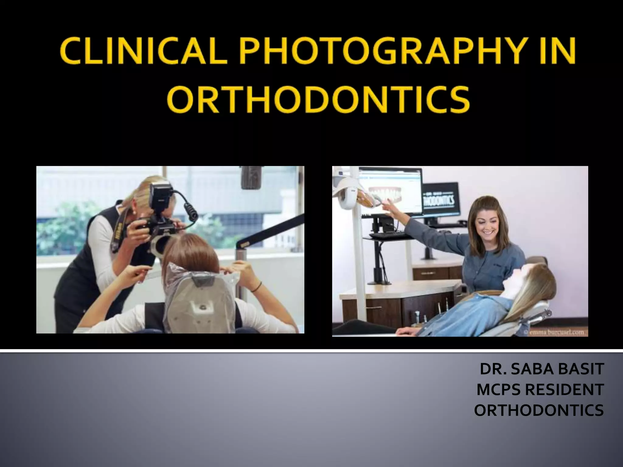


This document discusses photography in orthodontics, including intraoral and extraoral photography. It outlines the importance of photography for treatment planning, case discussions, aiding treatment, and marketing. Recent developments in digital cameras have made photography more convenient due to ease of use, ability to edit photos, and cost effectiveness. The document provides guidelines for various intraoral and extraoral photo techniques, including the use of retractors, mirrors, and flashes. It recommends a Dental Eye III camera system and describes proper patient positioning and angles for different standard orthodontic photos.


