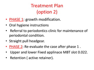
1. Case Presentation
Done By: Dr. Jumana Al-Abbadi
3rd year orthodontic resident
Supervised By:
Dr. Ahmad Al-Tarawneh
Dr. Jumana Tbeishat
Dr. Raed AlRabta
Dr. Bashar Al-Momani
Dr. Anwar Rahamneh
13. Smile esthatic
- On full smile the whole length of maxillary incisors show.
- Gummy smile.
- Upper incisors edges do not run parallel to the lower lip
(improper smile arc ).
- Upper midline is coincident with the facial midline.
- Lower midline is coincident with the facial midline .
- Average Buccal corridor.
14. Soft tissue Examination:-
- The lower third is slightly shorter than the middle third
.
-Upper lip in the upper third of the lower part .
-Lower lip in the lower two thirds of the lower part.
13
23
15. Soft tissue Examination:-
- Nasal tip deviated
to the left side.
- Interpupillary
distance is not equal to
mouth width.
- The width of the nose is slightly
wider than the central fifth.
- Potentially Competent lips.
19. Lower arch
- U-shaped arch and symmetric.
- Mild crowding in the lower labial segment.
- Partially erupted LT canine.
- Proclined lower incisors.
- Carious LR E.
- flat curve of spee.
20. Upper arch:-
- U-shaped arch and symmetric.
- Spaced upper arch.
- rotated UL centaral &
lateral..
- rotated UR 5.
- Flat curve of spee.
21. Teeth in occlusion
- Incisor relationship: classII div 1.
- Overjet: 8 mm
- Overbite: on both sides : increased, complete to the tooth,
atraumatic.
- Upper & lower midlines are coincident with each other & with
the facial midline.
22. Buccal segment in occlusion
-Molars:-
Rt: full unit class II
Lt: ½ unit class II
-Canines:
Rt & Lt: can’t be determined.
-UR 4 overerupted.
-Displacements: None detected.
23. Study model analysis in occlusion
Anterioposterior:
OJ: 8 mm
Right Left
-Molar: full unit Class II
-Canine: can’t be determined.
-Molar: ½ unit Class II
-Canine: can’t be determined.
30. Radiographical examination
R L
Orthopantomogram “OPT” Analysis
- No apperant pathology.
- All teeth are present but 7 3 3 5 7 still unerupted.
7 5 4 3 7
- The tooth buds of all 8 ‘s are present.
38. Diagnostic Summary :-
A.M is 12 years old male patient medically fit, C/O
( my upper teeth are sticking out “pointing to the
upper centrals”),
on examination he has class II div 1 incisor on
class II skeletal base, with increased
O.J(8mm),deep O.B, average LFH , convex profile,
complicated by: proclined upper incisors& lower
incisors, rotated (UL1, UL2,UR5), overerupted
UR4.
39. Problem List:-
Pathological Problem:-
1- poor oral hygiene.
2- carious LR E.
Patient’s concern:-
A.M. is complaining that “my upper teeth are sticking out “pointing to upper
incisors”
Skeletal Problems:-
1- Class II skeletal base.
Soft tissue problems :-
1- improper smile arc.
2- upper incisors are not close neither touching the lower lip.
3- obtuse frontonasal angle.
4- convex profile.
5- gummy smile.
40. Dental problems :
1- mild crowding in the lower labial segment.
2-rotated UL1, UL2, UR5.
3- proclined lower incisors.
4- proclined upper incisors.
5- overerupted UR4.
6- increased O.J
7- increased O.B
8- class II div 1 incisor relation.
9- full unit class II molar on the right side.
10- ½ unit class II molar on the left side.
41. Treatment Aims:-
• Improve oral hygiene.
• Stabilization of periodontal health.
• Restoraton of the carious LR E.
• Correct pt’s chief complaint ( protrusion ).
• Correct skeletal class II relationship.
• Correct slightly decreased LFH.
• Improve the pt’s smile.
• Improve facial profile.
• Achieve normal Overjet.(2-3 mm)
• Achieve normal Overbite.
• Achieve class 1 incisor relationship.
• Achieve class 1 molar relationship on both sides.
• Correct rotated teeth(UL1, UL2, UR5) .
• Correct the incisor inclination.
• Level and align the arches.
• Finishing and detailing of the occlusion.
• maintain the corrected results.
42. Treatment Plan
(option 1)
• Phase 1: growth modification (functional).
• Oral hygiene instructions
• Referral to periodontics clinic for maintenance of
periodontal condition.
• Medium Opening Activator.
• Phase 2: Re-evaluate the case after phase 1 .
• Upper and lower Fixed applinace MBT slot 0.022.
• Retention ( active retainer).
43. Retention Protocol
• After phase 1 : : inclined anterior bite plane (8
mm thickness , 70 degrees inclination.)
• After phase 2 :
• -Short term : upper & lower Hawley retainer
with inclined ABP,( full time wear for 6
months, night time wear for another 6
months).
• Long term: upper & lower bonded retainer from 3-3.
44. Treatment Plan
(option 2)
• PHASE 1: growth modification.
• Oral hygiene instructions
• Referral to periodontics clinic for maintenance of
periodontal condition.
• Straight pull headgear.
• PHASE 2: Re-evaluate the case after phase 1 .
• Upper and lower Fixed applinace MBT slot 0.022.
• Retention ( active retainer).
45. Retention Protocol
• After phase 1 : headgear night time wear
(250gm per side for 12 hours).
• After phase 2 :
• -Short term : upper & lower Hawley retainer
with inclined ABP, (full time wear for 6
months, night time wear for another 6
months).
• Long term: upper & lower bonded retainer.
46. MOA design
• • Lower incisal capping 3-3
• • Anterior palatal wire 3-3 in 1.0 mm S.S wire
• Adams crib 64/46 0.7 mm S.S wire
• • Acrylic connecting “sturts”/ vertical supports
• coffin spring 1.25 mm SS wire.
47. Justification
• Growth modification > functional appliance:
• growing patient.
• class II skeletal base.
• proclined upper incisors.
• Average MMPA with increased overbite.
• minimal crowded arches.
• Class II molar relationships.
• Increased overjet.
48. Justification
MOA :
1. Moderately displaces the mandible
2. Moderate bite opening
3. Robust.
4. Easy to repair
5. Can incorporate expansion screw
6. Capping lower incisors to prevent proclination
49. Justification
• Inclined anterior bite plane:
• to retain functional appliance results during
transition to fixed appliance .
• allow settling of the occlusion.
50. • Upper and lower Fixed applinace :
• We need 3D teeth movement.
• Alignment of rotated teeth.
• Closure of spaces.
• MBT prescription is preferred:
• It correct LLS and ULS inclination.
• Increased lingual crown torque of LLS which helps to
counteract the side effect of functional appliance.
• Zero degree tip of upper molars:
- prevent distal tipping of the root of the first molar against
the second molar which might cause root resorption when HG
is used.
- keep the molars upright while the future growth might
cause further mesial tip.
51. Treatment details
• Full records
• Take impression for MOA and functional bite
with 2mm separation at incisors (deep bite)
with the patient biting edge to edge.
• Delivery of the appliance and instructions.
• After finishing the functional phase deliver the
inclined anterior bite plane to retain
functional appliance results during transition
to fixed appliance.
52. • bonding of the fixed appliance MBT
prescription on the upper & the lower arch.
• Alignment with superelastic 0.016”niti wire.
The normal sequence of wires until reaching
rigid S.S 19*25 wires.
• TMA wire for finishing and settling of the
occlusion .
53. • Debonding and impression taking for upper
and lower Hawley retainer with inclined ABP.
• The delivary of upper and lower hawely
retainer.
54. Justification
• Growth modification (straight pull headgear):
• Growing patient.
• Class II skeletal.
• Normal MMPA.
• To restrain the maxillary growth ( prognathic
maxilla).
• Increased O.J (corrected by allowing autorotation
of mandible).
• Increased O.B ( corrected by allowing
autorotation of mandible).
55. Treatment details
• Full records.
• Separators around U6’s.
• Band selection & cementation.
• Fitting of facebow and headgear with
instructions.
• ( force 500-900 gm/ side for 12 - 14 hours).
56. • bonding of the fixed appliance MBT
prescription on the upper & the lower arch.
• Alignment with superelastic 0.016”niti wire.
The normal sequence of wires until reaching
rigid S.S 19*25 wires.
• TMA wire for finishing and settling of the
occlusion .