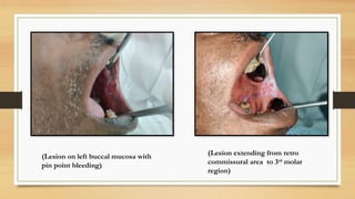
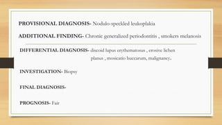
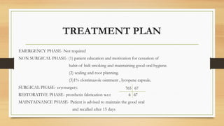
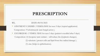
LEUKOPLAKIA
PRESENTED BY
ASHUTOSH YADAV
BDS 2017
UNDER THE GUIDANCE OF
DR.SUMIT GOEL
PROFESSOR
DEPARTMENT OF ORAL MEDICINE AND RADIOLOGY
ACKNOWLEDGEMENT
I WOULD LIKE TO EXPRESSMY SPECIAL THANKS AND SINCERE GRATITUDE TO
THE DEPARTMENT OF ORAL MEDICINE AND RADIOLOGY
FOR ALLOWING THIS PROJECT TO BE COMPLETED
FIRSTLY, I WOULD LIKE TO THANK DR.NAGARAJU KAMARTHI SIR,HEAD OF DEPARTMENT, ORAL
MEDICINE AND RADIOLOGY,FOR HIS PERCEPTIVENESS ANS UNDERSTANDING THROUGH OUT THE
COURSE OF THIS PROJECT.
I WOULD LIKE TO EXPRESS MY GRATITUDE AND SINCERE THANKS TO MY GUIDE, DR.SUMIT GOEL SIR,
FOR THE OPPOURTUNITY TO EXAMINE THE PATIENTS AND LEARN VISUAL CONNECT AND SUPPORT,
ENCOURAGEMENT AND MOTIVATIONAL IN ALL PHASES PF COMPLETION OF THIS CASE.
I WOULD ALSO LIKE TO THANK DR.SANGEETA MALIK MA’AM,DR.SWATI GUPTA MA’AM,DR.ABHINAV
SHARMA SIR, DR.KHUSHBOO BHALLA MA’AM,DR.ISHA MAHESHWARI MA’AM FOR THEIR NEVER ENDING
SUPPORT AND GUIDANCE.
I WOULD LIKE TO EXTEND MY THANK TO THE PATIENT FOR HELPING AND COOPERATING THROUGH
OUT THE MAKING OF THIS CASE.
CERTIFICATE BY
HEAD OF DEPARTMENT
ORAL MEDICINE AN RADIOLOGY
THIS IS TO CERTIFY THAT THE SPECIAL CASE ENTITLED
“LEUKOPLAKIA”
IS A GENUINE WORK OF
ASHUTOSH YADAV, BATCH 2017,
HE HAS BEEN AWARDED _ GRADE
DR.NAGARAJU K.
PROFESSOR AND HEAD OF DEPARTMENT
ORAL MEDICINE AND RADIOLOGY
CERTIFICATE BY THE
GUIDE
I HERE BY CERTIFY THAT THE SPECIAL CASE ENTITLED
“LEUKOPLAKIA”
HAS BEEN SATISFACTORILY COMPLETED BY
ASHUTOSH YADAV,BATCH 2017
DR.SUMIT GOEL
PROFESSOR
DEPARTMENT OF ORAL MEDICINE AND RADIOLOGY
CASE REPORT
PATIENT INFORMATION
NAME - MR.SATYAPRAKASH SHARMA
AGE/SEX - 51/MALE
OPD- 528642
ADDRESS-MAWANA MEERUT
OCCUPATION- FARMER
INCOME- 75,000/ANNUM
CHIEF COMPLAINT
PATIENT COMPLAINS OF CUT IN LEFT CHEECK REGION AND
BURNING SENSATION IN THAT AREA WHILE HAVING FOOD
SINCE 5 YEARS.
HE ALSO COMPLAINS OF REDUCED TASTE SENSATION.
HISTORY OF PRESENT ILLNESS
PATIENT WAS APPARENTLY ASYMPTOMATIC 5 YEARS BACK WHEN HE STARTED NOTICING A CUT
ON HIS LEFT BUCCAL MUCOSA ,THERE WAS NO PAIN IN THAT REGION BUT THERE WERE
BURNING SENSATION ON THE SAME SITE WHENEVER PATIENT EATS AND SUBSIDES ON ITS OWN
AFTER SOMETIME.
HE VISITED TO LOCAL DOCTOR IN THE TOWN FOR SEEKING THE TREATMENT OF THE SAME
WHERE HE WAS TOLD THAT THE CAUSE OF THE LESION IS SOME GIT DISORDER (UNSPECIFIED)
AND GOT MEDICATED WITH SOME INJECTION(UNSPECIFIED) BUT PATIENT WAS NOT SATISFIED
BY THE TREATMENT AS THE LESION REOCCURED ONCE THE INJECTIONS ARE STOPED.
THEN HE VISITED TO MULCHAND SHARBATI DEVI CHARITABLE E.N.T AND DENTAL HOSPITAL
MEEUT CITY FOR THE TREATMENT OF THE SAME WHERE IS WAS SUSPECTED WITH THE
PREMALIGNANT LESION AND REFFERD TO DEPARTMENT OF ORAL MEDICINE AND RADIOLOGY
SUBHARTI DENTAL COLLEGE AND HOSPITAL FOR THE CONFIRMATION AND TREATMENT.
PERSONAL HISTORY
MEDICAL HISTORY- Patient gave on significant medical history.
DENTAL HISTORY- Patient gave history of an amalgam restoration in 38.
PERSONAL HABIT- Patient gave the history of chronic BIDI smoking from last
20 years (1 packet/day).
ORAL HYGIENE- patient brushes once a day with tooth paste(DNX
tooth paste prescribed by the local doctor for the same lesion) and finger.
GENERAL EXAMINATION
GAIT- normal CLUBBING- absent
BUILT- mesomorphic CYANOSIS- absent
HEIGHT-5.5 inch PULSE RATE- 78 beats per minute
WEIGHT- 64 kilograms BLOOD PRESSURE- 124/84 mm of hg
ICTERUS- absent RESPIRATORY RATE- 18 breath per minute
PALLOR- absent PIGMENTATION- absent
LOCAL EXAMINATION
EXTRA ORAL EXAMINATION
FACIAL SYMMETRY- bilateral symmetry evident
TEMPOROMANDIBULAR JOINT-
• MOUTH OPENING- adequate mouth opening
• INSPECTION- no asymmetry, no swelling and ulceration ,no deflection and deviation evident.
• PALPATION- tenderness absent.
• AUSCULTATION- no abnormal sound heard.
MUSCLE OF MASTICATION- non tender.
SALIVARY GLANDS – no abnormality detected
SINUSES- non tender , trans-illumination positive
LYMPH NODES- non tender , non palpable
LOCAL EXAMINATION
INTRA-ORAL EXAMINATION
SOFT TISSUE EXAMINATION
BUCCAL MUCOSA- Right side-bluish pigmentation is evident.
Left side-white lesion with central erythema with white nodule is evident.
PALATAL MUCOSA- bluish pigmentation is evident
GINGIVA- soft , edematous and pigmented gingiva.
generalized gingival recession present.
ALVEOLAR MUCOSA- bluish pigmentation is present on edentulous site.
FLOOR OF MOUTH- no abnormality detected.
TONGUE- no abnormality detected.
SALIVA- nothing significant found.
LOCAL EXAMINATON
INTRA-ORAL EXAMINATION
HARD TISSUE EXAMINATION
TOOTH- tooth present 4321 12345
754321 123458
ANY OTHER- grade I mobility 1 1 , stains +++, calculus +++
OCCLUSION-
• MOLAR RELATIONSHIP- cannot be defined (because of absence of first molars)
• CANINE RELATIONSHIP- bilateral class I canine relationship.
• CROWDING- no crowding evident.
• TRAUMA FROM OCCLUSION- absent
TOOTH WEAR PATTERN-
• ATTRITION - absent
• ABRASION – absent
• EROSION-absent
EXAMINATION OF AREA OF
INTEREST
INSPECTION-A mixed red and white lesion seen on left retro-commissural,
measuring 2x3 cm anteriorly and extending posteriorly like a white keratotic line
up to retro-molar region. The periphery of lesion appears as white keratotic
plaque like. The central area is erythematous with multiple white nodules with
in it.
PALPATION-lesion is non-scrapable ,non tender and pin point bleeding was
there at angle of mouth on touching the lesion.
(Lesion on left buccal mucosa with
pin point bleeding)
(Lesion extending from retro
commissural area to 3rd
molar
region)
PROVISIONAL DIAGNOSIS- Nodulo-speckled leukoplakia
ADDITIONAL FINDING- Chronic generalized periodontitis , smokers melanosis
DIFFERENTIAL DIAGNOSIS- discoid lupus erythematosus , erosive lichen
planus , mosicatio buccarum, malignancy.
INVESTIGATION- Biopsy
FINAL DIAGNOSIS-
PROGNOSIS- Fair
TREATMENT PLAN
EMERGENCY PHASE- Not required
NON SURGICAL PHASE- (1) patient education and motivation for cessation of
habit of bidi smoking and maintaining good oral hygiene.
(2) scaling and root planning.
(3)1% clotrimazole ointment , lycopene capsule.
SURGICAL PHASE- cryosurgery.
RESTORATIVE PHASE- prosthesis fabrication w.r.t
MAINTAINANCE PHASE- Patient is advised to maintain the good oral
and recalled after 15 days
765 67
6 67
PRESCRIPTION
RX, DATE-08/03/2021
• OINTMENT CANDID -3 TIMES/DAY for next 5 days (topical application)
Composition: 1%clotrimazole (anti fungal action ).
• LYCORED SG- 2 TIMES /DAY for next 5 days (patient is recalled after 5 days)
Composition: (1) lycopene (anti oxidant – will reduce the dysplastic changes).
(2) selenium ( protect cells and lipid from free radical damage ).
(3) zinc (helps in epithelization).
FOLLOW UP
RX, DATE-13/03/2021
• RETINO-A CREAM – 2 times/day for next 10 days
(topical application)
Composition: tretinoin (vitamin A derivative) shows synergistic effect with other
antioxidant .
(to be mix with turmeric paste and apply to that area- better antioxidant action)
• CANDID – B-2 times/day for next 10 days
(topical application)
Composition: (1) 1%clotrimazole (anti fungal action)
(2) 0.025%beclometasone ( corticosteroid –for reducing the inflammation in
the erythematous area).
(patient is recalled after 10 days) Picture taken on -13/03/2021
FOLLOW UP
RX, DATE-23/03/2021
• OINTMENT –CANDID- 2 TIMES/DAY
(topical application)
Composition:1%clotrimazole (anti fungal action)
• OINTMENT KENACORT- 2 TIMES/DAY
(topical application)
Composition:0.1%triamcinalone acetonide (corticosteroid –for
reducing the inflammation)
*Patient is kept on online follow up due to covid 19 and kept on same
medication for next 3 month. Picture taken on- 23/03/2021
FOLLOW UP
DATE-02/07/2021
PATIENT VISITED TO DEPARTMENT FOR THE
FOLLOW UP
EXAMINATION OF THE LESION-
The central erythema of the lesion is markedly reduced
to half and its nodular presentation is lost.
Patient is advised for surgical excision of the remaining
lesion and for that he is referred to the department of
oral and maxillofacial surgery for further treatment. Picture taken on -02/07/2017
SURGICAL TREATMENT
DATE-02/07/2021
PATEINT VISITED TO THE DEPARTMENT OF ORAL AND
MAXILLOFACIAL SURGERY, WHERE IS WAS EXAMINED AND
ADVISED FOR
“BIOPSY”
BIOPSY
DATE -02/07/2021
SITE OF COLLECTION OF SAMPLE-
2 sample are taken:
 From Centre of the lesion
 From the superior margin of the lesion
with normal adjacent tissue
SAMPLE IS SENDED TO THE
DEPARTMENT OF ORAL PATHOLOGY
AND MICROBIOLOGY FOR THE
ANALYSIS.
(Sample collection for biopsy) ( two sample ae taken from
different sites around the
lesion)
BIOPSY REPORT
IN BIOPSY THE LESION COMES OUT TO BE A HYPERORTHOKERATOTIC
LESION.
FOLLOW UP
AS THE LESION COMES OUT TO HYPERKERATOTIC LESION
AND HE IS ADVISED TO UNDERGO
“CRYOSURGERY”
FOR THE FURTHER TREATMENT OF THE LESION
*PATIENT AGREAD TO UNDERGO FOR CRYOSURGERY AND SURGERY IS
SCHEDULED FOR 14/07/2021
CRYOSURGERY
DATE : 14/07/2017
Patient visited for the surgery in
the department of oral and
maxillofacial surgery,
Patient vitals are in the normal
range
PREOPERATIVE PICTURE OF LESION BEFORE
THE CRYOSURGERY
ARMAMENTARIUM USED
Cryogun
INTRA OPERATIVE
PICTURES
POST OPERATIVE
MEDICATION
RX,
• DOLOGEL-2 TIMES/DAY (topical application)
composition: (1) choline salicylate (NSAID-anti-inflammatory action)
(2) lidocaine (anesthetic agent)
• KENACORT-2 TIMES/DAY (topical application)
composition: 0.1% triamcinolone acetonide
• DYNA PLUS –S.O.S (as per need)
composition: diclofenac and paracetamol (NSAID –anti inflammatory action)
(patient is recalled after five days for follow up)
FOLLOW UP
Rx, 19/07/2021
• XYLOCAIN GEL
Composition: 2% lidocaine hydrochloride (local anesthetic agent)
• VITAMIN A THERAPY
1.AQUASOL-A CREAM- 2 TO 3TIMES/DAY
Composition: vitamin A(antioxidant action)
2.CAP. LYCOSTA -2 TIMES/DAY
Composition: lycopene and carotene(antioxidant)
• TANTUM MOUTHWASH -3 TIMES/DAY
Composition: benzydamine hydrochloride ( reduces pain ,swelling
and also have antibacterial action)
Patient is recalled on 31/07/2021
Showing cryonecrosis (sloughing) After removal of sloughing
Picture Taken on- 09/07/2021
FOLLOW UP
Patient is visited on 31/07/2021 for the follow
up.
Site appears to be slightly inflamed and swelled.
Healing is evident.
Patient is kept on same medication and recalled
after one week.
Picture taken on-31/08/2021
FOLLOW UP
 Patient visited on follow up on 07/07/2021.
 The lesion is completely healed in the retro molar
region .
 Slight inflammation is there on the anterior region.
 Patient is kept on same medication.
Picture taken on-07/08/2021
FOLLOW UP
Rx 09/10/2021
 VITAMIN A THERAPY
1.AQUASOL-A CREAM- 2 TO 3TIMES/DAY
Composition: vitamin A(antioxidant action)
2.CAP. LYCOSTA -2 TIMES/DAY
Composition: lycopene and carotene(antioxidant)
 KENACORT-2 TIMES/DAY (topical application)
composition: 0.1% triamcinolone acetonide
( TO BE MIXED TOGETHER AND APPLIED ON THE
AFFECTED AREA)
Picture take on-09/10/2021
FOLLOW UP
 Patient visited on 23/10/2021 for follow up.
 Lesion appear to be almost healed.
 There is no swelling, erythema is evident on the target
site.
 Patient do not have any complain of pain or burning
sensation over that area.
Picture taken on-23/10/2021
PATIENT CONSENT
DISCUSSION:
LEUKOPLAKIA
CONTENT
• INTRODUCTION
• CLINICAL FEATURES
• HISTOPATHOLOGIC FEATURES
• RADIOGRAPHIC FEATURES
• TREATMENT
INTRODUCTION
The term leukoplakia term derives from two Greek LEUKO means white, PLAKIA means patch.
Leukoplakia/white patch/leukokeratosis is the most common ,potentially malignant lesion of the oral
mucosa.
DEFINITION: “ a white patch or plaque that cannot be characterized ,clinically or pathologically as any
other disease” –by WHO in 1978
Redefined by Axell et al in 1984 as “ a whitish patch or plaque that cannot be characterized clinically or
pathologically as any other diseases and it is not associated with any physical or chemical causative agent
except the use of tobacco”
In 2005 WHO redefined leukoplakia as “a white plaque of questionable risk having excluded known
diseases or disorder that carry no increased risk of cancer”
EPIDIMEOLOGY
Over all prevalence rate of oral leukoplakia is in range of 1%-5% ,
With malignant transformation rate from 3%-17%
In India prevalence of this lesion is highest in Ernakulum district of Kerala,
17 per 1000.
Annual age adjusted incidence rate was 2.1 per 1000 among men and 1.3 per
1000 in women
ETIOLOGY
LOCAL FACTORS:
• TOBACCO
• ALCOHOL
• CHRONIC
IRRITATION
• CANDIDIASIS
• ELECTROMAGNE
TIC REACTION
AND GALVINISM
REGIONAL AND
SYSTEMIC FACTORS:
• SYPHILLIS
• VITAMIN
DEFICIENCY
• VIRUS
• HORMONES
• ACTINIC
RADIATION
• CONDITION
CAUSING
XEROSTOMIA
• DRUGS
• RENAL DISORDER
IDIOPATHIC
LEUKOPLAKIA
CLASSIFICATION
1.HOMOGENOUS : it is a completely white
lesion
- FLAT: it has smooth surface.
-CORRUGATED: like a beach at ebbing tide.
-PUMICE-LIKE: with a pattern of fine line.
-WRINKLED-like dry, cracked mud surface.
2.NON-HOMOGENOUS : well demarcated
raised white areas , interspersed with reddened
areas
-NODULAR OR SPECKLED- characterized by white specs or
nodules on erythematous base.
-VERRUCOUS –slow growing, papillary proliferations above the
mucosal surface that may be heavily keratinized.
-ULCERATED- lesion exhibits red areas at the periphery of
which white patches are present.
-ERYTHROLEUKOPLAKIA-leukoplakia is present in
association with erythroplakia.
ACCORDING TO CLINICAL DESCRIPTION:
Clinical features:
• Age- more common between 35 to 45 years.
• Sex- males are more affected because of more in tobacco use.
• Site- can be present anywhere in the oral cavity.
• Appearance- usually localized lesions of extensive white patches present a relatively consistent
pattern through out, sometimes it may appear as corrugated, with pattern of fine lines or
wrinkled or papillomatous surface.
• Size and margins- characterized by raised plaque formation consisting of single and groups of
plaque varying in size and irregular edges.
• Color- usually white in color but may be yellowish white or yellow.
• Symptoms-generally asymptomatic, patient may report the feeling of increased thickness of
mucosa. those with ulcerated and nodular type may complain of burning sensation.
Leukoplakia may present itself in form that is different from typical form
ULCERATED LEUKOPLAKIA: it is characterized by red areas ,which at times exhibits as yellowish
areas of fibrin , giving the appearance of ulceration. white patches at the periphery of the lesion. May
be associated with pigmentation.
NODULAR LEUKOPLAKIA: it is also called as leukoplakia erosive or speckled leukoplakia. It is mixed
re-white lesion which small keratotic nodules are scattered over a atrophic patch pf oral mucosa. Nodules
may be pinhead sized or even larger. it has got the malignant potential.
VERRUCOUS LEUKOPLAKIA: it is also called as verruciform leukoplakia or leukoplakia verrucosa. Its
is characterized by verrucous proliferations above the mucosal surface , demonstrate sharp or blunt
projection.
ERYTHROLEUKOPLAKIA: in lesion of leukoplakia ,red component is present . This intermixed lesion is
called as erythroleukoplakia .
PROLIFERATIVE VERRUCOUS LEKOPLAKIA:
• First described by Hansen et al. in 1985
• Aggressive form of oral idiopathic leukoplakia.
• Considerable morbidity and malignant transformation.
• Most common site – gingiva(lower) , buccal mucosa, tongue.
• There appears to be no correlation with alcohol , tobacco chewing, smoking or
HPV infection
Ulcerative leukoplakia
Verrucous leukoplakia
Proliferative verrucous leukoplakia
erythroleukoplakia
Nodular leukoplakia
Histological feature:
Microscopic (histologic) description
•Leukoplakia with dysplasia exhibits characteristic architectural and
cytological features of keratinizing dysplasia
• Features include hyperkeratosis / parakeratosis, epithelial
atrophy or hyperplasia with bulbous rete ridges, basal cell
hyperplasia with nuclear hyperchromasia or increased
nuclear cytoplasmic ratio, variable suprabasal or atypical
mitoses, dyskeratosis or glassy cytoplasm, dyscohesion
• Approximately 33% of dysplasias are characterized by an
inflammatory infiltrate and should not be misdiagnosed as
lichen planus
•Leukoplakia without dysplasia exhibits hyperkeratosis with no
histologic features of a frictional / reactive process but is otherwise less
well characterized
• Histologic features include
• Compact hyperkeratosis with hypergranulosis
• Subtle verrucoid architecture
• Epithelial atrophy or hyperplasia
• If the periphery of the leukoplakia is sampled,
hyperkeratosis will appear sharply demarcated
•Some leukoplakias without dysplasia may exhibit prominent verrucous
architecture and be reported as verrucous hyperplasia
Staging of leukoplakia
Based on size, clinical aspect and pathological feature:
Size is denoted by L
L1- size is less than 2cm.
L2-size is in the range of 2-4cm.
L3-size is more than 4cm.
Lx-size is not specified.
Clinical aspect is denoted by C
C1-homogenous
C2-non-homogenous
Cx-not specified
Pathological feature is denoted by P
P1-no dysplasia
P2-mild dysplasia
P3-moderate dysplasia
P4-severe dysplasia
Px0 not specified
Site is denoted by S
S1-all sites except floor of
mouth and tongue
S2-floor of mouth and
tongue
Sx-not specified
Stages
Stage1-any L,S1,C1,P1 or P2
Stages2-anyL,S1,C1,P1 or P2
anyL,S2,C1,P1 or P2
Stages3-anyL,S2,C2,P1 or P2
Stages4-any L, any S, any C, P3 or P4
DIFFERENTIAL DIAGNOSIS
Lichen palnus
Distinguished
by presence of
wickham’s straie
Discoid lupus erythematosus
Central atrophic area with radiating
white straie
Leukoedema
Disappear on
stretching
Verrucous carcinoma
Exophytic growth
Mosicatio –buccarum
Patient give history of
check biting
Hairy leukoplakia
Common on lateral border
on tongue
Caused by EBV
White sponge nevus
Familial pattern
Occur soon after birth on
bilateral side
Lichen planus
Discoid lupus erythematosus
leukoedema Verrucous carcinoma
Mosicatio-buccarum
White sponge nevus
Oral hairy leukoplakia
MANAGEMENT
 Elimination of etiological factors:
o prohibition from smoking: many cases of leukoplakia regress after ceasing the smoking.
o Removal of any chronic irritant: doctor should remove sharp or broken tooth.
o Elimination of other etiological factors: factors like syphilis, alcohol,
Restoration should be removed.
 Conservative management:
o Vitamin therapy: vitamin A is used topically.
It has protective effect on the epithelium.
daily requirement is 4000 IU FOR 3 Months.
o 13-cis-retonic acid: it is the synthetic analogue of vitamin A.
dose given is 1.5-2.0mg/kg bodyweight for 3 months.
o Antioxidant therapy: beta-carotene supplements.
tomato pury can me advised to patient to eat.
this reduces dysplasia.
o Nystatin therapy:given in candidal leukoplakia.
daily dose 500000 IU twice daily +2%glycerolor 1%gentian violet.
topical clotrimazole ointment.
o Others: topical bleomycin 1% (anti cancer drug)
 Surgical management:
o Conventional surgery- excising out the lesion with safe margins and after that approximate the free edges
o Cryosugery- exposing the target tissue to extreme cold to produce irreversible cell damage.
temperature used is -20 degree Celsius.
freeze time-1 minute and thaw time-5 minute.
depth of penetration-3 to 5 mm
mechanism-intra cellular crystal formation causing cellular damage.
altered electrolyte concentration leading to osmotic shock.
Change in microvasculature also lead to cell death.
o Electrosurgery: electrofulgration lead to destruction of the tissues by high voltage or coagulating the tissue.
o Depth of [enetration 0.2 to 0.3mm
Can be used in biopsy ,laser peel,laser ablation.
BIBILOGRAPHY
BURKET’S ORAL MEDICINE 13th
edition by Michel glick.
Textbook of oral medicine 3rd
edition by Anil govindrao ghom.
Shafer's textbook of oral pathology 9th
edition by shafer-hine-levy

LEUKOPLAKIA presentation for dental student

  • 1.
    LEUKOPLAKIA PRESENTED BY ASHUTOSH YADAV BDS2017 UNDER THE GUIDANCE OF DR.SUMIT GOEL PROFESSOR DEPARTMENT OF ORAL MEDICINE AND RADIOLOGY
  • 2.
    ACKNOWLEDGEMENT I WOULD LIKETO EXPRESSMY SPECIAL THANKS AND SINCERE GRATITUDE TO THE DEPARTMENT OF ORAL MEDICINE AND RADIOLOGY FOR ALLOWING THIS PROJECT TO BE COMPLETED FIRSTLY, I WOULD LIKE TO THANK DR.NAGARAJU KAMARTHI SIR,HEAD OF DEPARTMENT, ORAL MEDICINE AND RADIOLOGY,FOR HIS PERCEPTIVENESS ANS UNDERSTANDING THROUGH OUT THE COURSE OF THIS PROJECT. I WOULD LIKE TO EXPRESS MY GRATITUDE AND SINCERE THANKS TO MY GUIDE, DR.SUMIT GOEL SIR, FOR THE OPPOURTUNITY TO EXAMINE THE PATIENTS AND LEARN VISUAL CONNECT AND SUPPORT, ENCOURAGEMENT AND MOTIVATIONAL IN ALL PHASES PF COMPLETION OF THIS CASE. I WOULD ALSO LIKE TO THANK DR.SANGEETA MALIK MA’AM,DR.SWATI GUPTA MA’AM,DR.ABHINAV SHARMA SIR, DR.KHUSHBOO BHALLA MA’AM,DR.ISHA MAHESHWARI MA’AM FOR THEIR NEVER ENDING SUPPORT AND GUIDANCE. I WOULD LIKE TO EXTEND MY THANK TO THE PATIENT FOR HELPING AND COOPERATING THROUGH OUT THE MAKING OF THIS CASE.
  • 3.
    CERTIFICATE BY HEAD OFDEPARTMENT ORAL MEDICINE AN RADIOLOGY THIS IS TO CERTIFY THAT THE SPECIAL CASE ENTITLED “LEUKOPLAKIA” IS A GENUINE WORK OF ASHUTOSH YADAV, BATCH 2017, HE HAS BEEN AWARDED _ GRADE DR.NAGARAJU K. PROFESSOR AND HEAD OF DEPARTMENT ORAL MEDICINE AND RADIOLOGY
  • 4.
    CERTIFICATE BY THE GUIDE IHERE BY CERTIFY THAT THE SPECIAL CASE ENTITLED “LEUKOPLAKIA” HAS BEEN SATISFACTORILY COMPLETED BY ASHUTOSH YADAV,BATCH 2017 DR.SUMIT GOEL PROFESSOR DEPARTMENT OF ORAL MEDICINE AND RADIOLOGY
  • 5.
  • 6.
    PATIENT INFORMATION NAME -MR.SATYAPRAKASH SHARMA AGE/SEX - 51/MALE OPD- 528642 ADDRESS-MAWANA MEERUT OCCUPATION- FARMER INCOME- 75,000/ANNUM
  • 7.
    CHIEF COMPLAINT PATIENT COMPLAINSOF CUT IN LEFT CHEECK REGION AND BURNING SENSATION IN THAT AREA WHILE HAVING FOOD SINCE 5 YEARS. HE ALSO COMPLAINS OF REDUCED TASTE SENSATION.
  • 8.
    HISTORY OF PRESENTILLNESS PATIENT WAS APPARENTLY ASYMPTOMATIC 5 YEARS BACK WHEN HE STARTED NOTICING A CUT ON HIS LEFT BUCCAL MUCOSA ,THERE WAS NO PAIN IN THAT REGION BUT THERE WERE BURNING SENSATION ON THE SAME SITE WHENEVER PATIENT EATS AND SUBSIDES ON ITS OWN AFTER SOMETIME. HE VISITED TO LOCAL DOCTOR IN THE TOWN FOR SEEKING THE TREATMENT OF THE SAME WHERE HE WAS TOLD THAT THE CAUSE OF THE LESION IS SOME GIT DISORDER (UNSPECIFIED) AND GOT MEDICATED WITH SOME INJECTION(UNSPECIFIED) BUT PATIENT WAS NOT SATISFIED BY THE TREATMENT AS THE LESION REOCCURED ONCE THE INJECTIONS ARE STOPED. THEN HE VISITED TO MULCHAND SHARBATI DEVI CHARITABLE E.N.T AND DENTAL HOSPITAL MEEUT CITY FOR THE TREATMENT OF THE SAME WHERE IS WAS SUSPECTED WITH THE PREMALIGNANT LESION AND REFFERD TO DEPARTMENT OF ORAL MEDICINE AND RADIOLOGY SUBHARTI DENTAL COLLEGE AND HOSPITAL FOR THE CONFIRMATION AND TREATMENT.
  • 9.
    PERSONAL HISTORY MEDICAL HISTORY-Patient gave on significant medical history. DENTAL HISTORY- Patient gave history of an amalgam restoration in 38. PERSONAL HABIT- Patient gave the history of chronic BIDI smoking from last 20 years (1 packet/day). ORAL HYGIENE- patient brushes once a day with tooth paste(DNX tooth paste prescribed by the local doctor for the same lesion) and finger.
  • 10.
    GENERAL EXAMINATION GAIT- normalCLUBBING- absent BUILT- mesomorphic CYANOSIS- absent HEIGHT-5.5 inch PULSE RATE- 78 beats per minute WEIGHT- 64 kilograms BLOOD PRESSURE- 124/84 mm of hg ICTERUS- absent RESPIRATORY RATE- 18 breath per minute PALLOR- absent PIGMENTATION- absent
  • 11.
    LOCAL EXAMINATION EXTRA ORALEXAMINATION FACIAL SYMMETRY- bilateral symmetry evident TEMPOROMANDIBULAR JOINT- • MOUTH OPENING- adequate mouth opening • INSPECTION- no asymmetry, no swelling and ulceration ,no deflection and deviation evident. • PALPATION- tenderness absent. • AUSCULTATION- no abnormal sound heard. MUSCLE OF MASTICATION- non tender. SALIVARY GLANDS – no abnormality detected SINUSES- non tender , trans-illumination positive LYMPH NODES- non tender , non palpable
  • 12.
    LOCAL EXAMINATION INTRA-ORAL EXAMINATION SOFTTISSUE EXAMINATION BUCCAL MUCOSA- Right side-bluish pigmentation is evident. Left side-white lesion with central erythema with white nodule is evident. PALATAL MUCOSA- bluish pigmentation is evident GINGIVA- soft , edematous and pigmented gingiva. generalized gingival recession present. ALVEOLAR MUCOSA- bluish pigmentation is present on edentulous site. FLOOR OF MOUTH- no abnormality detected. TONGUE- no abnormality detected. SALIVA- nothing significant found.
  • 13.
    LOCAL EXAMINATON INTRA-ORAL EXAMINATION HARDTISSUE EXAMINATION TOOTH- tooth present 4321 12345 754321 123458 ANY OTHER- grade I mobility 1 1 , stains +++, calculus +++ OCCLUSION- • MOLAR RELATIONSHIP- cannot be defined (because of absence of first molars) • CANINE RELATIONSHIP- bilateral class I canine relationship. • CROWDING- no crowding evident. • TRAUMA FROM OCCLUSION- absent TOOTH WEAR PATTERN- • ATTRITION - absent • ABRASION – absent • EROSION-absent
  • 14.
    EXAMINATION OF AREAOF INTEREST INSPECTION-A mixed red and white lesion seen on left retro-commissural, measuring 2x3 cm anteriorly and extending posteriorly like a white keratotic line up to retro-molar region. The periphery of lesion appears as white keratotic plaque like. The central area is erythematous with multiple white nodules with in it. PALPATION-lesion is non-scrapable ,non tender and pin point bleeding was there at angle of mouth on touching the lesion.
  • 15.
    (Lesion on leftbuccal mucosa with pin point bleeding) (Lesion extending from retro commissural area to 3rd molar region)
  • 16.
    PROVISIONAL DIAGNOSIS- Nodulo-speckledleukoplakia ADDITIONAL FINDING- Chronic generalized periodontitis , smokers melanosis DIFFERENTIAL DIAGNOSIS- discoid lupus erythematosus , erosive lichen planus , mosicatio buccarum, malignancy. INVESTIGATION- Biopsy FINAL DIAGNOSIS- PROGNOSIS- Fair
  • 17.
    TREATMENT PLAN EMERGENCY PHASE-Not required NON SURGICAL PHASE- (1) patient education and motivation for cessation of habit of bidi smoking and maintaining good oral hygiene. (2) scaling and root planning. (3)1% clotrimazole ointment , lycopene capsule. SURGICAL PHASE- cryosurgery. RESTORATIVE PHASE- prosthesis fabrication w.r.t MAINTAINANCE PHASE- Patient is advised to maintain the good oral and recalled after 15 days 765 67 6 67
  • 18.
    PRESCRIPTION RX, DATE-08/03/2021 • OINTMENTCANDID -3 TIMES/DAY for next 5 days (topical application) Composition: 1%clotrimazole (anti fungal action ). • LYCORED SG- 2 TIMES /DAY for next 5 days (patient is recalled after 5 days) Composition: (1) lycopene (anti oxidant – will reduce the dysplastic changes). (2) selenium ( protect cells and lipid from free radical damage ). (3) zinc (helps in epithelization).
  • 19.
    FOLLOW UP RX, DATE-13/03/2021 •RETINO-A CREAM – 2 times/day for next 10 days (topical application) Composition: tretinoin (vitamin A derivative) shows synergistic effect with other antioxidant . (to be mix with turmeric paste and apply to that area- better antioxidant action) • CANDID – B-2 times/day for next 10 days (topical application) Composition: (1) 1%clotrimazole (anti fungal action) (2) 0.025%beclometasone ( corticosteroid –for reducing the inflammation in the erythematous area). (patient is recalled after 10 days) Picture taken on -13/03/2021
  • 20.
    FOLLOW UP RX, DATE-23/03/2021 •OINTMENT –CANDID- 2 TIMES/DAY (topical application) Composition:1%clotrimazole (anti fungal action) • OINTMENT KENACORT- 2 TIMES/DAY (topical application) Composition:0.1%triamcinalone acetonide (corticosteroid –for reducing the inflammation) *Patient is kept on online follow up due to covid 19 and kept on same medication for next 3 month. Picture taken on- 23/03/2021
  • 21.
    FOLLOW UP DATE-02/07/2021 PATIENT VISITEDTO DEPARTMENT FOR THE FOLLOW UP EXAMINATION OF THE LESION- The central erythema of the lesion is markedly reduced to half and its nodular presentation is lost. Patient is advised for surgical excision of the remaining lesion and for that he is referred to the department of oral and maxillofacial surgery for further treatment. Picture taken on -02/07/2017
  • 22.
    SURGICAL TREATMENT DATE-02/07/2021 PATEINT VISITEDTO THE DEPARTMENT OF ORAL AND MAXILLOFACIAL SURGERY, WHERE IS WAS EXAMINED AND ADVISED FOR “BIOPSY”
  • 23.
    BIOPSY DATE -02/07/2021 SITE OFCOLLECTION OF SAMPLE- 2 sample are taken:  From Centre of the lesion  From the superior margin of the lesion with normal adjacent tissue SAMPLE IS SENDED TO THE DEPARTMENT OF ORAL PATHOLOGY AND MICROBIOLOGY FOR THE ANALYSIS. (Sample collection for biopsy) ( two sample ae taken from different sites around the lesion)
  • 24.
    BIOPSY REPORT IN BIOPSYTHE LESION COMES OUT TO BE A HYPERORTHOKERATOTIC LESION.
  • 25.
    FOLLOW UP AS THELESION COMES OUT TO HYPERKERATOTIC LESION AND HE IS ADVISED TO UNDERGO “CRYOSURGERY” FOR THE FURTHER TREATMENT OF THE LESION *PATIENT AGREAD TO UNDERGO FOR CRYOSURGERY AND SURGERY IS SCHEDULED FOR 14/07/2021
  • 26.
    CRYOSURGERY DATE : 14/07/2017 Patientvisited for the surgery in the department of oral and maxillofacial surgery, Patient vitals are in the normal range PREOPERATIVE PICTURE OF LESION BEFORE THE CRYOSURGERY
  • 27.
  • 28.
  • 29.
    POST OPERATIVE MEDICATION RX, • DOLOGEL-2TIMES/DAY (topical application) composition: (1) choline salicylate (NSAID-anti-inflammatory action) (2) lidocaine (anesthetic agent) • KENACORT-2 TIMES/DAY (topical application) composition: 0.1% triamcinolone acetonide • DYNA PLUS –S.O.S (as per need) composition: diclofenac and paracetamol (NSAID –anti inflammatory action) (patient is recalled after five days for follow up)
  • 30.
    FOLLOW UP Rx, 19/07/2021 •XYLOCAIN GEL Composition: 2% lidocaine hydrochloride (local anesthetic agent) • VITAMIN A THERAPY 1.AQUASOL-A CREAM- 2 TO 3TIMES/DAY Composition: vitamin A(antioxidant action) 2.CAP. LYCOSTA -2 TIMES/DAY Composition: lycopene and carotene(antioxidant) • TANTUM MOUTHWASH -3 TIMES/DAY Composition: benzydamine hydrochloride ( reduces pain ,swelling and also have antibacterial action) Patient is recalled on 31/07/2021 Showing cryonecrosis (sloughing) After removal of sloughing Picture Taken on- 09/07/2021
  • 31.
    FOLLOW UP Patient isvisited on 31/07/2021 for the follow up. Site appears to be slightly inflamed and swelled. Healing is evident. Patient is kept on same medication and recalled after one week. Picture taken on-31/08/2021
  • 32.
    FOLLOW UP  Patientvisited on follow up on 07/07/2021.  The lesion is completely healed in the retro molar region .  Slight inflammation is there on the anterior region.  Patient is kept on same medication. Picture taken on-07/08/2021
  • 33.
    FOLLOW UP Rx 09/10/2021 VITAMIN A THERAPY 1.AQUASOL-A CREAM- 2 TO 3TIMES/DAY Composition: vitamin A(antioxidant action) 2.CAP. LYCOSTA -2 TIMES/DAY Composition: lycopene and carotene(antioxidant)  KENACORT-2 TIMES/DAY (topical application) composition: 0.1% triamcinolone acetonide ( TO BE MIXED TOGETHER AND APPLIED ON THE AFFECTED AREA) Picture take on-09/10/2021
  • 34.
    FOLLOW UP  Patientvisited on 23/10/2021 for follow up.  Lesion appear to be almost healed.  There is no swelling, erythema is evident on the target site.  Patient do not have any complain of pain or burning sensation over that area. Picture taken on-23/10/2021
  • 35.
  • 36.
  • 37.
    CONTENT • INTRODUCTION • CLINICALFEATURES • HISTOPATHOLOGIC FEATURES • RADIOGRAPHIC FEATURES • TREATMENT
  • 38.
    INTRODUCTION The term leukoplakiaterm derives from two Greek LEUKO means white, PLAKIA means patch. Leukoplakia/white patch/leukokeratosis is the most common ,potentially malignant lesion of the oral mucosa. DEFINITION: “ a white patch or plaque that cannot be characterized ,clinically or pathologically as any other disease” –by WHO in 1978 Redefined by Axell et al in 1984 as “ a whitish patch or plaque that cannot be characterized clinically or pathologically as any other diseases and it is not associated with any physical or chemical causative agent except the use of tobacco” In 2005 WHO redefined leukoplakia as “a white plaque of questionable risk having excluded known diseases or disorder that carry no increased risk of cancer”
  • 39.
    EPIDIMEOLOGY Over all prevalencerate of oral leukoplakia is in range of 1%-5% , With malignant transformation rate from 3%-17% In India prevalence of this lesion is highest in Ernakulum district of Kerala, 17 per 1000. Annual age adjusted incidence rate was 2.1 per 1000 among men and 1.3 per 1000 in women
  • 40.
    ETIOLOGY LOCAL FACTORS: • TOBACCO •ALCOHOL • CHRONIC IRRITATION • CANDIDIASIS • ELECTROMAGNE TIC REACTION AND GALVINISM REGIONAL AND SYSTEMIC FACTORS: • SYPHILLIS • VITAMIN DEFICIENCY • VIRUS • HORMONES • ACTINIC RADIATION • CONDITION CAUSING XEROSTOMIA • DRUGS • RENAL DISORDER IDIOPATHIC LEUKOPLAKIA
  • 41.
    CLASSIFICATION 1.HOMOGENOUS : itis a completely white lesion - FLAT: it has smooth surface. -CORRUGATED: like a beach at ebbing tide. -PUMICE-LIKE: with a pattern of fine line. -WRINKLED-like dry, cracked mud surface. 2.NON-HOMOGENOUS : well demarcated raised white areas , interspersed with reddened areas -NODULAR OR SPECKLED- characterized by white specs or nodules on erythematous base. -VERRUCOUS –slow growing, papillary proliferations above the mucosal surface that may be heavily keratinized. -ULCERATED- lesion exhibits red areas at the periphery of which white patches are present. -ERYTHROLEUKOPLAKIA-leukoplakia is present in association with erythroplakia. ACCORDING TO CLINICAL DESCRIPTION:
  • 42.
    Clinical features: • Age-more common between 35 to 45 years. • Sex- males are more affected because of more in tobacco use. • Site- can be present anywhere in the oral cavity. • Appearance- usually localized lesions of extensive white patches present a relatively consistent pattern through out, sometimes it may appear as corrugated, with pattern of fine lines or wrinkled or papillomatous surface. • Size and margins- characterized by raised plaque formation consisting of single and groups of plaque varying in size and irregular edges. • Color- usually white in color but may be yellowish white or yellow. • Symptoms-generally asymptomatic, patient may report the feeling of increased thickness of mucosa. those with ulcerated and nodular type may complain of burning sensation.
  • 43.
    Leukoplakia may presentitself in form that is different from typical form ULCERATED LEUKOPLAKIA: it is characterized by red areas ,which at times exhibits as yellowish areas of fibrin , giving the appearance of ulceration. white patches at the periphery of the lesion. May be associated with pigmentation. NODULAR LEUKOPLAKIA: it is also called as leukoplakia erosive or speckled leukoplakia. It is mixed re-white lesion which small keratotic nodules are scattered over a atrophic patch pf oral mucosa. Nodules may be pinhead sized or even larger. it has got the malignant potential. VERRUCOUS LEUKOPLAKIA: it is also called as verruciform leukoplakia or leukoplakia verrucosa. Its is characterized by verrucous proliferations above the mucosal surface , demonstrate sharp or blunt projection. ERYTHROLEUKOPLAKIA: in lesion of leukoplakia ,red component is present . This intermixed lesion is called as erythroleukoplakia .
  • 44.
    PROLIFERATIVE VERRUCOUS LEKOPLAKIA: •First described by Hansen et al. in 1985 • Aggressive form of oral idiopathic leukoplakia. • Considerable morbidity and malignant transformation. • Most common site – gingiva(lower) , buccal mucosa, tongue. • There appears to be no correlation with alcohol , tobacco chewing, smoking or HPV infection
  • 45.
    Ulcerative leukoplakia Verrucous leukoplakia Proliferativeverrucous leukoplakia erythroleukoplakia Nodular leukoplakia
  • 46.
    Histological feature: Microscopic (histologic)description •Leukoplakia with dysplasia exhibits characteristic architectural and cytological features of keratinizing dysplasia • Features include hyperkeratosis / parakeratosis, epithelial atrophy or hyperplasia with bulbous rete ridges, basal cell hyperplasia with nuclear hyperchromasia or increased nuclear cytoplasmic ratio, variable suprabasal or atypical mitoses, dyskeratosis or glassy cytoplasm, dyscohesion • Approximately 33% of dysplasias are characterized by an inflammatory infiltrate and should not be misdiagnosed as lichen planus •Leukoplakia without dysplasia exhibits hyperkeratosis with no histologic features of a frictional / reactive process but is otherwise less well characterized • Histologic features include • Compact hyperkeratosis with hypergranulosis • Subtle verrucoid architecture • Epithelial atrophy or hyperplasia • If the periphery of the leukoplakia is sampled, hyperkeratosis will appear sharply demarcated •Some leukoplakias without dysplasia may exhibit prominent verrucous architecture and be reported as verrucous hyperplasia
  • 47.
    Staging of leukoplakia Basedon size, clinical aspect and pathological feature: Size is denoted by L L1- size is less than 2cm. L2-size is in the range of 2-4cm. L3-size is more than 4cm. Lx-size is not specified. Clinical aspect is denoted by C C1-homogenous C2-non-homogenous Cx-not specified Pathological feature is denoted by P P1-no dysplasia P2-mild dysplasia P3-moderate dysplasia P4-severe dysplasia Px0 not specified Site is denoted by S S1-all sites except floor of mouth and tongue S2-floor of mouth and tongue Sx-not specified Stages Stage1-any L,S1,C1,P1 or P2 Stages2-anyL,S1,C1,P1 or P2 anyL,S2,C1,P1 or P2 Stages3-anyL,S2,C2,P1 or P2 Stages4-any L, any S, any C, P3 or P4
  • 48.
    DIFFERENTIAL DIAGNOSIS Lichen palnus Distinguished bypresence of wickham’s straie Discoid lupus erythematosus Central atrophic area with radiating white straie Leukoedema Disappear on stretching Verrucous carcinoma Exophytic growth Mosicatio –buccarum Patient give history of check biting Hairy leukoplakia Common on lateral border on tongue Caused by EBV White sponge nevus Familial pattern Occur soon after birth on bilateral side
  • 49.
    Lichen planus Discoid lupuserythematosus leukoedema Verrucous carcinoma Mosicatio-buccarum White sponge nevus Oral hairy leukoplakia
  • 50.
    MANAGEMENT  Elimination ofetiological factors: o prohibition from smoking: many cases of leukoplakia regress after ceasing the smoking. o Removal of any chronic irritant: doctor should remove sharp or broken tooth. o Elimination of other etiological factors: factors like syphilis, alcohol, Restoration should be removed.  Conservative management: o Vitamin therapy: vitamin A is used topically. It has protective effect on the epithelium. daily requirement is 4000 IU FOR 3 Months. o 13-cis-retonic acid: it is the synthetic analogue of vitamin A. dose given is 1.5-2.0mg/kg bodyweight for 3 months. o Antioxidant therapy: beta-carotene supplements. tomato pury can me advised to patient to eat. this reduces dysplasia. o Nystatin therapy:given in candidal leukoplakia. daily dose 500000 IU twice daily +2%glycerolor 1%gentian violet. topical clotrimazole ointment. o Others: topical bleomycin 1% (anti cancer drug)
  • 51.
     Surgical management: oConventional surgery- excising out the lesion with safe margins and after that approximate the free edges o Cryosugery- exposing the target tissue to extreme cold to produce irreversible cell damage. temperature used is -20 degree Celsius. freeze time-1 minute and thaw time-5 minute. depth of penetration-3 to 5 mm mechanism-intra cellular crystal formation causing cellular damage. altered electrolyte concentration leading to osmotic shock. Change in microvasculature also lead to cell death. o Electrosurgery: electrofulgration lead to destruction of the tissues by high voltage or coagulating the tissue. o Depth of [enetration 0.2 to 0.3mm Can be used in biopsy ,laser peel,laser ablation.
  • 52.
    BIBILOGRAPHY BURKET’S ORAL MEDICINE13th edition by Michel glick. Textbook of oral medicine 3rd edition by Anil govindrao ghom. Shafer's textbook of oral pathology 9th edition by shafer-hine-levy