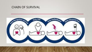
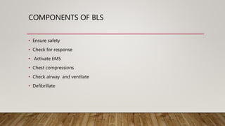
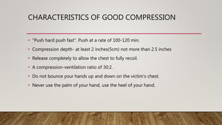
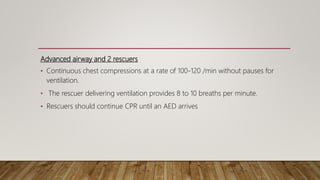
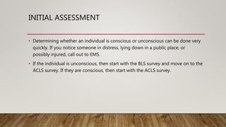
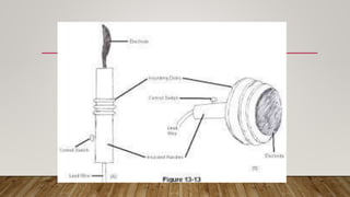
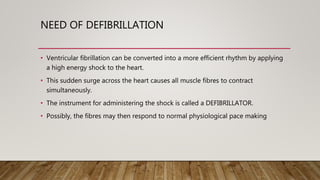
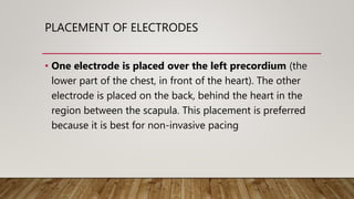
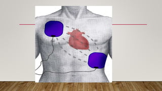
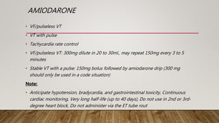
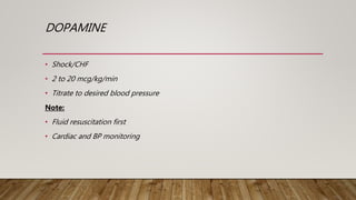
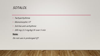
Advanced cardiac life support, or advanced cardiovascular life support, often referred to by its acronym, "ACLS", refers to a set of clinical algorithms for the urgent treatment of cardiac arrest, stroke, myocardial infarction (also known as a heart attack), and other life-threatening cardiovascular emergencies.
Advanced cardiac life support or advanced cardiovascular life support (ACLS) refers to a set of clinical interventions for the urgent treatment of cardiac arrest, stroke and other life-threatening medical emergencies, as well as the knowledge and skills to deploy those interventions.
1. A 65-year-old male presents with syncope and chest pain, with a BP of 70/33 and HR of 128.
2. A 45-year-old woman presents with palpitations, lightheadedness and a HR of 150.
3. A 56-year-old diabetic woman presents with dizziness and chest pain, with a BP of 80/60. Her ECG shows a shockable rhythm.
4. A 40-year-old man is found unconscious with no pulse.
Pacemakers are electronic devices that can be used to initiate a heartbeat when the heart's intrinsic electrical system cannot effectively generate an adequate heart rate. There are temporary pacemakers, which are used until the underlying condition resolves, and permanent pacemakers. A pacemaker system consists of a pulse generator and pacing leads. The pulse generator delivers electrical pulses through the leads to stimulate the heart. Pacemakers can pace one or both chambers of the heart and are programmed with settings for rate, output, and sensitivity. Nurses monitor for pacemaker function and complications and educate patients on pacemaker care.
The document discusses temporary pacemakers, including their uses, common settings, potential complications, and care/maintenance. Temporary pacemakers are used to stimulate the heart in the absence of an intrinsic rhythm or to supplement an inadequate rhythm. Common settings include AAI, VVI, and DDD. Complications include failure to pace or sense, and care involves cleaning and securing insertion sites and leads while monitoring for proper pacing.
1) Intra-aortic balloon counterpulsation (IABP) provides systolic unloading and diastolic augmentation to improve cardiac output.
2) IABP is indicated for cardiogenic shock, high-risk PCI/CABG, and mechanical complications.
3) Potential complications include limb ischemia, infection, bleeding, and aortic injury.
4) Optimal IABP waveform analysis and timing are important to maximize hemodynamic support.
ABG analysis is the measurement of the dissolved oxygen and carbon dioxide in the arterial blood to reveal the acid base balance and how well the oxygen is being carried to the body.
This document summarizes guidelines for emergency medicine training in resuscitation. It discusses adult cardiopulmonary resuscitation, including a focus on high-quality chest compressions. It also reviews the treatment of cardiac arrest, including defibrillation and drug therapy. Additional sections cover bradycardia, tachycardia, cardiac rhythms like atrial fibrillation and flutter, and management approaches.
The major physiological effects of counterpulsation include:
A) increased coronary artery perfusion, increased preload, decreased after load, decreased myocardial oxygen consumption
A Holter monitor is a portable device worn for 24-48 hours that continuously records heart activity through electrodes placed on the skin. It is used to diagnose conditions like valvular heart disease, CAD, arrhythmias, and pacemaker malfunctions. Proper nursing management during the procedure includes documenting attachment/removal times, encouraging activity logging, and avoiding interference. Risks are generally low but may include skin irritation.
This document provides an overview of Advanced Cardiac Life Support (ACLS). It discusses interventions to prevent cardiac arrest, treat cardiac arrest, and improve outcomes for those who experience return of spontaneous circulation after cardiac arrest. The key elements of the adult chain of survival are recognition, early CPR, rapid defibrillation, effective advanced life support, and post-cardiac arrest care. Causes of cardiac arrest include ventricular fibrillation, pulseless ventricular tachycardia, pulseless electric activity, and asystole. Drug therapies and their dosages are outlined, including epinephrine, vasopressin, amiodarone, and lidocaine. Key changes from 2005 ACLS guidelines include the use of continuous quantitative
The document provides information about code blue cardiac nursing care. It outlines the objectives of presenting code blue protocols which include describing the purpose of code language, mechanisms of cardiac arrest, and the treatment hierarchy. It then details the primary components of responding to a code blue including performing CABD (Circulation, Airway, Breathing, Defibrillation) surveys and administering appropriate medications. The document provides details on cardiac arrest mechanisms, contributing factors, medications used, documentation, and post-resuscitation care with the goal of successful resuscitation.
Continue CPR immediately after shock for 2
minutes before reanalysis
Operator: Thank you for the reminder. Yes, CPR should be
resumed immediately after shock delivery for 2 minutes
before reanalysis.
BLS(basic life support) & ACLS with PALS by Dr. ShailendraShailendra Satpute
This document provides information on Basic Life Support (BLS) and Advanced Cardiac Life Support (ACLS). It defines cardiac arrest, outlines its causes and types including ventricular fibrillation, pulseless ventricular tachycardia, asystole, and pulseless electrical activity. It describes the signs and symptoms of cardiac arrest. It also summarizes the steps of BLS including chest compressions, airway management, rescue breathing, and defibrillation. Advanced techniques like intubation, use of laryngeal mask airways, endotracheal tubes, and automated external defibrillators are also outlined.
This document discusses using echocardiography to measure stroke volume and cardiac output in ICU patients. It explains that stroke volume and cardiac output can provide early warning of circulation failure beyond what mean arterial pressure indicates. The document outlines how to obtain echocardiography images of the heart and left ventricle outflow tract to measure stroke volume using pulsed-wave Doppler. Stroke volume combined with heart rate can then be used to calculate cardiac output. Measuring stroke volume allows assessment of the effectiveness of fluid challenges, inotropes, or other therapeutic maneuvers.
The document outlines changes to BLS and ACLS guidelines for 2010, including emphasizing continuous chest compressions for cardiac arrest victims who are unresponsive and not breathing normally before rescue breathing. It recommends performing high-quality chest compressions at a rate of 100 per minute with full chest recoil and minimizing interruptions. The guidelines also stress applying an AED as soon as available and continuing CPR until the victim shows signs of life.
Trans-esophageal echocardiography (TEE) uses ultrasound to obtain high-quality images of the heart and surrounding structures. It involves inserting a probe with an ultrasound transducer at the tip through the mouth and esophagus. TEE provides clearer images than transthoracic echocardiography as the esophagus is directly behind the heart. A TEE exam involves systematically imaging the heart in various planes as the transducer is advanced and manipulated. Standard views include the mid-esophageal four-chamber, two-chamber, aortic, and RV inflow-outflow views. Real-time 3D TEE can provide en face views of structures.
The document provides an overview of basic pacing concepts including:
- Types of pacemakers such as single chamber, dual chamber, and triple chamber systems.
- Components of a pacemaker system including the pulse generator, leads, and electrical concepts such as voltage, current, and impedance.
- Factors that can affect pacing thresholds and how to test the pacemaker circuit including identifying high and low impedance conditions.
The document summarizes information about the intra-aortic balloon pump (IABP), which is a circulatory assist device used to support the left ventricle through counterpulsation. It describes how the IABP works by inflating and deflating a balloon catheter timed to the cardiac cycle to displace aortic blood. It provides details on patient criteria, device set-up, monitoring, complications, and weaning from the IABP.
ET Intubation- Definition, Anatomy of Respiratory Track, Types Of Tubes, Measurement of Tube, Measurement of mouth, Position, procedure, Tray Preparation, Education of Pts, Fixations, Testing of tube, Advantages, Disadvantages.
This document discusses pressure changes that can occur during coronary angiography, specifically damping and ventricularization. Damping is defined as a significant decrease in pressure at the coronary ostium when the catheter is placed, accompanied by the disappearance of pressure waveforms, suggesting no antegrade flow. Ventricularization occurs when blood circulates within a coronary artery like a closed system, deforming the aortic pressure waveform. The document emphasizes the importance of the operator recognizing abnormal pressure changes to avoid complications, and provides solutions like catheter replacement or intracoronary nitroglycerin to address issues.
The document provides information about intra-aortic balloon pumps (IABP). It discusses that IABPs were first described in 1958 and have since improved. IABPs provide temporary left ventricular support by displacing blood in the aorta. They work by inflating in diastole and deflating before systole to increase cardiac output and coronary perfusion pressure while decreasing workload. IABPs are used for cardiac failure, unstable angina, postoperative complications, and as a bridge to transplantation. Complications include limb ischemia, bleeding, thrombosis, and infection.
This document discusses hemodynamic monitoring components used to evaluate the cardiovascular system. It describes how components such as heart rate, blood pressure, cardiac output, stroke volume, central venous pressure and pulmonary artery pressures are measured and used to establish baseline values, evaluate trends, determine dysfunction, and guide interventions. Factors that influence hemodynamics like preload, afterload, contractility and resistance are also explained. Normal ranges for various measurement values are provided.
This document discusses pulse oximetry, which measures the amount of oxygenated hemoglobin in the blood. It describes how the first oximeter was introduced in 1935 and improved upon in 1972. A pulse oximeter uses light-emitting diodes and photodetectors to measure how much red and infrared light is absorbed by oxygenated and deoxygenated hemoglobin, allowing it to calculate blood oxygen saturation levels in a non-invasive manner based on Beer-Lambert law. The principles, components, procedure, factors affecting readings, and purposes of pulse oximetry are outlined.
Defibrillation uses electrical shocks to restore a normal heart rhythm. It is used for ventricular fibrillation and asystole. Biphasic defibrillators are preferred over monophasic as they cause less damage and have higher success rates. Defibrillators include automated external defibrillators for public use, semi-automated defibrillators for paramedics, and implantable defibrillators. Adhesive patches are now commonly used instead of paddles. Defibrillation procedures involve assessing rhythm, applying pads or paddles, delivering shock, and resuming CPR if needed. Causes of failure include patient condition, prolonged arrest, inadequate CPR, and technical issues.
This document provides an overview of basic life support (BLS). It defines BLS and explains its key steps and components, including the chain of survival and use of a defibrillator. BLS procedures like CPR can provide oxygenated blood to victims' brains and hearts after cardiac or respiratory arrest, increasing survival chances until emergency medical care arrives. The document outlines the ABCs of BLS (airway, breathing, circulation) and emphasizes starting chest compressions immediately for cardiac arrests before assessing airway and breathing. It provides guidance on high-quality chest compressions, ventilation, and use of an automated external defibrillator for defibrillation. The overall goal of BLS is to restore oxygenated blood circulation until a
Advanced Cardiovascular Life Support (ACLS).pptxRebilHeiru2
discusses the basic and Advanced Life support according to the AHA guidelines.
ACLS, BLS, defibrillation and Advanced medications at Adama Hospital medical college ICU
An automated external defibrillator (AED) delivers an electric shock to the heart to stop ventricular fibrillation and allow a normal rhythm to resume following sudden cardiac arrest; AEDs guide lay rescuers through the process of applying pads to the patient's chest, analyzing their heart rhythm, and delivering a shock if needed to restore a normal rhythm; It is critical to place AED pads correctly on the patient's chest, follow all prompts, and continue CPR between shocks to improve chances of survival from sudden cardiac arrest.
Basic Life Support (BLS) refers to emergency care including CPR, use of an AED, and clearing obstructed airways. BLS aims to maintain circulation and breathing until emergency help arrives. It involves assessing for responsiveness, calling for help, providing chest compressions at 100-120 per minute and rescue breaths in a 30:2 ratio. An AED should be used as soon as available to analyze rhythms and deliver shocks if indicated. CPR techniques differ between adults and children, such as using two fingers to compress the chest of an infant or child. Foreign object airway obstructions are managed through back blows, chest thrusts and abdominal thrusts depending on responsiveness and age of the victim.
DEFIBRILLATOR AND CARDIOVERSION - NURSES RESPONSIBILITYssuser002e70
A defibrillator uses electrical energy to temporarily depolarize the heart muscle and allow the heart's natural pacemaker to regain control of the heartbeat. There are several types of defibrillators, including manual external defibrillators, automated external defibrillators (AEDs), manual internal defibrillators, and implantable cardioverter-defibrillators (ICDs). The procedure for defibrillation involves charging the defibrillator, placing pads on the patient's chest, clearing everyone from contact, and delivering an electric shock to restore a normal heart rhythm.
This document summarizes guidelines for cardiopulmonary resuscitation (CPR). It discusses:
1) The sequence of steps for CPR has changed and is now CAB for all ages, even children and infants, with an emphasis on minimizing delays to chest compressions.
2) Recommendations for compressions in children include pushing hard and fast at a rate of 100-120 per minute to a depth of at least one third the chest diameter.
3) An automated external defibrillator (AED) can now be used for infants, starting with an initial shock of 2-4 joules.
4) Hypothermia treatment is recommended for comatose cardiac arrest patients
This document summarizes a presentation on basic and advanced cardiac life support. It discusses key concepts in BLS including recognition of cardiac arrest, activating emergency services, performing chest compressions, minimizing interruptions, monitoring compression quality, ventilation, and use of an automated external defibrillator. It then covers ACLS, including treatment algorithms, airway management, defibrillation procedures, medications used during CPR, monitoring techniques, and management of specific arrhythmias like ventricular fibrillation, asystole, and pulseless electrical activity. The goal of BLS and ACLS is to provide immediate life-saving interventions for cardiac arrest patients until the underlying cause can be addressed.
cardiopulmonary resuscitation for studentsShahnaali
This document provides information on cardiopulmonary resuscitation (CPR) including its history, current statistics on cardiac arrest, the goals and steps of CPR, and guidelines from the American Heart Association (AHA). It details the adult BLS sequence of assessing for responsiveness, activating emergency services, providing chest compressions, opening the airway, rescue breathing, and use of an automated external defibrillator. Advanced cardiac life support techniques like defibrillation, intubation, and use of resuscitation drugs are also summarized. New recommendations from the AHA on compression rate, ventilation volume, and capnography monitoring are highlighted.
This document provides information on cardiovascular emergencies and procedures for manual defibrillation, cardioversion, artificial pacemakers, and cardiac arrest treatment algorithms. It discusses defibrillation, synchronized cardioversion, manual vs automated defibrillation, safety measures, energy levels, and indications. Cardiac arrest treatment involves assessing CABs, starting CPR, attaching an AED/defibrillator, analyzing rhythms, delivering shocks if indicated, administering medications, and considering termination of resuscitation or transport depending on the patient's condition and response to treatment.
Sudden cardiac death is defined as an abrupt loss of consciousness within one hour of the onset of symptoms due to a cardiac cause. The main risk factors include age, race, sex, hereditary factors, lifestyle like smoking and obesity, left ventricular dysfunction, and ventricular arrhythmias. The most common causes are coronary artery disease, cardiomyopathies, acute heart failure, and electrophysiological abnormalities. Management of cardiac arrest focuses on continuous cardiopulmonary support, early defibrillation if needed, advanced life support including intubation, medications, and post-cardiac arrest care like therapeutic hypothermia. The goal is to restore spontaneous circulation and hemodynamic stability through these interventions.
This document provides information about Advanced Cardiac Life Support (ACLS). It begins by defining ACLS as a set of clinical interventions for urgently treating cardiac arrest and other life-threatening emergencies, as well as the knowledge and skills to perform those interventions. The document then discusses the American Heart Association protocols that are considered the gold standard for ACLS and how ACLS builds upon the foundation of basic life support. It also reviews the adult and pediatric chains of survival and components of high-quality CPR in BLS before providing details on ACLS interventions like defibrillation, airway management, ventilation, pharmacotherapy, synchronized cardioversion, and post-cardiac arrest care.
The document discusses cardiopulmonary resuscitation (CPR) and emergency cardiac care procedures. It defines CPR, basic life support (BLS), advanced cardiac life support (ACLS), and the indications and contraindications for their use. Key aspects of BLS covered include chest compressions, airway management, breathing, and defibrillation. Commonly used medications in ACLS like epinephrine, atropine, and amiodarone are also outlined. The document provides details on the steps of BLS, complications to watch for, and a nurse's responsibilities in caring for a patient after resuscitation.
Nursing encompasses autonomous and collaborative care of individuals of all ages in all settings, including promoting health, preventing illness, and caring for those who are ill, disabled, or dying. Advanced Cardiovascular Life Support (ACLS) refers to clinical guidelines for urgently treating life-threatening cardiac conditions that cause or can cause cardiac arrest, using advanced medical procedures, medications, and techniques. The ACLS algorithms address airway management, ventilation, chest compressions, defibrillation, and medications to treat dangerous arrhythmias and cardiac arrest.
This lecture provides an introduction to RECOVER, a campaign to standardize veterinary cardiopulmonary resuscitation (CPR) guidelines. The goals of RECOVER and this lecture are to establish evidence-based best practices for veterinary CPR with the aim of improving survival rates. The lecture covers the basic chain of survival, including preparation, basic life support, advanced life support, monitoring, and post-resuscitation care. Key recommendations include rapid initiation of chest compressions and ventilation, use of epinephrine and vasopressin, biphasic defibrillation when indicated, and monitoring end-tidal carbon dioxide to guide resuscitation efforts.
The document provides guidelines for performing basic life support, including checking for response, calling for help, opening the airway, checking for breathing, performing 30 chest compressions followed by 2 rescue breaths, and continuing cycles of compressions and breaths until emergency services arrive or the victim starts breathing on their own. Early CPR and defibrillation are critical for survival from cardiac arrest, with survival rates declining rapidly without intervention in the first few minutes. Bystander CPR can double or triple a victim's chance of survival.
Sudden cardiac arrest (SCA)&Sudden cardiac death (SCD)Abdullah Ansari
INTRODUCTION
SCD : Definition
Epidemiology
Etiology
THE INITIAL ASSESSMENT
BASIC LIFE SUPPORT
CPR Steps
SELF-ASSESSMENT FOR BLS
ADVANCED CARDIAC LIFE SUPPORT
PRINCIPLES OF EARLY DEFIBRILLATION
AUTOMATED EXTERNAL DEFIBRILLATOR
SELF-ASSESSMENT FOR ACLS
Basic Life Support, or BLS, generally refers to the type of care that first-responders, healthcare providers and public safety professionals provide to anyone who is experiencing cardiac arrest, respiratory distress or an obstructed airway. It requires knowledge and skills in cardiopulmonary resuscitation (CPR), using automated external defibrillators (AED) and relieving airway obstructions in patients of every age.
Here is the updated list of Top Best Ayurvedic medicine for Gas and Indigestion and those are Gas-O-Go Syp for Dyspepsia | Lavizyme Syrup for Acidity | Yumzyme Hepatoprotective Capsules etc
Rasamanikya is a excellent preparation in the field of Rasashastra, it is used in various Kushtha Roga, Shwasa, Vicharchika, Bhagandara, Vatarakta, and Phiranga Roga. In this article Preparation& Comparative analytical profile for both Formulationon i.e Rasamanikya prepared by Kushmanda swarasa & Churnodhaka Shodita Haratala. The study aims to provide insights into the comparative efficacy and analytical aspects of these formulations for enhanced therapeutic outcomes.
Local Advanced Lung Cancer: Artificial Intelligence, Synergetics, Complex Sys...Oleg Kshivets
Overall life span (LS) was 1671.7±1721.6 days and cumulative 5YS reached 62.4%, 10 years – 50.4%, 20 years – 44.6%. 94 LCP lived more than 5 years without cancer (LS=2958.6±1723.6 days), 22 – more than 10 years (LS=5571±1841.8 days). 67 LCP died because of LC (LS=471.9±344 days). AT significantly improved 5YS (68% vs. 53.7%) (P=0.028 by log-rank test). Cox modeling displayed that 5YS of LCP significantly depended on: N0-N12, T3-4, blood cell circuit, cell ratio factors (ratio between cancer cells-CC and blood cells subpopulations), LC cell dynamics, recalcification time, heparin tolerance, prothrombin index, protein, AT, procedure type (P=0.000-0.031). Neural networks, genetic algorithm selection and bootstrap simulation revealed relationships between 5YS and N0-12 (rank=1), thrombocytes/CC (rank=2), segmented neutrophils/CC (3), eosinophils/CC (4), erythrocytes/CC (5), healthy cells/CC (6), lymphocytes/CC (7), stick neutrophils/CC (8), leucocytes/CC (9), monocytes/CC (10). Correct prediction of 5YS was 100% by neural networks computing (error=0.000; area under ROC curve=1.0).
Osteoporosis - Definition , Evaluation and Management .pdfJim Jacob Roy
Osteoporosis is an increasing cause of morbidity among the elderly.
In this document , a brief outline of osteoporosis is given , including the risk factors of osteoporosis fractures , the indications for testing bone mineral density and the management of osteoporosis
8 Surprising Reasons To Meditate 40 Minutes A Day That Can Change Your Life.pptxHolistified Wellness
We’re talking about Vedic Meditation, a form of meditation that has been around for at least 5,000 years. Back then, the people who lived in the Indus Valley, now known as India and Pakistan, practised meditation as a fundamental part of daily life. This knowledge that has given us yoga and Ayurveda, was known as Veda, hence the name Vedic. And though there are some written records, the practice has been passed down verbally from generation to generation.
Muktapishti is a traditional Ayurvedic preparation made from Shoditha Mukta (Purified Pearl), is believed to help regulate thyroid function and reduce symptoms of hyperthyroidism due to its cooling and balancing properties. Clinical evidence on its efficacy remains limited, necessitating further research to validate its therapeutic benefits.
These lecture slides, by Dr Sidra Arshad, offer a quick overview of the physiological basis of a normal electrocardiogram.
Learning objectives:
1. Define an electrocardiogram (ECG) and electrocardiography
2. Describe how dipoles generated by the heart produce the waveforms of the ECG
3. Describe the components of a normal electrocardiogram of a typical bipolar lead (limb II)
4. Differentiate between intervals and segments
5. Enlist some common indications for obtaining an ECG
6. Describe the flow of current around the heart during the cardiac cycle
7. Discuss the placement and polarity of the leads of electrocardiograph
8. Describe the normal electrocardiograms recorded from the limb leads and explain the physiological basis of the different records that are obtained
9. Define mean electrical vector (axis) of the heart and give the normal range
10. Define the mean QRS vector
11. Describe the axes of leads (hexagonal reference system)
12. Comprehend the vectorial analysis of the normal ECG
13. Determine the mean electrical axis of the ventricular QRS and appreciate the mean axis deviation
14. Explain the concepts of current of injury, J point, and their significance
Study Resources:
1. Chapter 11, Guyton and Hall Textbook of Medical Physiology, 14th edition
2. Chapter 9, Human Physiology - From Cells to Systems, Lauralee Sherwood, 9th edition
3. Chapter 29, Ganong’s Review of Medical Physiology, 26th edition
4. Electrocardiogram, StatPearls - https://www.ncbi.nlm.nih.gov/books/NBK549803/
5. ECG in Medical Practice by ABM Abdullah, 4th edition
6. Chapter 3, Cardiology Explained, https://www.ncbi.nlm.nih.gov/books/NBK2214/
7. ECG Basics, http://www.nataliescasebook.com/tag/e-c-g-basics
ABDOMINAL TRAUMA in pediatrics part one.drhasanrajab
Abdominal trauma in pediatrics refers to injuries or damage to the abdominal organs in children. It can occur due to various causes such as falls, motor vehicle accidents, sports-related injuries, and physical abuse. Children are more vulnerable to abdominal trauma due to their unique anatomical and physiological characteristics. Signs and symptoms include abdominal pain, tenderness, distension, vomiting, and signs of shock. Diagnosis involves physical examination, imaging studies, and laboratory tests. Management depends on the severity and may involve conservative treatment or surgical intervention. Prevention is crucial in reducing the incidence of abdominal trauma in children.
TEST BANK For Community Health Nursing A Canadian Perspective, 5th Edition by...Donc Test
TEST BANK For Community Health Nursing A Canadian Perspective, 5th Edition by Stamler, Verified Chapters 1 - 33, Complete Newest Version Community Health Nursing A Canadian Perspective, 5th Edition by Stamler, Verified Chapters 1 - 33, Complete Newest Version Community Health Nursing A Canadian Perspective, 5th Edition by Stamler Community Health Nursing A Canadian Perspective, 5th Edition TEST BANK by Stamler Test Bank For Community Health Nursing A Canadian Perspective, 5th Edition Pdf Chapters Download Test Bank For Community Health Nursing A Canadian Perspective, 5th Edition Pdf Download Stuvia Test Bank For Community Health Nursing A Canadian Perspective, 5th Edition Study Guide Test Bank For Community Health Nursing A Canadian Perspective, 5th Edition Ebook Download Stuvia Test Bank For Community Health Nursing A Canadian Perspective, 5th Edition Questions and Answers Quizlet Test Bank For Community Health Nursing A Canadian Perspective, 5th Edition Studocu Test Bank For Community Health Nursing A Canadian Perspective, 5th Edition Quizlet Test Bank For Community Health Nursing A Canadian Perspective, 5th Edition Stuvia Community Health Nursing A Canadian Perspective, 5th Edition Pdf Chapters Download Community Health Nursing A Canadian Perspective, 5th Edition Pdf Download Course Hero Community Health Nursing A Canadian Perspective, 5th Edition Answers Quizlet Community Health Nursing A Canadian Perspective, 5th Edition Ebook Download Course hero Community Health Nursing A Canadian Perspective, 5th Edition Questions and Answers Community Health Nursing A Canadian Perspective, 5th Edition Studocu Community Health Nursing A Canadian Perspective, 5th Edition Quizlet Community Health Nursing A Canadian Perspective, 5th Edition Stuvia Community Health Nursing A Canadian Perspective, 5th Edition Test Bank Pdf Chapters Download Community Health Nursing A Canadian Perspective, 5th Edition Test Bank Pdf Download Stuvia Community Health Nursing A Canadian Perspective, 5th Edition Test Bank Study Guide Questions and Answers Community Health Nursing A Canadian Perspective, 5th Edition Test Bank Ebook Download Stuvia Community Health Nursing A Canadian Perspective, 5th Edition Test Bank Questions Quizlet Community Health Nursing A Canadian Perspective, 5th Edition Test Bank Studocu Community Health Nursing A Canadian Perspective, 5th Edition Test Bank Quizlet Community Health Nursing A Canadian Perspective, 5th Edition Test Bank Stuvia
2. ADVANCED LIFE SUPPORT IN PERSPECTIVE
Chain of survival:
Early access to emergency services(102).
Early Basic life support(by hand only).
Early defibrillation.
Early advanced life support.
3. INTRODUCTION
• According to recent statistics sudden cardiac arrest is rapidly becoming the
leading cause of death.
• Once the heart ceases to function, a healthy human brain may survive without
oxygen for up to 4 minutes without suffering any permanent damage.
• Unfortunately, a typical EMS response may take 6, 8 or even 10 minutes
4. • The goal of advanced cardiovascular life support is to achieve the best possible
outcome for individual who are experiencing a life- threating event.
• These ACLS protocols have been developed through research, patient case
student, clinical studies and opinion of experts in the field.
• All ACLS providers are perfumed capable of performing BLS correctly.
5. • While ACLS providers should always be mindful of timeliness.
• It is important to provide the intervention that most appropriately fits the needs
of the individual.
• Proper utilization of ACLS requires rapid and accurate assessment of an
individual’s condition.
7. WHAT IS BLS ??
• Basic Life Support (BLS) refers to the care healthcare providers and public safety
professionals provide to patients who are experiencing respiratory arrest, cardiac
arrest or airway obstruction.
• BLS includes psychomotor skills for performing high-quality cardiopulmonary
resuscitation (CPR), using an automated external defibrillator (AED) and relieving
an obstructed airway for patients of all ages
8. COMPONENTS OF BLS
• Ensure safety
• Check for response
• Activate EMS
• Chest compressions
• Check airway and ventilate
• Defibrillate
9. CHARACTERISTICS OF GOOD COMPRESSION
• “Push hard push fast”. Push at a rate of 100-120 min.
• Compression depth- at least 2 inches(5cm) not more than 2.5 inches
• Release completely to allow the chest to fully recoil.
• A compression-ventilation ratio of 30:2 .
• Do not bounce your hands up and down on the victim's chest.
• Never use the palm of your hand, use the heel of your hand.
10. When 2 or more rescuers available,
• Switch the compressor about every 2 minutes (or after 5 cycles of compressions
and ventilations at a ratio of 30:2).
• Accomplish this switch in ≤5 seconds.
11. Advanced airway and 2 rescuers
• Continuous chest compressions at a rate of 100-120 /min without pauses for
ventilation.
• The rescuer delivering ventilation provides 8 to 10 breaths per minute.
• Rescuers should continue CPR until an AED arrives
12. INITIAL ASSESSMENT
• Determining whether an individual is conscious or unconscious can be done very
quickly. If you notice someone in distress, lying down in a public place, or
possibly injured, call out to EMS.
• If the individual is unconscious, then start with the BLS survey and move on to the
ACLS survey. If they are conscious, then start with the ACLS survey.
13. ACLS
• Understanding normal cardiac anatomy and physiology is an important
component of performing ACLS.
• The heart is hollow muscle comprised of four chambers surrounded by thick wall
of tissue(septum). The atria are two upper chambers and ventricles are two lower
chambers.
• The left ands right valves of heart work together to pump the blood throughout
the body.
14. THE ACLS SURVEY
AIRWAY :-
Maintain airway in unconscious patient
Consider advanced airway
Monitor advanced airway if placed with quantitative waveform capnography.
15. BREATHING
• Give 100% oxygen.
• Assess effective ventilation with quantitative waveform capnography.
• Do not over-ventilate.
16. CIRCULATION
• Evaluate rhythm and pulse.
• Defibrillation/Cardioversion
• Obtain IV/IO access
• Give rhythm- specific medication
• Give IV/IO fluids if needed
17. DIFFERENTIAL DIAGNOSIS
• Identify and treat reversible causes.
• Cardiac rhythm and patient history are the keys to differential diagnosis.
• Assess when to shock versus medicate.
18. DEFIBRILLATION
• Defibrillation is a process in which an electronic device sends an electric shock to
the heart to stop an extremely rapid, irregular heartbeat, and restore the normal
heart rhythm.
• Defibrillation is a common treatment for life threatening cardiac dysrhythmias,
ventricular fibrillation, and pulse less ventricular tachycardia.
19.
20. HISTORY OF DEFIBRILLATION
• Defibrillation was invented in____ by Prevost and Batelli, two Italian physiologists.
They discovered that electric shocks could convert ventricular fibrillation to sinus
rhythm in dogs.The first case of a human life saved by defibrillation was reported
by Beck in 1947 .
21. TYPES OF DEFIBRILLATION
a) Internal defibrillator
• Electrodes placed directly to the heart e.g..-Pacemaker
b) External defibrillator
• Electrodes placed directly on the heart e.g..-AED
22. TYPES OF DEFIBRILLATOR ELECTRODES:-
a) Spoon shaped electrode
• Applied directly to the heart
b) Paddle type electrode
• Applied against the chest wall
c) Pad type electrode
• Applied directly on chest wall
23.
24. NEED OF DEFIBRILLATION
• Ventricular fibrillation can be converted into a more efficient rhythm by applying
a high energy shock to the heart.
• This sudden surge across the heart causes all muscle fibres to contract
simultaneously.
• The instrument for administering the shock is called a DEFIBRILLATOR.
• Possibly, the fibres may then respond to normal physiological pace making
25. PLACEMENT OF ELECTRODES
• One electrode is placed over the left precordium (the
lower part of the chest, in front of the heart). The other
electrode is placed on the back, behind the heart in the
region between the scapula. This placement is preferred
because it is best for non-invasive pacing
26.
27. PURPOSE OF DEFIBRILLATION
• Defibrillation is performed to correct life threatening fibrillations of the heart,
which could result in cardiac arrest. It should be performed immediately after
identifying that the patient is experiencing a cardiac emergency, has no pulse,
and is unresponsive
28. PRINCIPLE OF DEFIBRILLATION
• Energy storage capacitor is charged at relatively slow rate .
• Energy stored in capacitor is then delivered at a relatively rapid rate to chest of
the patient.
• Simple arrangement involve the discharge of capacitor energy through the
patient’s own resistance.
29. MECHANISM
• Fibrillations cause the heart to stop pumping blood, leading to brain damage.
• Defibrillators deliver a brief electric shock to the heart, which enables the heart's
natural pacemaker to regain control and establish a normal heart rhythm.
30. STRENGHT DURING CURVE
• operator selects energy delivered: 50-360 joules, depends on:
– intrinsic characteristics of patient
– patient’s disease
– duration of arrhythmia
– patient’s age
– type of arrhythmia (more energy required for v. fib.)
31. CLASSES OF DISCHARGING WAVWFORM
• There are two general classes of waveforms:
A) mono-phasic waveform
Energy delivered in one direction through the patient’s heart
B) Biphasic waveform
Energy delivered in both direction through the patient’s heart
32. • A monophasic type, give a high-energy shock, up to 360 to 400 joules due to
which increased cardiac injury and in burns the chest around the shock pad sites.
• • A biphasic type, give two sequential lower energy shocks of 120 - 200 joules,
with each shock moving in an opposite polarity between the pads.
33. PRECAUTION
• The paddles used in the procedure should not be placed:-
• on a woman's breasts
• over an internal pacemaker patients.
• Before the paddle is used, a gel must be applied to the patient's skin
34. RISK IN DEFIBRILLATION
• Skin burns from the defibrillator paddles are the most common complication of
defibrillation.
• Other risks include injury to the heart muscle, abnormal heart rhythms, and blood
clots.
35. DRUGS USED IN ACLS
Adenosine
• Narrow PSVT/SVT
• Wide QRS tachycardia, avoid adenosine in irregular wide QRS
• 6 mg IV bolus, may repeat with 12 mg in 1 to 2 min.
Note:
• Rapid IV push close to the hub, followed by a saline bolus
• Continuous cardiac monitoring during administration
• Causes flushing and chest heaviness
36. AMIODARONE
• VF/pulseless VT
• VT with pulse
• Tachycardia rate control
• VF/pulseless VT: 300mg dilute in 20 to 30ml., may repeat 150mg every 3 to 5
minutes
• Stable VT with a pulse: 150mg bolus followed by amiodarone drip (300 mg
should only be used in a code situation)
Note:
• Anticipate hypotension, bradycardia, and gastrointestinal toxicity, Continuous
cardiac monitoring, Very long half-life (up to 40 days), Do not use in 2nd or 3rd-
degree heart block, Do not administer via the ET tube rout
37. DOPAMINE
• Shock/CHF
• 2 to 20 mcg/kg/min
• Titrate to desired blood pressure
Note:
• Fluid resuscitation first
• Cardiac and BP monitoring