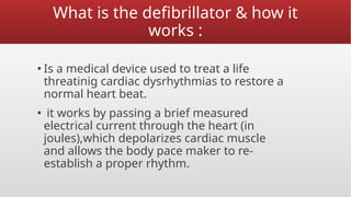
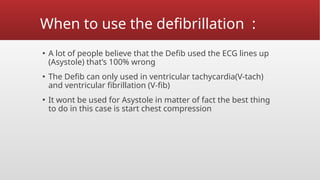
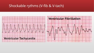
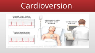
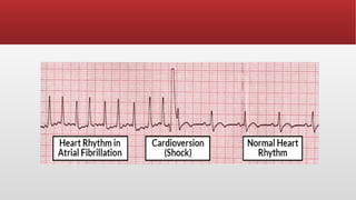
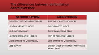
DEFEBRELATION
&CAREDIOVERSION
BY: MOHAMMAD AMRO &
MOHAMMAD SHALASH
learning objective defibrillation:
▪ Definition of defibrillation
▪ Mechanism of defibrillation
▪ Indication
▪ Types of defibrillation
▪ Paddles placement
What is the defibrillator & how it
works :
▪ Is a medical device used to treat a life
threatinig cardiac dysrhythmias to restore a
normal heart beat.
▪ it works by passing a brief measured
electrical current through the heart (in
joules),which depolarizes cardiac muscle
and allows the body pace maker to re-
establish a proper rhythm.
When to use the defibrillation :
▪ A lot of people believe that the Defib used the ECG lines up
(Asystole) that’s 100% wrong
▪ The Defib can only used in ventricular tachycardia(V-tach)
and ventricular fibrillation (V-fib)
▪ It wont be used for Asystole in matter of fact the best thing
to do in this case is start chest compression
Steps to use the defibrillator :
▪ Turn on the Defib and make sure is on the Defib mode
▪ Make sure the patient is shockable
▪ Charge the paddles
▪ Make sure no one touching the patient (clear)
▪ Shock the patient
▪ Immediately start chest compression
Shockable rythms (V-fib & V-tach)
Non shockable rhythm (Asystole &PEA)
• Types of defibrillators:
▪ There are different types of defibrillators including :
• Automated external defibrillators (AEDs):
Portable device found in public places, designed for use by
people without medical training .
cont;
• Implantable cardioverter defibrillators (ICDs):
Devices implanted in the body that can automatically detect and treat
arrythmias.
Cont;
• Wearable cardioverter defibrillator :
Worn like a vest these monitor heart rhythm and can deliver a shock if needed.
Paddle placement :
▪ Antero lateral placement
• Place one paddle on Rt. Of sternum below clavicle (2nd
or 3rd
ICS)
• The other paddle on Lt 4th
or 5th
ICS or mid axillary line
Cont;:
▪ Anterior posterior placement :
• Anterior paddle to the left side of lower part of sternum and
posterior paddle below the tip of the left scapula.
• often used in children with adult paddles .
Classes of discharge waveform :
▪ Monophasic wave form(one direction) usually in AEDs 360J
delivered through the patient heart .
▪ Biphasic wave form (120-200J) delivered in both direction
through the patient heart .
The difference between the monophasic and
biphasic wave form :
MONOPHASIC BIPHASIC
Current travels on one
direction
Deliver current in 2 direction
Requires more electrical
energy
Less electrical energy
More chance of burn Fewer chance of burn
Cause more myocardial
damage
Cause less myocardial damage
First shock success rate60% First shock success rate90%
Conclousion :
• Defibrillators are crucial treating life –threatening arrythmias like ventricular
fibrillation & ventricular tachycardia ,which can lead to sudden cardiac arrest .
They are often used in conjunction with CPR to improve the chances of survival .
Cardioversion
Cardioversion learning objective:
▪ Indication
▪ Types
▪ Preparing
▪ Procedure
▪ Nursing role
▪ Complication
Cardioversion:
▪ Is a medical procedure that uses quick ,low energy shocks to
restore regular heart rhythm
▪ It is particularly effective for treating conditions like atrial
fibrillation (A-fib).
▪ Joules vary from (50-200)Joules .
Indications for
cardioversion:
Atrial fibrillation.
Atrial flutter.
Supraventricular tachycardia.
Images:
Methods :
▪ Electric cardioversion:
This methods involves using machine and electrodes to
deliver quick ,low energy shocks to the chest .
It allows health care providers to instantly assess whether a
typical heart beat has been restored .
▪ Chemical(pharmacological) cardioversion :
Instead of electrical shocks medication are used to restore the
heart beat rhythm .
This method takes longer to work than electric cardioversion
Chemical cardioversion :
Is using medication to restore your heart rhythm from
abnormal to normal one .
Some medication that used in chemical cardioversion :
1. Adenosine.
2. Diltiazem.
3. Amiodarone.
4. Digoxin.
Risk & consideration of electrical
cardioversion :
• Dislodged blood clots:
Some people who have irregular heartbeats, such as A-fib,
have blood clots form in the heart. Shocking the heart can
cause these blood clots to move to other parts of the body.
This can cause life-threatening complications, such as a stroke
or a blood clot traveling to lungs.
A health care provider may order tests to check for blood clots
before doing cardioversion. Some people may be given blood
thinners before the procedure.
cont;:
• Irregular heart rhythms (arrhythmia):
Rarely, some people develop other irregular heartbeats
during or after cardioversion. If it happens, it usually occurs
minutes after the procedure. Medications or additional shocks
can be given to correct the heart's rhythm.
• Skin burns:
Rarely, some people get minor burns on their skin from the
sensors (electrodes).
Preparing for cardioversion :
▪ NPO for 8 hrs at least .
▪ The patient receives moderate sedation IV as well as
analgesic medication or anaesthesia.
▪ Anticoagulant may be given to prevent clots from moving to
the heart .
▪ Stop digoxin 48 hrs prior to the procedure .
▪ Do not apply any lotions or ointment to chest or backs as
this may interfere with the adhesiveness of shocking pads .
Procedure:
▪ To perform cardioversion follow (ACLS) guidelines :
• Turn the defibrillator
• Select the appropriate energy level
• Activate the synchronize mode by pressing the synchronized
button
• Check to verify that the machine is correctly sensing R wave
• Charge machine to ordered energy (50-200)
Nursing role :
▪ Explain the procedure and obtain the consent form
▪ Restrict patient food and drinks for 8 hrs at least (unless the
cardioversion is emergent )
▪ Record ECG and vital sign
▪ Prepare all necessary resuscitation equipment
▪ Evaluate and supplies potassium and magnesium levels
▪ Sedate the patient (unless anesthetis is there)
▪ Turn the defibrillator and attach the monitoring electriods to
the patient chest
The differences between defibrillation
&cardioversion:
DEFIBRILLATION CARDIOVERSION
EMERGENCY LIFE SAVING PROCEDURE ELECTIVE PLANNED PROCEDURE
UN –SYNCHRONIZED SHOCK SYNCHRONIZED SHOCK
NO DELAY, IMMEDIATE THERE CAN BE SOME DELAY
NO ANTICOAGULATION NEEDED ANTI CO AGULATION NEEDED
MORE DAMAGE TO MYOCARDIUM LESS DAMAGE TO MYOCARDIUM
USED IN VT/VF USED IN MOST OF THE MOST ARRYTHMIAS
EXCEPT VF/VT

DEFEBRELATION &CAREDIOVERSION Made EASY FOR YOU

  • 1.
  • 2.
    learning objective defibrillation: ▪Definition of defibrillation ▪ Mechanism of defibrillation ▪ Indication ▪ Types of defibrillation ▪ Paddles placement
  • 3.
    What is thedefibrillator & how it works : ▪ Is a medical device used to treat a life threatinig cardiac dysrhythmias to restore a normal heart beat. ▪ it works by passing a brief measured electrical current through the heart (in joules),which depolarizes cardiac muscle and allows the body pace maker to re- establish a proper rhythm.
  • 4.
    When to usethe defibrillation : ▪ A lot of people believe that the Defib used the ECG lines up (Asystole) that’s 100% wrong ▪ The Defib can only used in ventricular tachycardia(V-tach) and ventricular fibrillation (V-fib) ▪ It wont be used for Asystole in matter of fact the best thing to do in this case is start chest compression
  • 5.
    Steps to usethe defibrillator : ▪ Turn on the Defib and make sure is on the Defib mode ▪ Make sure the patient is shockable ▪ Charge the paddles ▪ Make sure no one touching the patient (clear) ▪ Shock the patient ▪ Immediately start chest compression
  • 6.
  • 7.
    Non shockable rhythm(Asystole &PEA)
  • 8.
    • Types ofdefibrillators: ▪ There are different types of defibrillators including : • Automated external defibrillators (AEDs): Portable device found in public places, designed for use by people without medical training .
  • 9.
    cont; • Implantable cardioverterdefibrillators (ICDs): Devices implanted in the body that can automatically detect and treat arrythmias.
  • 10.
    Cont; • Wearable cardioverterdefibrillator : Worn like a vest these monitor heart rhythm and can deliver a shock if needed.
  • 11.
    Paddle placement : ▪Antero lateral placement • Place one paddle on Rt. Of sternum below clavicle (2nd or 3rd ICS) • The other paddle on Lt 4th or 5th ICS or mid axillary line
  • 12.
    Cont;: ▪ Anterior posteriorplacement : • Anterior paddle to the left side of lower part of sternum and posterior paddle below the tip of the left scapula. • often used in children with adult paddles .
  • 13.
    Classes of dischargewaveform : ▪ Monophasic wave form(one direction) usually in AEDs 360J delivered through the patient heart . ▪ Biphasic wave form (120-200J) delivered in both direction through the patient heart .
  • 14.
    The difference betweenthe monophasic and biphasic wave form : MONOPHASIC BIPHASIC Current travels on one direction Deliver current in 2 direction Requires more electrical energy Less electrical energy More chance of burn Fewer chance of burn Cause more myocardial damage Cause less myocardial damage First shock success rate60% First shock success rate90%
  • 15.
    Conclousion : • Defibrillatorsare crucial treating life –threatening arrythmias like ventricular fibrillation & ventricular tachycardia ,which can lead to sudden cardiac arrest . They are often used in conjunction with CPR to improve the chances of survival .
  • 16.
  • 17.
    Cardioversion learning objective: ▪Indication ▪ Types ▪ Preparing ▪ Procedure ▪ Nursing role ▪ Complication
  • 18.
    Cardioversion: ▪ Is amedical procedure that uses quick ,low energy shocks to restore regular heart rhythm ▪ It is particularly effective for treating conditions like atrial fibrillation (A-fib). ▪ Joules vary from (50-200)Joules .
  • 19.
  • 20.
  • 21.
    Methods : ▪ Electriccardioversion: This methods involves using machine and electrodes to deliver quick ,low energy shocks to the chest . It allows health care providers to instantly assess whether a typical heart beat has been restored . ▪ Chemical(pharmacological) cardioversion : Instead of electrical shocks medication are used to restore the heart beat rhythm . This method takes longer to work than electric cardioversion
  • 22.
    Chemical cardioversion : Isusing medication to restore your heart rhythm from abnormal to normal one . Some medication that used in chemical cardioversion : 1. Adenosine. 2. Diltiazem. 3. Amiodarone. 4. Digoxin.
  • 23.
    Risk & considerationof electrical cardioversion : • Dislodged blood clots: Some people who have irregular heartbeats, such as A-fib, have blood clots form in the heart. Shocking the heart can cause these blood clots to move to other parts of the body. This can cause life-threatening complications, such as a stroke or a blood clot traveling to lungs. A health care provider may order tests to check for blood clots before doing cardioversion. Some people may be given blood thinners before the procedure.
  • 24.
    cont;: • Irregular heartrhythms (arrhythmia): Rarely, some people develop other irregular heartbeats during or after cardioversion. If it happens, it usually occurs minutes after the procedure. Medications or additional shocks can be given to correct the heart's rhythm. • Skin burns: Rarely, some people get minor burns on their skin from the sensors (electrodes).
  • 25.
    Preparing for cardioversion: ▪ NPO for 8 hrs at least . ▪ The patient receives moderate sedation IV as well as analgesic medication or anaesthesia. ▪ Anticoagulant may be given to prevent clots from moving to the heart . ▪ Stop digoxin 48 hrs prior to the procedure . ▪ Do not apply any lotions or ointment to chest or backs as this may interfere with the adhesiveness of shocking pads .
  • 26.
    Procedure: ▪ To performcardioversion follow (ACLS) guidelines : • Turn the defibrillator • Select the appropriate energy level • Activate the synchronize mode by pressing the synchronized button • Check to verify that the machine is correctly sensing R wave • Charge machine to ordered energy (50-200)
  • 27.
    Nursing role : ▪Explain the procedure and obtain the consent form ▪ Restrict patient food and drinks for 8 hrs at least (unless the cardioversion is emergent ) ▪ Record ECG and vital sign ▪ Prepare all necessary resuscitation equipment ▪ Evaluate and supplies potassium and magnesium levels ▪ Sedate the patient (unless anesthetis is there) ▪ Turn the defibrillator and attach the monitoring electriods to the patient chest
  • 29.
    The differences betweendefibrillation &cardioversion: DEFIBRILLATION CARDIOVERSION EMERGENCY LIFE SAVING PROCEDURE ELECTIVE PLANNED PROCEDURE UN –SYNCHRONIZED SHOCK SYNCHRONIZED SHOCK NO DELAY, IMMEDIATE THERE CAN BE SOME DELAY NO ANTICOAGULATION NEEDED ANTI CO AGULATION NEEDED MORE DAMAGE TO MYOCARDIUM LESS DAMAGE TO MYOCARDIUM USED IN VT/VF USED IN MOST OF THE MOST ARRYTHMIAS EXCEPT VF/VT