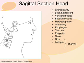
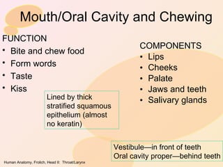
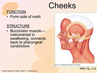
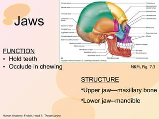
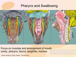
HEAD/NECK II:  Throat/ Pharynx Overview:  Sagittal view of nose/mouth/throat Nasal Cavity and Breathing Mouth and Chewing Throat and Swallowing Larynx and Singing
Sagittal Section Head Cranial cavity Brain/Spinal cord Vertebral bodies Epaxial muscles Hard/soft palate Oral cavity Esophagus Trachea Epiglottis Naso- Oro- Laringo- pharynx
Nose/Nasal Cavity and Breathing Function: Inlet for air to lung Warm/filter air (mucous membranes on  ethmoid   conchae ) Smell (nerve endings on nasal membranes) M&M, Fig. 21.1
Conchae of Ethmod Bone Scroll-like bones Covered in mucous membrane for Smell Filter air Warm air M&M, Fig. 7.10
Sinuses All connected to nasal cavity All lined with mucous membranes Cold/allergies—fill with mucous=sinus headache Maxillary Ethmoid Frontal Sphenoid M&M, Fig. 7.11
Mouth/Oral Cavity and Chewing COMPONENTS Lips Cheeks Palate Jaws and teeth Salivary glands FUNCTION Bite and chew food Form words Taste Kiss Vestibule—in front of teeth Oral cavity proper—behind teeth Lined by thick stratified squamous epithelium (almost no keratin)
Lips FUNCTION Close mouth Keep food in Make speech sounds Tactile STRUCTURE Core of sphinchter-shape skeletal muscle (orbicularis oris) ā€œ Red marginā€ transition from keratinized skin to oral mucosa Red because clear color lets underlying vessels show through No sweat or sebaceous glands, thus needs to be wet (or lip balm) M&M, Fig. 11.6
Cheeks FUNCTION Form side of moth STRUCTURE Buccinator muscle—instrumental in swallowing, connects back to pharyngeal constrictors M&M, Fig. 11.6
Palate Hard palate anterior Maxilla Palatine Soft palate is posterior extension, soft tissue Palatoglossal arch (palate to tongue)  Palatopharyngeal arch (palate to pharynx) Tonsils between arches Uvula…??? M&M, Fig. 22.7
Jaws FUNCTION Hold teeth Occlude in chewing STRUCTURE Upper jaw—maxillary bone Lower jaw--mandible M&M, Fig. 7.3
Teeth Deciduous teethā€”ā€milkā€ or ā€œbabyā€ teeth Emerge 6 mos. – 2 yrs. Replaced by permanent teeth 6-12 yrs. Wisdom teeth (3 rd  molar) erupts 17-25 yrs or remains in jaw Key to healthy teeth and gums:  Flossing Visiting dentist regularly (every 6 mos.) and starting at young age (3-4 yrs.)
Structure of individual tooth—it’s alive!!
Jaw muscles Masseter, temporalis—elevate mandible (ā€œclose jawā€) Medial pterygoid—lateral (side-to-side) chewing Lateral pterygoid—translates mandible anteriorly (part of opening) Digastric (not shown)—depresses mandible (ā€œopens jawā€) Chewing is circular motion M&M, Fig. 11.7
Tongue FUNCTION Position food between teeth Form words in speech STRUCTURE Intrinsic muscles (allow for shape change with fibers in various directions) Extrinsic muscles—attach tongue to skeleton Genioglossus hyoglossus M&M, Fig. 22.7
Salivary glands Intrinsic—all over mucous membranes of tongue, palate, lips, lining of cheek Extrinsic—secrete more saliva when eating (or anticipating) Parotid Submandibular sublingual M&M, Fig. 22.10
Saliva Moistens mouth Dissolves food to be tasted Wets and binds food Contains amylase to start starch digestion (saltine to sugar experiment) Contains bicarbonate to neutralize cavity-causing acids produced by bacteria Contains anti-bacterial and anti-viral enzymes and cyanide-like compound to kill harmful micro-organisms Contains proteins that stimulate growth of beneficial bacteria in the mouth
Throat/Pharynx and Swallowing Back of mouth to larynx Food and air mix and cross When it goes wrong—choking!! STRUCTURES Entry—pharyngeal arches Exit—glottis (epiglottis is lid) Pharyngeal constrictors squeeze food through Food PHARYNX Air Trachea Esophagus
Pharynx and Swallowing Focus on muscles and arrangement of mouth cavity, pharynx, larynx, epiglottis, trachea Pharynx/Swallowing in detail!!
Descent of the larynx
Larynx and Singing FUNCTION Channel air out of trachea Vibrate to produce sound for speech/song STRUCTURES External skeleton or frame (cartilage) Internal vocal cords and associated muscles
Skeleton of larynx Cricothyroid ligament is usual site of emergency tracheotomy (feel on self—SURFACE ANATOMY) M&M, Fig. 21.5
Vocal cords M&M, Fig. 21.6
COMING SOON!! Head/Neck III: Special Senses Head/Neck IV:  Cranial Nerves

Head neck 2nd pharynx

  • 1.
    HEAD/NECK II: Throat/ Pharynx Overview: Sagittal view of nose/mouth/throat Nasal Cavity and Breathing Mouth and Chewing Throat and Swallowing Larynx and Singing
  • 2.
    Sagittal Section HeadCranial cavity Brain/Spinal cord Vertebral bodies Epaxial muscles Hard/soft palate Oral cavity Esophagus Trachea Epiglottis Naso- Oro- Laringo- pharynx
  • 3.
    Nose/Nasal Cavity andBreathing Function: Inlet for air to lung Warm/filter air (mucous membranes on ethmoid conchae ) Smell (nerve endings on nasal membranes) M&M, Fig. 21.1
  • 4.
    Conchae of EthmodBone Scroll-like bones Covered in mucous membrane for Smell Filter air Warm air M&M, Fig. 7.10
  • 5.
    Sinuses All connectedto nasal cavity All lined with mucous membranes Cold/allergies—fill with mucous=sinus headache Maxillary Ethmoid Frontal Sphenoid M&M, Fig. 7.11
  • 6.
    Mouth/Oral Cavity andChewing COMPONENTS Lips Cheeks Palate Jaws and teeth Salivary glands FUNCTION Bite and chew food Form words Taste Kiss Vestibule—in front of teeth Oral cavity proper—behind teeth Lined by thick stratified squamous epithelium (almost no keratin)
  • 7.
    Lips FUNCTION Closemouth Keep food in Make speech sounds Tactile STRUCTURE Core of sphinchter-shape skeletal muscle (orbicularis oris) ā€œ Red marginā€ transition from keratinized skin to oral mucosa Red because clear color lets underlying vessels show through No sweat or sebaceous glands, thus needs to be wet (or lip balm) M&M, Fig. 11.6
  • 8.
    Cheeks FUNCTION Formside of moth STRUCTURE Buccinator muscle—instrumental in swallowing, connects back to pharyngeal constrictors M&M, Fig. 11.6
  • 9.
    Palate Hard palateanterior Maxilla Palatine Soft palate is posterior extension, soft tissue Palatoglossal arch (palate to tongue) Palatopharyngeal arch (palate to pharynx) Tonsils between arches Uvula…??? M&M, Fig. 22.7
  • 10.
    Jaws FUNCTION Holdteeth Occlude in chewing STRUCTURE Upper jaw—maxillary bone Lower jaw--mandible M&M, Fig. 7.3
  • 11.
    Teeth Deciduous teethā€”ā€milkā€or ā€œbabyā€ teeth Emerge 6 mos. – 2 yrs. Replaced by permanent teeth 6-12 yrs. Wisdom teeth (3 rd molar) erupts 17-25 yrs or remains in jaw Key to healthy teeth and gums: Flossing Visiting dentist regularly (every 6 mos.) and starting at young age (3-4 yrs.)
  • 12.
    Structure of individualtooth—it’s alive!!
  • 13.
    Jaw muscles Masseter,temporalis—elevate mandible (ā€œclose jawā€) Medial pterygoid—lateral (side-to-side) chewing Lateral pterygoid—translates mandible anteriorly (part of opening) Digastric (not shown)—depresses mandible (ā€œopens jawā€) Chewing is circular motion M&M, Fig. 11.7
  • 14.
    Tongue FUNCTION Positionfood between teeth Form words in speech STRUCTURE Intrinsic muscles (allow for shape change with fibers in various directions) Extrinsic muscles—attach tongue to skeleton Genioglossus hyoglossus M&M, Fig. 22.7
  • 15.
    Salivary glands Intrinsic—allover mucous membranes of tongue, palate, lips, lining of cheek Extrinsic—secrete more saliva when eating (or anticipating) Parotid Submandibular sublingual M&M, Fig. 22.10
  • 16.
    Saliva Moistens mouthDissolves food to be tasted Wets and binds food Contains amylase to start starch digestion (saltine to sugar experiment) Contains bicarbonate to neutralize cavity-causing acids produced by bacteria Contains anti-bacterial and anti-viral enzymes and cyanide-like compound to kill harmful micro-organisms Contains proteins that stimulate growth of beneficial bacteria in the mouth
  • 17.
    Throat/Pharynx and SwallowingBack of mouth to larynx Food and air mix and cross When it goes wrong—choking!! STRUCTURES Entry—pharyngeal arches Exit—glottis (epiglottis is lid) Pharyngeal constrictors squeeze food through Food PHARYNX Air Trachea Esophagus
  • 18.
    Pharynx and SwallowingFocus on muscles and arrangement of mouth cavity, pharynx, larynx, epiglottis, trachea Pharynx/Swallowing in detail!!
  • 19.
  • 20.
    Larynx and SingingFUNCTION Channel air out of trachea Vibrate to produce sound for speech/song STRUCTURES External skeleton or frame (cartilage) Internal vocal cords and associated muscles
  • 21.
    Skeleton of larynxCricothyroid ligament is usual site of emergency tracheotomy (feel on self—SURFACE ANATOMY) M&M, Fig. 21.5
  • 22.
  • 23.
    COMING SOON!! Head/NeckIII: Special Senses Head/Neck IV: Cranial Nerves