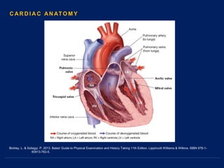
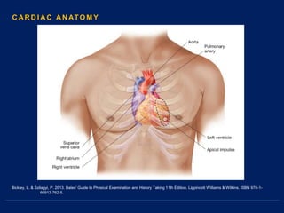
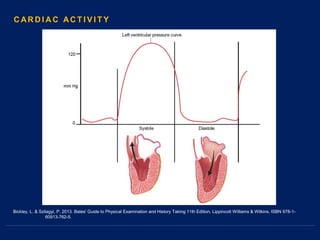
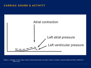
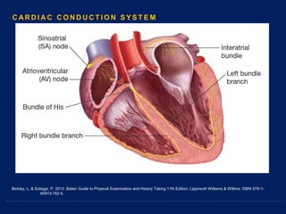
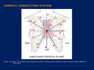
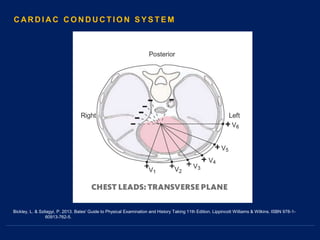
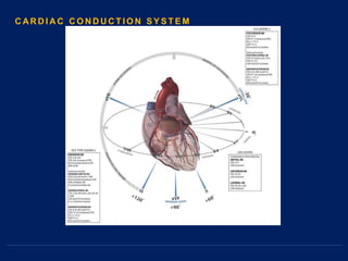
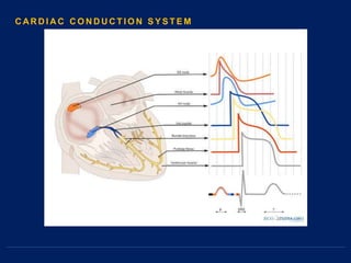
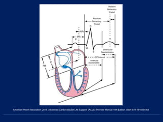
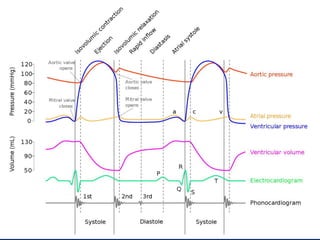
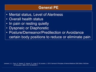
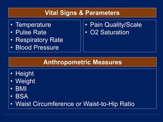
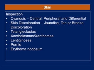
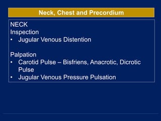
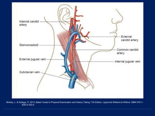
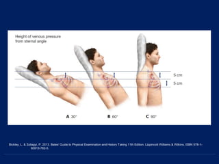
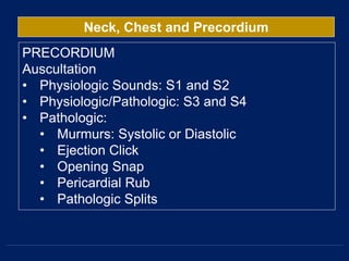
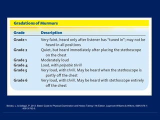
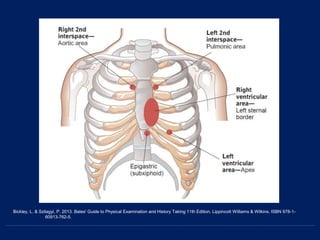
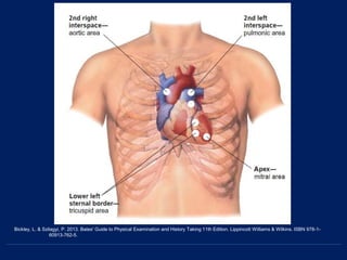
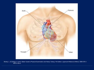
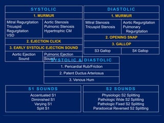
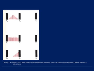
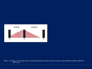
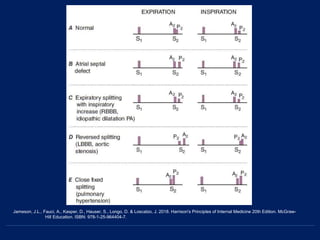
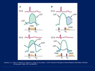
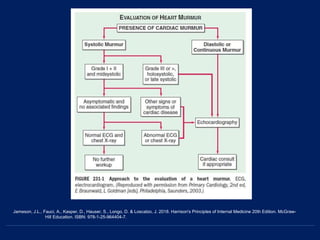
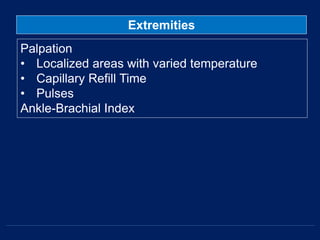
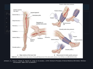
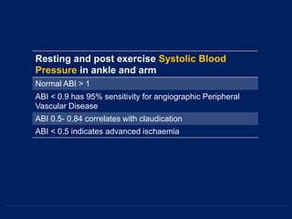
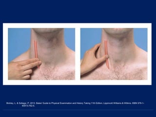
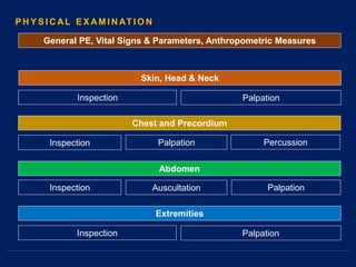
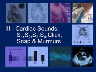
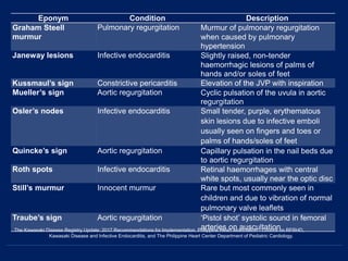
Cardiovascular
Physical Examination
Elino, Meccar Moniem H.
First Year Resident 2019
The Department of Internal Medicine
Southern Philippines Medical Center
7th January 2019
Objective Points
I. Introduction
A. Significance
B. Cardiac Anatomy
C. Cardiac Sound Activity, Cycle & Conduction System
D. Relevant History
II. Physical Examination in Cardiovascular Perspective
A. General
B. Regional/Systemic
C. Neck, Chest, Precordium
III. Cardiac Sounds: S1,S2, S3, S4, Click, Snap & Murmurs
IV. Commonly Used Cardiac Findings/Eponyms
Main
References:
1 Jameson, J.L., Fauci, A., Kasper, D., Hauser, S., Longo, D. & Loscalzo, J.
2018. Harrison's Principles of Internal Medicine 20th Edition. McGraw-
Hill Education. ISBN: 978-1-25-964404-7.
2 Kasper, D., Fauci, A., Hauser, S., Longo, D., Jameson, J.L., & Loscalzo, J.
2015. Harrison's Principles of Internal Medicine 19th Edition. McGraw-
Hill Education. ISBN: 978-0-07-180216-1.
3 Bickley, L. & Szilagyi, P. 2013. Bates' Guide to Physical Examination and
History Taking 11th Edition. Lippincott Williams & Wilkins. ISBN 978-1-
60913-762-5.
4 Camm, A.J., Luscher, T.F., Maurer, & Serruys, P. 2018. The ESC Textbook of
Cardiovascular Medicine 3rd Edition. European Society of Cardiology.
The Oxford University Press. ISBN 978-0198784906. Chapter 1.
1 2 3 4
Minor
References:
Heart Sounds
1 University of Michigan. 2018. Heart Sound and Murmur Library. Webpage.
Retrieved last January 6, 2018 at
http://www.med.umich.edu/lrc/psb_open/html/repo/primer_heartsound/
primer_heartsound.html
Images
2 Porter, R.S. 2018. The Merck Manual of Diagnosis and Therapy 20th Edition.
Merck Publishing. ISBN 978-0911910421
3 American Heart Association. 2016. Advanced Cardiovascular Life Support (ACLS)
Provider Manual 16th Edition. ISBN 978-1616694005
4 Godara, H., Hirbe, A., Nassif, M., Otepka, H. & Rosenstock, A. 2014. The
Washington Manual of Medical Therapeutics 34th Edition.
Department of Medicine Washinton University School of Medicine St.
Louis, Missouri. Lippincott Williams & Wilkins. ISBN: 978-1-4511-8851-6.
1 2 3 4
I - Introduction
Kasper, D., Fauci, A., Hauser, S., Longo, D., Jameson, J.L., & Loscalzo, J. 2015. Harrison's Principles of Internal Medicine 19th Edition. McGraw-
Hill Education. ISBN: 978-0-07-180216-1.
Jameson, J.L., Fauci, A., Kasper, D., Hauser, S., Longo, D. & Loscalzo, J. 2018. Harrison's Principles of Internal Medicine 20th Edition. McGraw-
Hill Education. ISBN: 978-1-25-964404-7.
Bickley, L. & Szilagyi, P. 2013. Bates' Guide to Physical Examination and History Taking 11th Edition. Lippincott Williams & Wilkins. ISBN 978-1-
60913-762-5.
C AR D I A C AN ATO M Y
Bickley, L. & Szilagyi, P. 2013. Bates' Guide to Physical Examination and History Taking 11th Edition. Lippincott Williams & Wilkins. ISBN 978-1-
60913-762-5.
C AR D I A C AN ATO M Y
Bickley, L. & Szilagyi, P. 2013. Bates' Guide to Physical Examination and History Taking 11th Edition. Lippincott Williams & Wilkins. ISBN 978-1-
60913-762-5.
C AR D I A C AC T I V I T Y
Bickley, L. & Szilagyi, P. 2013. Bates' Guide to Physical Examination and History Taking 11th Edition. Lippincott Williams & Wilkins. ISBN 978-1-
60913-762-5.
C AR D I A C S O U N D & AC T I V I T Y
Bickley, L. & Szilagyi, P. 2013. Bates' Guide to Physical Examination and History Taking 11th Edition. Lippincott Williams & Wilkins. ISBN 978-1-
60913-762-5.
C AR D I A C S O U N D & AC T I V I T Y
Bickley, L. & Szilagyi, P. 2013. Bates' Guide to Physical Examination and History Taking 11th Edition. Lippincott Williams & Wilkins. ISBN 978-1-
60913-762-5.
C AR D I A C S O U N D & AC T I V I T Y
Bickley, L. & Szilagyi, P. 2013. Bates' Guide to Physical Examination and History Taking 11th Edition. Lippincott Williams & Wilkins. ISBN 978-1-
60913-762-5.
C AR D I A C S O U N D & AC T I V I T Y
Bickley, L. & Szilagyi, P. 2013. Bates' Guide to Physical Examination and History Taking 11th Edition. Lippincott Williams & Wilkins. ISBN 978-1-
60913-762-5.
C AR D I A C S O U N D & AC T I V I T Y
Bickley, L. & Szilagyi, P. 2013. Bates' Guide to Physical Examination and History Taking 11th Edition. Lippincott Williams & Wilkins. ISBN 978-1-
60913-762-5.
C AR D I A C S O U N D & AC T I V I T Y
Bickley, L. & Szilagyi, P. 2013. Bates' Guide to Physical Examination and History Taking 11th Edition. Lippincott Williams & Wilkins. ISBN 978-1-
60913-762-5.
C AR D I A C S O U N D & AC T I V I T Y
Bickley, L. & Szilagyi, P. 2013. Bates' Guide to Physical Examination and History Taking 11th Edition. Lippincott Williams & Wilkins. ISBN 978-1-
60913-762-5.
C AR D I A C S O U N D & AC T I V I T Y
Bickley, L. & Szilagyi, P. 2013. Bates' Guide to Physical Examination and History Taking 11th Edition. Lippincott Williams & Wilkins. ISBN 978-1-
60913-762-5.
C AR D I A C C O N D U C T I O N S Y S T E M
Bickley, L. & Szilagyi, P. 2013. Bates' Guide to Physical Examination and History Taking 11th Edition. Lippincott Williams & Wilkins. ISBN 978-1-
60913-762-5.
C AR D I A C C O N D U C T I O N S Y S T E M
Bickley, L. & Szilagyi, P. 2013. Bates' Guide to Physical Examination and History Taking 11th Edition. Lippincott Williams & Wilkins. ISBN 978-1-
60913-762-5.
C AR D I A C C O N D U C T I O N S Y S T E M
American Heart Association. 2016. Advanced Cardiovascular Life Support (ACLS) Provider Manual 16th Edition. ISBN 978-1616694005
Past and family history
Risk factors
Employment: may be affected by the presence of cardiac problems, e.g.
professional drivers, pilots, divers etc.
Chest pain ± radiation
Shortness of breath/Cough/Orthopnea/Paroxysmal Nocturnal Dyspnea
Palpitations: awareness of irregular heartbeat
‘Dizziness’ and unsteadiness
Syncope and falls
Fatigue
Ankle edema (a symptom and a sign)
Less common symptoms:
Abdominal pain
Vomiting with acute MI
Polyuria associated with tachycardia
Pulsating in the neck associated with tachycardia and with tricuspid
regurgitation
Abdominal swelling — ascites
Weakness of legs
Back pain
R E L E VAN T H I S TO RY
Always establish some basics for all symptoms
Nature and severity of symptom
Duration of symptom:
When did it start?
How long did it last?
How often does it occur?
Precipitating and relieving factors
Similarity to previous incidents
Impact on daily life and job
The Kawasaki Disease Registry Update: 2017 Recommendations for Implementation. Philippine Heart Association - Council on RFRHD,
Kawasaki Disease and Infective Endocarditis, and The Philippine Heart Center Department of Pediatric Cardiology.
II- Physical
Examination
Inspection
Skin, Head & Neck
Inspection
Inspection
Palpation
Chest and Precordium
Palpation Percussion
Abdomen
Auscultation Palpation
Extremities
Inspection Palpation
P H Y S I C AL E X AM I N AT I O N
General PE, Vital Signs & Parameters, Anthropometric Measures
General PE
• Mental status, Level of Alertness
• Overall health status
• In pain or resting quietly
• Dyspneic or Diaphoretic
• Posture/Demeanor/Predilection or Avoidance
certain body positions to reduce or eliminate pain
Jameson, J.L., Fauci, A., Kasper, D., Hauser, S., Longo, D. & Loscalzo, J. 2018. Harrison's Principles of Internal Medicine 20th Edition. McGraw-
Hill Education. ISBN: 978-1-25-964404-7.
Vital Signs & Parameters
• Temperature
• Pulse Rate
• Respiratory Rate
• Blood Pressure
• Height
• Weight
• BMI
• BSA
• Waist Circumference or Waist-to-Hip Ratio
Anthropometric Measures
• Pain Quality/Scale
• O2 Saturation
Eight Joint National Committee on Prevention, Detection, Evaluation, and Treatment (JNC-8) guidelines on Hypertension
American Heart Association 2018 & American Stroke Association 2018
SBP DBP AHA/ACC JNC 8
<120 & <80 Normal Normal
120-129 & <80 Elevated BP Prehypertension
130-139 & 80-89 Hypertension Stage
I
Prehypertension
140-159 & 90-99 Hypertension Stage
II
Hypertension
Stage I
≥ 160 & ≥ 100 Hypertension Stage
II
Hypertension
Stage II
≥ 180 & ≥ 120 Hypertensive Crisis
Skin
Inspection
• Cyanosis – Central, Peripheral and Differential
• Skin Discoloration – Jaundice, Tan or Bronze
Discoloration
• Telangiectasias
• Xanthelasmas/Xanthomas
• Lentiginoses
• Pernio
• Erythema nodosum
Skin
Inspection
• Cyanosis – Central, Peripheral and Differential
• Skin Discoloration – Jaundice, Tan or Bronze
Discoloration
• Telangiectasias
• Xanthelasmas/Xanthomas
• Lentiginoses
• Pernio
• Erythema nodosum
Central Cyanosis Differential Cyanosis Peripheral Cyanosis
Telangiectasia Xanthelasma
Porter, R.S. 2018. The Merck Manual of Diagnosis and Therapy 20th Edition. Merck
Publishing. ISBN 978-0911910421
Skin
Inspection
• Cyanosis – Central, Peripheral and Differential
• Skin Discoloration – Jaundice, Tan or Bronze
Discoloration
• Telangiectasias
• Xanthelasmas/Xanthomas
• Lentiginoses
• Pernio
• Erythema nodosum
Lentiginosis
Erythema nodosum Pernio
Porter, R.S. 2018. The Merck Manual of Diagnosis and Therapy 20th Edition. Merck
Publishing. ISBN 978-0911910421
Jaundice
Head & Neck
Inspection
• Eye and Fundoscopic Examination
• Blue sclerae
• Arcus senilis pattern
• Papilledema, Hollenhorst Plaque, Retinal
Occlusion
• Oral Examination
• High-arched Palate, Bifid Uvula, Orange
Tonsils
• Dysmorphisms
• Hypertelorism, Low-set ears, or Micrognathia
• Saddle-nose deformity, Webbed Neck
• Inflamed Pinna
Blue Sclerae Arcus Senilis
Hollen Horst Plaque Saddle Deformity
Porter, R.S. 2018. The Merck Manual of Diagnosis and Therapy 20th Edition. Merck
Publishing. ISBN 978-0911910421
Head & Neck
Palpation
• Lymphadenopathies
Neck, Chest and Precordium
NECK
Inspection
• Jugular Venous Distention
Palpation
• Carotid Pulse – Bisfriens, Anacrotic, Dicrotic
Pulse
• Jugular Venous Pressure Pulsation
Bickley, L. & Szilagyi, P. 2013. Bates' Guide to Physical Examination and History Taking 11th Edition. Lippincott Williams & Wilkins. ISBN 978-1-
60913-762-5.
Bickley, L. & Szilagyi, P. 2013. Bates' Guide to Physical Examination and History Taking 11th Edition. Lippincott Williams & Wilkins. ISBN 978-1-
60913-762-5.
Bickley, L. & Szilagyi, P. 2013. Bates' Guide to Physical Examination and History Taking 11th Edition. Lippincott Williams & Wilkins. ISBN 978-1-
60913-762-5.
Bickley, L. & Szilagyi, P. 2013. Bates' Guide to Physical Examination and History Taking 11th Edition. Lippincott Williams & Wilkins. ISBN 978-1-
60913-762-5.
Bickley, L. & Szilagyi, P. 2013. Bates' Guide to Physical Examination and History Taking 11th Edition. Lippincott Williams & Wilkins. ISBN 978-1-
60913-762-5.
Bickley, L. & Szilagyi, P. 2013. Bates' Guide to Physical Examination and History Taking 11th Edition. Lippincott Williams & Wilkins. ISBN 978-1-
60913-762-5.
Neck, Chest and Precordium
CHEST
Inspection
• Midline sternotomy, left posterolateral
thoracotomy, or infraclavicular scars
• Prominent venous collateral pattern
• Pectus carinatum and pectus excavatum
• Barrel chest deformity
• Severe kyphosis and compensatory lumbar,
pelvic, and knee flexion of ankylosing spondylitis
• Straight back syndrome
• Asymmetric, with anterior displacement of the
left hemithorax
Pectus Excavatum Pectus Carinatum
Straight Back Syndrome
Porter, R.S. 2018. The Merck Manual of Diagnosis and Therapy 20th Edition. Merck
Publishing. ISBN 978-0911910421
Neck, Chest and Precordium
PRECORDIUM
“portion of the anterior chest wall with the cardiac
area of dullness; or simply anterior chest area that
overlays heart”
Inspection
• Adynamic or Dynamic
Palpation
• Apical Impulse
• Point of Maximal Impulse
• Thrills
• Heaves
Bickley, L. & Szilagyi, P. 2013. Bates' Guide to Physical Examination and History Taking 11th Edition. Lippincott Williams & Wilkins. ISBN 978-1-
60913-762-5.
Neck, Chest and Precordium
PRECORDIUM
Auscultation
• Physiologic Sounds: S1 and S2
• Physiologic/Pathologic: S3 and S4
• Pathologic:
• Murmurs: Systolic or Diastolic
• Ejection Click
• Opening Snap
• Pericardial Rub
• Pathologic Splits
Bickley, L. & Szilagyi, P. 2013. Bates' Guide to Physical Examination and History Taking 11th Edition. Lippincott Williams & Wilkins. ISBN 978-1-
60913-762-5.
Bickley, L. & Szilagyi, P. 2013. Bates' Guide to Physical Examination and History Taking 11th Edition. Lippincott Williams & Wilkins. ISBN 978-1-
60913-762-5.
Bickley, L. & Szilagyi, P. 2013. Bates' Guide to Physical Examination and History Taking 11th Edition. Lippincott Williams & Wilkins. ISBN 978-1-
60913-762-5.
Bickley, L. & Szilagyi, P. 2013. Bates' Guide to Physical Examination and History Taking 11th Edition. Lippincott Williams & Wilkins. ISBN 978-1-
60913-762-5.
S Y S TO L I C
1. MURMUR
Mitral Regurgitation
Tricuspid
Regurgitation
VSD
Aortic Stenosis
Pulmonic Stenosis
Hypertrophic CM
2. EJECTION CLICK
3. EARLY SYSTOLIC EJECTION SOUND
Aortic Ejection
Sound
Pulmonic Ejection
Sound
D I AS TO L I C
1. MURMUR
Mitral Stenosis
Tricuspid Stenosis
Aortic Regurgitation
Pulmonary
Regurgitation
2. OPENING SNAP
3. GALLOP
S3 Gallop S4 Gallop
S Y S TO L I C & D I AS TO L I C
1. Pericardial Rub/Friction
2. Patent Ductus Arteriosus
3. Venous Hum
S 1 S O U N D S
Accentuated S1
Diminished S1
Varying S1
Split S1
S 2 S O U N D S
Physiologic S2 Splitting
Pathologic Wide S2 Splitting
Pathologic Fixed S2 Splitting
Paradoxical Reversed S2 Splitting
Bickley, L. & Szilagyi, P. 2013. Bates' Guide to Physical Examination and History Taking 11th Edition. Lippincott Williams & Wilkins. ISBN 978-1-
60913-762-5.
Bickley, L. & Szilagyi, P. 2013. Bates' Guide to Physical Examination and History Taking 11th Edition. Lippincott Williams & Wilkins. ISBN 978-1-
60913-762-5.
Bickley, L. & Szilagyi, P. 2013. Bates' Guide to Physical Examination and History Taking 11th Edition. Lippincott Williams & Wilkins. ISBN 978-1-
60913-762-5.
Jameson, J.L., Fauci, A., Kasper, D., Hauser, S., Longo, D. & Loscalzo, J. 2018. Harrison's Principles of Internal Medicine 20th Edition. McGraw-
Hill Education. ISBN: 978-1-25-964404-7.
Jameson, J.L., Fauci, A., Kasper, D., Hauser, S., Longo, D. & Loscalzo, J. 2018. Harrison's Principles of Internal Medicine 20th Edition. McGraw-
Hill Education. ISBN: 978-1-25-964404-7.
Jameson, J.L., Fauci, A., Kasper, D., Hauser, S., Longo, D. & Loscalzo, J. 2018. Harrison's Principles of Internal Medicine 20th Edition. McGraw-
Hill Education. ISBN: 978-1-25-964404-7.
The Kawasaki Disease Registry Update: 2017 Recommendations for Implementation. Philippine Heart Association - Council on RFRHD,
Kawasaki Disease and Infective Endocarditis, and The Philippine Heart Center Department of Pediatric Cardiology.
Abdomen
Inspection
• Ascites
• Caput Medusae
Auscultation
• Arterial bruit over abdomen
Palpation
• PMI in the epigastrium
• Splenomegaly
• Hepatomegaly
• Pulsatile/Expansile Mass
• Fluid Shifting
Abdomen
Inspection
• Ascites
• Caput Medusae
Auscultation
• Arterial bruit over abdomen
Palpation
• PMI in the epigastrium
• Splenomegaly
• Hepatomegaly
• Pulsatile/Expansile Mass
• Fluid Shifting
Porter, R.S. 2018. The Merck Manual of Diagnosis and Therapy 20th Edition. Merck
Publishing. ISBN 978-0911910421
Ascites Caput Medusa
Abdomen
Inspection
• Ascites
• Caput Medusae
Auscultation
• Arterial bruit over abdomen
Palpation
• PMI in the epigastrium
• Splenomegaly
• Hepatomegaly
• Pulsatile/Expansile Mass
• Fluid Shifting
Bickley, L. & Szilagyi, P. 2013. Bates' Guide to Physical
Examination and History Taking 11th
Edition. Lippincott Williams & Wilkins.
ISBN 978-1-60913-762-5.
Extremities
Inspection
• Clubbing
• Fingerized Thumb
• Janeway lesions
• Osler's nodes
• Splinter Hemorrhages
• Homan’s sign
• Muscular atrophy
• Lipodystrophy
• Claudication
• Nicotine staining
Extremities
Inspection
• Clubbing
• Fingerized Thumb
• Janeway lesions
• Osler's nodes
• Splinter Hemorrhages
• Homan’s sign
• Muscular atrophy
• Lipodystrophy
• Claudication
• Nicotine staining
Porter, R.S. 2018. The Merck Manual of Diagnosis and Therapy 20th Edition. Merck
Publishing. ISBN 978-0911910421
Janeway Lesions Osler Nodes
Clubbing of Fingers
Splinter Hemorrhages
Fingerized Thumb in Holt-Oram
Syndrome
Nicotine Staining
Homan’s Sign
Extremities
Palpation
• Localized areas with varied temperature
• Capillary Refill Time
• Pulses
Ankle-Brachial Index
Extremities
Palpation
• Localized areas with varied temperature
• Capillary Refill Time
• Pulses
Ankle-Brachial Index
Jameson, J.L., Fauci, A., Kasper, D., Hauser, S., Longo, D. & Loscalzo, J. 2018. Harrison's Principles of Internal Medicine 20th Edition. McGraw-
Hill Education. ISBN: 978-1-25-964404-7.
Resting and post exercise Systolic Blood
Pressure in ankle and arm
Normal ABI > 1
ABI < 0.9 has 95% sensitivity for angiographic Peripheral
Vascular Disease
ABI 0.5- 0.84 correlates with claudication
ABI < 0.5 indicates advanced ischaemia
Bickley, L. & Szilagyi, P. 2013. Bates' Guide to Physical Examination and History Taking 11th Edition. Lippincott Williams & Wilkins. ISBN 978-1-
60913-762-5.
Jameson, J.L., Fauci, A., Kasper, D., Hauser, S., Longo, D. & Loscalzo, J. 2018. Harrison's Principles of Internal Medicine 20th Edition. McGraw-
Hill Education. ISBN: 978-1-25-964404-7.
Jameson, J.L., Fauci, A., Kasper, D., Hauser, S., Longo, D. & Loscalzo, J. 2018. Harrison's Principles of Internal Medicine 20th Edition. McGraw-
Hill Education. ISBN: 978-1-25-964404-7.
Jameson, J.L., Fauci, A., Kasper, D., Hauser, S., Longo, D. & Loscalzo, J. 2018. Harrison's Principles of Internal Medicine 20th Edition. McGraw-
Hill Education. ISBN: 978-1-25-964404-7.
Jameson, J.L., Fauci, A., Kasper, D., Hauser, S., Longo, D. & Loscalzo, J. 2018. Harrison's Principles of Internal Medicine 20th Edition. McGraw-
Hill Education. ISBN: 978-1-25-964404-7.
Inspection
Skin, Head & Neck
Inspection
Inspection
Palpation
Chest and Precordium
Palpation Percussion
Abdomen
Auscultation Palpation
Extremities
Inspection Palpation
P H Y S I C AL E X AM I N AT I O N
General PE, Vital Signs & Parameters, Anthropometric Measures
III - Cardiac Sounds:
S1,S2,S3,S4,Click,
Snap & Murmurs
S Y S TO L I C
1. MURMUR
Mitral Regurgitation
Tricuspid
Regurgitation
VSD
Aortic Stenosis
Pulmonic Stenosis
HCM
2. EJECTION CLICK
3. EARLY SYSTOLIC EJECTION SOUND
Aortic Ejection
Sound
Pulmonic Ejection
Sound
D I AS TO L I C
1. MURMUR
Mitral Stenosis
Tricuspid Stenosis
Aortic Regurgitation
Pulmonary
Regurgitation
2. OPENING SNAP
3. GALLOP
S3 Gallop S4 Gallop
S Y S TO L I C & D I AS TO L I C
1. Pericardial Rub/Friction
2. Patent Ductus Arteriosus
3. Venous Hum
S 1 S O U N D S
Accentuated S1
Diminished S1
Varying S1
Split S1
S 2 S O U N D S
Physiologic S2 Splitting
Pathologic Wide S2 Splitting
Pathologic Fixed S2 Splitting
Paradoxical Reversed S2 Splitting
Mitral Regurgitation Tricuspid
Regurgitation
Ventricular Septal
Defect
Location Apex Lower Left Sternal
Border
3rd,4th,5th Left
Interspaces
Radiation To Left Axilla/Left
Sternal Border
To Right of the
sternum, xiphoid area,
left midclavicular
wide
Intensity Soft to loud with an
apical thrill
Variable Very loud with thrill
Pitch Medium to High Medium High, Holosystolic
Quality Harsh Holosystolic Blowing Holosystolic Harsh Holosystolic
Position/ Cycle of
Respiration
-- Increase during
Inspiration
--
SYSTOLIC : HOLOSYSTOLIC/PANSYSTOLIC MURMUR
Bickley, L. & Szilagyi, P. 2013. Bates' Guide to Physical Examination and History Taking 11th Edition. Lippincott Williams & Wilkins. ISBN 978-1-
60913-762-5.
Innocent/Physiologic
Murmur
Location 2nd to 4th Left
Interspace between
the left sternal border
and the apex
Radiation Little
Intensity Grade 1-2 possibly 3
Pitch Soft to Medium
Quality Variable
Position/ Cycle of
Respiration
Usually decreases or
disappears on sitting
SYSTOLIC : MIDSYSTOLIC MURMUR
Bickley, L. & Szilagyi, P. 2013. Bates' Guide to Physical Examination and History Taking 11th Edition. Lippincott Williams & Wilkins. ISBN 978-1-
60913-762-5.
Aortic Stenosis Hypertrophic
Cardiomyopathy
Pulmonic Stenosis
Location Right 2nd Interspace 3rd and 4th Left
Interspace
2nd and 3rd Left
interspace
Radiation To the carotids, Left
Sternal Border, apex
Left Sternal Border,
apex, But not to the
Carotids
Left Shoulder and
Neck
Intensity Soft to loud with a thrill Variable Soft to loud with a thrill
Pitch Medium, Harsh,
Crescendo-
Decrescendo
Medium Medium, Harsh,
Crescendo-
Decrescendo
Quality Musical at the apex Harsh Harsh
Position/ Cycle of
Respiration
Heard Best with
patient sitting or
leaning forward
Increase during
Valsalva Straining, and
Standing
SYSTOLIC : MIDSYSTOLIC MURMUR
Bickley, L. & Szilagyi, P. 2013. Bates' Guide to Physical Examination and History Taking 11th Edition. Lippincott Williams & Wilkins. ISBN 978-1-
60913-762-5.
Bickley, L. & Szilagyi, P. 2013. Bates' Guide to Physical Examination and History Taking 11th Edition. Lippincott Williams & Wilkins. ISBN 978-1-
60913-762-5.
Early systolic ejection sounds
• occur shortly after S1,
• coincident with opening of the aortic
and pulmonic valves. They are
relatively high in pitch, have a sharp,
clicking quality, and are heard better
with the diaphragm of the
stethoscope.
Aortic Ejection Sound: louder at Apex
• accompany a dilated aorta, or aortic
valve disease from congenital
stenosis or a bicuspid aortic valve.
Pulmonic ejection sound:
• 2nd and 3rd left interspaces.
• Its intensity often decreases with
inspiration. Causes include dilatation
of the pulmonary artery, pulmonary
hypertension, and pulmonic stenosis.
SYSTOLIC : EARLY SYSTOLIC EJECTION SOUND
Systolic Ejection Click
• usually caused by mitral valve prolapse—an
abnormal systolic ballooning of part of the
mitral valve into the left atrium from both leaflet
redundancy and elongation of the chordae
tendineae.
• The clicks are usually mid- or late systolic.
Prolapse of the mitral valve is a common
cardiac condition, affecting about 2% to 3% of
the general population.
• Squatting may Delay the Click (first image) due
to Increased Left Ventricular volume from
Increased venous return to heart compared to
standing (second image)
SYSTOLIC : SYSTOLIC EJECTION CLICK
Bickley, L. & Szilagyi, P. 2013. Bates' Guide to Physical Examination and History Taking 11th Edition. Lippincott Williams & Wilkins. ISBN 978-1-
60913-762-5.
S Y S TO L I C
1. MURMUR
Mitral Regurgitation
Tricuspid
Regurgitation
VSD
Aortic Stenosis
Pulmonic Stenosis
HCM
2. EJECTION CLICK
3. EARLY SYSTOLIC EJECTION SOUND
Aortic Ejection
Sound
Pulmonic Ejection
Sound
D I AS TO L I C
1. MURMUR
Mitral Stenosis
Tricuspid Stenosis
Aortic Regurgitation
Pulmonary
Regurgitation
2. OPENING SNAP
3. GALLOP
S3 Gallop S4 Gallop
S Y S TO L I C & D I AS TO L I C
1. Pericardial Rub/Friction
2. Patent Ductus Arteriosus
3. Venous Hum
S 1 S O U N D S
Accentuated S1
Diminished S1
Varying S1
Split S1
S 2 S O U N D S
Physiologic S2 Splitting
Pathologic Wide S2 Splitting
Pathologic Fixed S2 Splitting
Paradoxical Reversed S2 Splitting
Aortic Regurgitation Mitral Stenosis
Location 2nd to 4th Left Interspaces Usually limited to Apex
Radiation To the apex or to the
right sternal border
Little or none
Intensity Grade 1 to 3 Grade 1 to 4
Pitch High low pitch
Quality Early Diastolic
Decresendo
Late Diastolic
Decresendo
Position/ Cycle of
Respiration
Heard Best with patient
sitting or leaning forward
Best Heard in Left
Lateral Decubitus; and
During Exhalation
DIASTOLIC : MURMURS
Bickley, L. & Szilagyi, P. 2013. Bates' Guide to Physical Examination and History Taking 11th Edition. Lippincott Williams & Wilkins. ISBN 978-1-
60913-762-5.
Bickley, L. & Szilagyi, P. 2013. Bates' Guide to Physical Examination and History Taking 11th Edition. Lippincott Williams & Wilkins. ISBN 978-1-
60913-762-5.
Opening Snap
• very early diastolic sound
• produced by the opening of a
stenotic mitral valve.
• Location: medial to the apex and
along the lower left sternal
border.
• Radiation: to the apex and to the
pulmonic area, where it may be
mistaken for the pulmonic
component of a split S2.
• high pitch and snapping quality
help to distinguish it from an S2.
DIASTOLIC
DIASTOLIC : OPENING SNAP
Bickley, L. & Szilagyi, P. 2013. Bates' Guide to Physical Examination and History Taking 11th Edition. Lippincott Williams & Wilkins. ISBN 978-1-
60913-762-5.
S3 Gallop
Physiologic S3 :
• children and in young adults last
trimester of pregnancy.
• Early Diastole
Pathologic S3:
• adults over age 40 is usually
pathologic, arising from high
pressures and abrupt
deceleration of inflow across the
mitral valve at the end of the
rapid filling phase of diastole.
DIASTOLIC : GALLOP
Bickley, L. & Szilagyi, P. 2013. Bates' Guide to Physical Examination and History Taking 11th Edition. Lippincott Williams & Wilkins. ISBN 978-1-
60913-762-5.
S4 Gallop
• also known as Atrial Gallop
• occurs before S1
Physiologic S4
• occasionally normal, especially
in trained athletes and older age
groups.
Pathologic S4
• More commonly, it is due to
increased resistance to
ventricular filling following atrial
contraction. This increased
resistance is related to
decreased compliance
DIASTOLIC : GALLOP
S Y S TO L I C
1. MURMUR
Mitral Regurgitation
Tricuspid
Regurgitation
VSD
Aortic Stenosis
Pulmonic Stenosis
HCM
2. EJECTION CLICK
3. EARLY SYSTOLIC EJECTION SOUND
Aortic Ejection
Sound
Pulmonic Ejection
Sound
D I AS TO L I C
1. MURMUR
Mitral Stenosis
Tricuspid Stenosis
Aortic Regurgitation
Pulmonary
Regurgitation
2. OPENING SNAP
3. GALLOP
S3 Gallop S4 Gallop
S Y S TO L I C & D I AS TO L I C
1. Pericardial Rub/Friction
2. Patent Ductus Arteriosus
3. Venous Hum
S 1 S O U N D S
Accentuated S1
Diminished S1
Varying S1
Split S1
S 2 S O U N D S
Physiologic S2 Splitting
Pathologic Wide S2 Splitting
Pathologic Fixed S2 Splitting
Paradoxical Reversed S2 Splitting
Venous Hum Pericardial Friction
Rub
Patent Ductus
Arteriosus
Timing Continuous Murmur
without a silent
interval; Loudest in
Diastole
Three components
due to Friction from
cardiac movement in
the pericardial sac: 1.
Atrial Systole 2.
Ventricular Sytole 3.
Ventricular Diastole
Continuous Murmur
Location Medial Third of the
clavicles especially on
the right
Variable; but usually
heard best in 3rd
interspace to the left of
sternum
Left 2nd Interspace
Radiation 1st and 2nd Interspaces Little Toward the Left
Clavicle
Intensity Soft to loud with a thrill Variable Soft to loud with a thrill
Pitch Low High Medium
Quality Humming Roaring Scratchy Scraping Harsh, Machinery Like
Position/ Cycle of
Respiration
Heard Best with
patient sitting or
leaning forward
Increase during
Valsalva Straining, and
Standing
SYSTOLIC & DIASTOLIC
Bickley, L. & Szilagyi, P. 2013. Bates' Guide to Physical Examination and History Taking 11th Edition. Lippincott Williams & Wilkins. ISBN 978-1-
60913-762-5.
Venous Hum
Timing Continuous Murmur without
a silent interval; Loudest in
Diastole
Location Medial Third of the clavicles
especially on the right
Radiation 1st and 2nd Interspaces
Intensity Soft to loud with a thrill
Pitch Low
Quality Humming Roaring
Position/
Cycle of
Respiration
Heard Best with patient
sitting or leaning forward
Bickley, L. & Szilagyi, P. 2013. Bates' Guide to Physical Examination and History Taking 11th Edition. Lippincott Williams & Wilkins. ISBN 978-1-
60913-762-5.
Pericardial Friction Rub
Timing Three components due to
Friction from cardiac
movement in the
pericardial sac: 1. Atrial
Systole 2. Ventricular
Sytole 3. Ventricular
Diastole
Location Variable; but usually heard
best in 3rd interspace to
the left of sternum
Radiation Little
Intensity Variable
Pitch High
Quality Scratchy Scraping
Position/
Cycle of
Respiration
Increase during Valsalva
Straining, and Standing
Bickley, L. & Szilagyi, P. 2013. Bates' Guide to Physical Examination and History Taking 11th Edition. Lippincott Williams & Wilkins. ISBN 978-1-
60913-762-5.
Patent Ductus
Arteriosus
Timing Continuous Murmur
Location Left 2nd Interspace
Radiation Toward the Left Clavicle
Intensity Soft to loud with a thrill
Pitch Medium
Quality Harsh, Machinery Like
Position/ Cycle of
Respiration
S Y S TO L I C
1. MURMUR
Mitral Regurgitation
Tricuspid
Regurgitation
VSD
Aortic Stenosis
Pulmonic Stenosis
HCM
2. EJECTION CLICK
3. EARLY SYSTOLIC EJECTION SOUND
Aortic Ejection
Sound
Pulmonic Ejection
Sound
D I AS TO L I C
1. MURMUR
Mitral Stenosis
Tricuspid Stenosis
Aortic Regurgitation
Pulmonary
Regurgitation
2. OPENING SNAP
3. GALLOP
S3 Gallop S4 Gallop
S Y S TO L I C & D I AS TO L I C
1. Pericardial Rub/Friction
2. Patent Ductus Arteriosus
3. Venous Hum
S 1 S O U N D S
Accentuated S1
Diminished S1
Varying S1
Split S1
S 2 S O U N D S
Physiologic S2 Splitting
Pathologic Wide S2 Splitting
Pathologic Fixed S2 Splitting
Paradoxical Reversed S2 Splitting
Bickley, L. & Szilagyi, P. 2013. Bates' Guide to Physical Examination and History Taking 11th Edition. Lippincott Williams & Wilkins. ISBN 978-1-
60913-762-5.
S1 is softer than S2 at
the base (right and left
2nd interspaces).
S1 is often but not
always louder than S2
at the apex.
Normal Variation
S1 SOUND
Bickley, L. & Szilagyi, P. 2013. Bates' Guide to Physical Examination and History Taking 11th Edition. Lippincott Williams & Wilkins. ISBN 978-1-
60913-762-5.
S1 is accentuated in (1) tachycardia,
rhythms with a short PR interval, and
high cardiac output states (e.g.,
exercise, anemia, hyperthyroidism) and
(2) mitral stenosis. In these conditions,
the mitral valve is still open wide at the
onset of ventricular systole and then
closes quickly.
S1 is diminished in first-degree heart block
(delayed conduction from atria to ventricles).
Here the mitral valve has had time after atrial
contraction to float back into an almost
closed position before ventricular contraction
shuts it. It closes more quietly.
S1 SOUND
Bickley, L. & Szilagyi, P. 2013. Bates' Guide to Physical Examination and History Taking 11th Edition. Lippincott Williams & Wilkins. ISBN 978-1-
60913-762-5.
S1 varies in intensity
(1) in complete heart block, when atria and ventricles are beating
independently of each other and
(2) in any totally irregular rhythm (e.g., atrial fibrillation). In these
situations, the mitral valve is in varying positions before being
shut by ventricular contraction. Its closure sound, therefore,
varies in loudness.
S1 SOUND
Bickley, L. & Szilagyi, P. 2013. Bates' Guide to Physical Examination and History Taking 11th Edition. Lippincott Williams & Wilkins. ISBN 978-1-
60913-762-5.
S1 may be split normally along the
lower left sternal border where the
tricuspid component, often too faint
to be heard, becomes audible. This
split may sometimes be heard at
the apex, but consider also an S4,
an aortic ejection sound, and an
early systolic click. Abnormal
splitting of both heart sounds may
be heard in right bundle branch
block and in premature ventricular
contractions.
S1 SOUND
S Y S TO L I C
1. MURMUR
Mitral Regurgitation
Tricuspid
Regurgitation
VSD
Aortic Stenosis
Pulmonic Stenosis
HCM
2. EJECTION CLICK
3. EARLY SYSTOLIC EJECTION SOUND
Aortic Ejection
Sound
Pulmonic Ejection
Sound
D I AS TO L I C
1. MURMUR
Mitral Stenosis
Tricuspid Stenosis
Aortic Regurgitation
Pulmonary
Regurgitation
2. OPENING SNAP
3. GALLOP
S3 Gallop S4 Gallop
S Y S TO L I C & D I AS TO L I C
1. Pericardial Rub/Friction
2. Patent Ductus Arteriosus
3. Venous Hum
S 1 S O U N D S
Accentuated S1
Diminished S1
Varying S1
Split S1
S 2 S O U N D S
Physiologic S2 Splitting
Pathologic Wide S2 Splitting
Pathologic Fixed S2 Splitting
Paradoxical Reversed S2 Splitting
Bickley, L. & Szilagyi, P. 2013. Bates' Guide to Physical Examination and History Taking 11th Edition. Lippincott Williams & Wilkins. ISBN 978-1-
60913-762-5.
Normal splitting is accentuated by inspiration and usually
disappears on expiration. In some patients, especially younger
ones, S2 may not become single on expiration. It may merge
when the patient sits up.
S2 SOUND
Bickley, L. & Szilagyi, P. 2013. Bates' Guide to Physical Examination and History Taking 11th Edition. Lippincott Williams & Wilkins. ISBN 978-1-
60913-762-5.
Wide splitting of S2
• an increase in the usual splitting that
persists throughout the respiratory
cycle;
• can be caused by delayed closure of the
pulmonic valve (as in pulmonic stenosis
or right bundle branch block).
Fixed splitting wide splitting
• does not vary with respiration
• occurs in atrial septal defect and right
ventricular failure.
Paradoxical or reversed splitting
• appears on expiration and disappears
on inspiration. Closure of the aortic
• valve is abnormally delayed
• most common cause of paradoxical
splitting is left bundle branch block.
S2 SOUND
S Y S TO L I C
1. MURMUR
Mitral Regurgitation
Tricuspid
Regurgitation
VSD
Aortic Stenosis
Pulmonic Stenosis
HCM
2. EJECTION CLICK
3. EARLY SYSTOLIC EJECTION SOUND
Aortic Ejection
Sound
Pulmonic Ejection
Sound
D I AS TO L I C
1. MURMUR
Mitral Stenosis
Tricuspid Stenosis
Aortic Regurgitation
Pulmonary
Regurgitation
2. OPENING SNAP
3. GALLOP
S3 Gallop S4 Gallop
S Y S TO L I C & D I AS TO L I C
1. Pericardial Rub/Friction
2. Patent Ductus Arteriosus
3. Venous Hum
S 1 S O U N D S
Accentuated S1
Diminished S1
Varying S1
Split S1
S 2 S O U N D S
Physiologic S2 Splitting
Pathologic Wide S2 Splitting
Pathologic Fixed S2 Splitting
Paradoxical Reversed S2 Splitting
MRSA = SYSTOLIC
MURMUR
TRSP = SYSTOLIC
MURMUR
JUST FOR MNEMONICS  Methicillin Resistant Staphylococcus Aureus !!
SYSTOLIC !!
MSRA = DIASTOLIC
MURMUR
TSRP = DIASTOLIC
MURMUR
IV- Cardiac
Findings &
Eponyms
The Kawasaki Disease Registry Update: 2017 Recommendations for Implementation. Philippine Heart Association - Council on RFRHD,
Kawasaki Disease and Infective Endocarditis, and The Philippine Heart Center Department of Pediatric Cardiology.
Eponym Condition Description
Austin Flint murmur Aortic regurgitation Mid-diastolic murmur due to partial
closure of mitral valve by jet of aortic
regurgitation
Becker’s sign Aortic regurgitation Pulsation of the retinal vessels on
fundoscopy
Broadbent’s sign Constrictive pericarditis Intercostal indrawing during systole
Carey–Coombs
murmur
Rheumatic fever Early diastolic murmur associated
with acute mitral valve infl ammation
Carvallo’s sign Tricuspid regurgitation Increasing systolic murmur intensity
with inspiration
Cheyne–Stokes
respiration
Heart failure Periodic or cyclic respiration pattern
Corrigan’s pulse Aortic regurgitation Collapsing pulse with rapid upstroke
and decline, typical of aortic
regurgitation
De Musset’s sign Aortic regurgitation ‘Head nodding’ sign in time with
cardiac cycle associated with
excessive pulsation from aortic
regurgitation
Duroziez’s sign Aortic regurgitation Diastolic murmur heard over femoral
pulses when partly occluded below
the stethoscope
The Kawasaki Disease Registry Update: 2017 Recommendations for Implementation. Philippine Heart Association - Council on RFRHD,
Kawasaki Disease and Infective Endocarditis, and The Philippine Heart Center Department of Pediatric Cardiology.
Eponym Condition Description
Graham Steell
murmur
Pulmonary regurgitation Murmur of pulmonary regurgitation
when caused by pulmonary
hypertension
Janeway lesions Infective endocarditis Slightly raised, non-tender
haemorrhagic lesions of palms of
hands and/or soles of feet
Kussmaul’s sign Constrictive pericarditis Elevation of the JVP with inspiration
Mueller’s sign Aortic regurgitation Cyclic pulsation of the uvula in aortic
regurgitation
Osler’s nodes Infective endocarditis Small tender, purple, erythematous
skin lesions due to infective emboli
usually seen on fingers and toes or
palms of hands/soles of feet
Quincke’s sign Aortic regurgitation Capillary pulsation in the nail beds due
to aortic regurgitation
Roth spots Infective endocarditis Retinal haemorrhages with central
white spots, usually near the optic disc
Still’s murmur Innocent murmur Rare but most commonly seen in
children and due to vibration of normal
pulmonary valve leaflets
Traube’s sign Aortic regurgitation ‘Pistol shot’ systolic sound in femoral
arteries on auscultation
The findings on
Physical Examination
are integrated with the
symptoms previously
elicited with
a careful history to
construct an appropriate
differential diagnosis
and proceed with
indicated imaging
and laboratory
assessment.
Furthermore,
Physical Examination
is an irreplaceable
component of the
diagnostic algorithm,
can inform prognosis
which can even treat
or reduced
mortality…
…cost effectively.

Cardiovascular pe 2019 spmc

  • 1.
    Cardiovascular Physical Examination Elino, MeccarMoniem H. First Year Resident 2019 The Department of Internal Medicine Southern Philippines Medical Center 7th January 2019
  • 2.
    Objective Points I. Introduction A.Significance B. Cardiac Anatomy C. Cardiac Sound Activity, Cycle & Conduction System D. Relevant History II. Physical Examination in Cardiovascular Perspective A. General B. Regional/Systemic C. Neck, Chest, Precordium III. Cardiac Sounds: S1,S2, S3, S4, Click, Snap & Murmurs IV. Commonly Used Cardiac Findings/Eponyms
  • 3.
    Main References: 1 Jameson, J.L.,Fauci, A., Kasper, D., Hauser, S., Longo, D. & Loscalzo, J. 2018. Harrison's Principles of Internal Medicine 20th Edition. McGraw- Hill Education. ISBN: 978-1-25-964404-7. 2 Kasper, D., Fauci, A., Hauser, S., Longo, D., Jameson, J.L., & Loscalzo, J. 2015. Harrison's Principles of Internal Medicine 19th Edition. McGraw- Hill Education. ISBN: 978-0-07-180216-1. 3 Bickley, L. & Szilagyi, P. 2013. Bates' Guide to Physical Examination and History Taking 11th Edition. Lippincott Williams & Wilkins. ISBN 978-1- 60913-762-5. 4 Camm, A.J., Luscher, T.F., Maurer, & Serruys, P. 2018. The ESC Textbook of Cardiovascular Medicine 3rd Edition. European Society of Cardiology. The Oxford University Press. ISBN 978-0198784906. Chapter 1. 1 2 3 4
  • 4.
    Minor References: Heart Sounds 1 Universityof Michigan. 2018. Heart Sound and Murmur Library. Webpage. Retrieved last January 6, 2018 at http://www.med.umich.edu/lrc/psb_open/html/repo/primer_heartsound/ primer_heartsound.html Images 2 Porter, R.S. 2018. The Merck Manual of Diagnosis and Therapy 20th Edition. Merck Publishing. ISBN 978-0911910421 3 American Heart Association. 2016. Advanced Cardiovascular Life Support (ACLS) Provider Manual 16th Edition. ISBN 978-1616694005 4 Godara, H., Hirbe, A., Nassif, M., Otepka, H. & Rosenstock, A. 2014. The Washington Manual of Medical Therapeutics 34th Edition. Department of Medicine Washinton University School of Medicine St. Louis, Missouri. Lippincott Williams & Wilkins. ISBN: 978-1-4511-8851-6. 1 2 3 4
  • 5.
  • 6.
    Kasper, D., Fauci,A., Hauser, S., Longo, D., Jameson, J.L., & Loscalzo, J. 2015. Harrison's Principles of Internal Medicine 19th Edition. McGraw- Hill Education. ISBN: 978-0-07-180216-1.
  • 7.
    Jameson, J.L., Fauci,A., Kasper, D., Hauser, S., Longo, D. & Loscalzo, J. 2018. Harrison's Principles of Internal Medicine 20th Edition. McGraw- Hill Education. ISBN: 978-1-25-964404-7.
  • 8.
    Bickley, L. &Szilagyi, P. 2013. Bates' Guide to Physical Examination and History Taking 11th Edition. Lippincott Williams & Wilkins. ISBN 978-1- 60913-762-5. C AR D I A C AN ATO M Y
  • 9.
    Bickley, L. &Szilagyi, P. 2013. Bates' Guide to Physical Examination and History Taking 11th Edition. Lippincott Williams & Wilkins. ISBN 978-1- 60913-762-5. C AR D I A C AN ATO M Y
  • 10.
    Bickley, L. &Szilagyi, P. 2013. Bates' Guide to Physical Examination and History Taking 11th Edition. Lippincott Williams & Wilkins. ISBN 978-1- 60913-762-5. C AR D I A C AC T I V I T Y
  • 11.
    Bickley, L. &Szilagyi, P. 2013. Bates' Guide to Physical Examination and History Taking 11th Edition. Lippincott Williams & Wilkins. ISBN 978-1- 60913-762-5. C AR D I A C S O U N D & AC T I V I T Y
  • 12.
    Bickley, L. &Szilagyi, P. 2013. Bates' Guide to Physical Examination and History Taking 11th Edition. Lippincott Williams & Wilkins. ISBN 978-1- 60913-762-5. C AR D I A C S O U N D & AC T I V I T Y
  • 13.
    Bickley, L. &Szilagyi, P. 2013. Bates' Guide to Physical Examination and History Taking 11th Edition. Lippincott Williams & Wilkins. ISBN 978-1- 60913-762-5. C AR D I A C S O U N D & AC T I V I T Y
  • 14.
    Bickley, L. &Szilagyi, P. 2013. Bates' Guide to Physical Examination and History Taking 11th Edition. Lippincott Williams & Wilkins. ISBN 978-1- 60913-762-5. C AR D I A C S O U N D & AC T I V I T Y
  • 15.
    Bickley, L. &Szilagyi, P. 2013. Bates' Guide to Physical Examination and History Taking 11th Edition. Lippincott Williams & Wilkins. ISBN 978-1- 60913-762-5. C AR D I A C S O U N D & AC T I V I T Y
  • 16.
    Bickley, L. &Szilagyi, P. 2013. Bates' Guide to Physical Examination and History Taking 11th Edition. Lippincott Williams & Wilkins. ISBN 978-1- 60913-762-5. C AR D I A C S O U N D & AC T I V I T Y
  • 17.
    Bickley, L. &Szilagyi, P. 2013. Bates' Guide to Physical Examination and History Taking 11th Edition. Lippincott Williams & Wilkins. ISBN 978-1- 60913-762-5. C AR D I A C S O U N D & AC T I V I T Y
  • 18.
    Bickley, L. &Szilagyi, P. 2013. Bates' Guide to Physical Examination and History Taking 11th Edition. Lippincott Williams & Wilkins. ISBN 978-1- 60913-762-5. C AR D I A C S O U N D & AC T I V I T Y
  • 19.
    Bickley, L. &Szilagyi, P. 2013. Bates' Guide to Physical Examination and History Taking 11th Edition. Lippincott Williams & Wilkins. ISBN 978-1- 60913-762-5. C AR D I A C C O N D U C T I O N S Y S T E M
  • 20.
    Bickley, L. &Szilagyi, P. 2013. Bates' Guide to Physical Examination and History Taking 11th Edition. Lippincott Williams & Wilkins. ISBN 978-1- 60913-762-5. C AR D I A C C O N D U C T I O N S Y S T E M
  • 21.
    Bickley, L. &Szilagyi, P. 2013. Bates' Guide to Physical Examination and History Taking 11th Edition. Lippincott Williams & Wilkins. ISBN 978-1- 60913-762-5. C AR D I A C C O N D U C T I O N S Y S T E M
  • 22.
    C AR DI A C C O N D U C T I O N S Y S T E M
  • 23.
    C AR DI A C C O N D U C T I O N S Y S T E M
  • 24.
    American Heart Association.2016. Advanced Cardiovascular Life Support (ACLS) Provider Manual 16th Edition. ISBN 978-1616694005
  • 26.
    Past and familyhistory Risk factors Employment: may be affected by the presence of cardiac problems, e.g. professional drivers, pilots, divers etc. Chest pain ± radiation Shortness of breath/Cough/Orthopnea/Paroxysmal Nocturnal Dyspnea Palpitations: awareness of irregular heartbeat ‘Dizziness’ and unsteadiness Syncope and falls Fatigue Ankle edema (a symptom and a sign) Less common symptoms: Abdominal pain Vomiting with acute MI Polyuria associated with tachycardia Pulsating in the neck associated with tachycardia and with tricuspid regurgitation Abdominal swelling — ascites Weakness of legs Back pain R E L E VAN T H I S TO RY
  • 27.
    Always establish somebasics for all symptoms Nature and severity of symptom Duration of symptom: When did it start? How long did it last? How often does it occur? Precipitating and relieving factors Similarity to previous incidents Impact on daily life and job
  • 28.
    The Kawasaki DiseaseRegistry Update: 2017 Recommendations for Implementation. Philippine Heart Association - Council on RFRHD, Kawasaki Disease and Infective Endocarditis, and The Philippine Heart Center Department of Pediatric Cardiology.
  • 29.
  • 30.
    Inspection Skin, Head &Neck Inspection Inspection Palpation Chest and Precordium Palpation Percussion Abdomen Auscultation Palpation Extremities Inspection Palpation P H Y S I C AL E X AM I N AT I O N General PE, Vital Signs & Parameters, Anthropometric Measures
  • 31.
    General PE • Mentalstatus, Level of Alertness • Overall health status • In pain or resting quietly • Dyspneic or Diaphoretic • Posture/Demeanor/Predilection or Avoidance certain body positions to reduce or eliminate pain Jameson, J.L., Fauci, A., Kasper, D., Hauser, S., Longo, D. & Loscalzo, J. 2018. Harrison's Principles of Internal Medicine 20th Edition. McGraw- Hill Education. ISBN: 978-1-25-964404-7.
  • 32.
    Vital Signs &Parameters • Temperature • Pulse Rate • Respiratory Rate • Blood Pressure • Height • Weight • BMI • BSA • Waist Circumference or Waist-to-Hip Ratio Anthropometric Measures • Pain Quality/Scale • O2 Saturation
  • 33.
    Eight Joint NationalCommittee on Prevention, Detection, Evaluation, and Treatment (JNC-8) guidelines on Hypertension
  • 34.
    American Heart Association2018 & American Stroke Association 2018
  • 35.
    SBP DBP AHA/ACCJNC 8 <120 & <80 Normal Normal 120-129 & <80 Elevated BP Prehypertension 130-139 & 80-89 Hypertension Stage I Prehypertension 140-159 & 90-99 Hypertension Stage II Hypertension Stage I ≥ 160 & ≥ 100 Hypertension Stage II Hypertension Stage II ≥ 180 & ≥ 120 Hypertensive Crisis
  • 36.
    Skin Inspection • Cyanosis –Central, Peripheral and Differential • Skin Discoloration – Jaundice, Tan or Bronze Discoloration • Telangiectasias • Xanthelasmas/Xanthomas • Lentiginoses • Pernio • Erythema nodosum
  • 37.
    Skin Inspection • Cyanosis –Central, Peripheral and Differential • Skin Discoloration – Jaundice, Tan or Bronze Discoloration • Telangiectasias • Xanthelasmas/Xanthomas • Lentiginoses • Pernio • Erythema nodosum Central Cyanosis Differential Cyanosis Peripheral Cyanosis Telangiectasia Xanthelasma Porter, R.S. 2018. The Merck Manual of Diagnosis and Therapy 20th Edition. Merck Publishing. ISBN 978-0911910421
  • 38.
    Skin Inspection • Cyanosis –Central, Peripheral and Differential • Skin Discoloration – Jaundice, Tan or Bronze Discoloration • Telangiectasias • Xanthelasmas/Xanthomas • Lentiginoses • Pernio • Erythema nodosum Lentiginosis Erythema nodosum Pernio Porter, R.S. 2018. The Merck Manual of Diagnosis and Therapy 20th Edition. Merck Publishing. ISBN 978-0911910421 Jaundice
  • 39.
    Head & Neck Inspection •Eye and Fundoscopic Examination • Blue sclerae • Arcus senilis pattern • Papilledema, Hollenhorst Plaque, Retinal Occlusion • Oral Examination • High-arched Palate, Bifid Uvula, Orange Tonsils • Dysmorphisms • Hypertelorism, Low-set ears, or Micrognathia • Saddle-nose deformity, Webbed Neck • Inflamed Pinna
  • 40.
    Head & Neck Inspection •Eye and Fundoscopic Examination • Blue sclerae • Arcus senilis pattern • Papilledema, Hollenhorst Plaque, Retinal Occlusion • Oral Examination • High-arched Palate, Bifid Uvula, Orange Tonsils • Dysmorphisms • Hypertelorism, Low-set ears, or Micrognathia • Saddle-nose deformity, Webbed Neck • Inflamed Pinna Blue Sclerae Arcus Senilis Hollen Horst Plaque Saddle Deformity Porter, R.S. 2018. The Merck Manual of Diagnosis and Therapy 20th Edition. Merck Publishing. ISBN 978-0911910421
  • 41.
    Head & Neck Palpation •Lymphadenopathies
  • 42.
    Neck, Chest andPrecordium NECK Inspection • Jugular Venous Distention Palpation • Carotid Pulse – Bisfriens, Anacrotic, Dicrotic Pulse • Jugular Venous Pressure Pulsation
  • 43.
    Bickley, L. &Szilagyi, P. 2013. Bates' Guide to Physical Examination and History Taking 11th Edition. Lippincott Williams & Wilkins. ISBN 978-1- 60913-762-5.
  • 44.
    Bickley, L. &Szilagyi, P. 2013. Bates' Guide to Physical Examination and History Taking 11th Edition. Lippincott Williams & Wilkins. ISBN 978-1- 60913-762-5.
  • 45.
    Bickley, L. &Szilagyi, P. 2013. Bates' Guide to Physical Examination and History Taking 11th Edition. Lippincott Williams & Wilkins. ISBN 978-1- 60913-762-5.
  • 46.
    Bickley, L. &Szilagyi, P. 2013. Bates' Guide to Physical Examination and History Taking 11th Edition. Lippincott Williams & Wilkins. ISBN 978-1- 60913-762-5.
  • 47.
    Bickley, L. &Szilagyi, P. 2013. Bates' Guide to Physical Examination and History Taking 11th Edition. Lippincott Williams & Wilkins. ISBN 978-1- 60913-762-5.
  • 48.
    Bickley, L. &Szilagyi, P. 2013. Bates' Guide to Physical Examination and History Taking 11th Edition. Lippincott Williams & Wilkins. ISBN 978-1- 60913-762-5.
  • 49.
    Neck, Chest andPrecordium CHEST Inspection • Midline sternotomy, left posterolateral thoracotomy, or infraclavicular scars • Prominent venous collateral pattern • Pectus carinatum and pectus excavatum • Barrel chest deformity • Severe kyphosis and compensatory lumbar, pelvic, and knee flexion of ankylosing spondylitis • Straight back syndrome • Asymmetric, with anterior displacement of the left hemithorax
  • 50.
    Neck, Chest andPrecordium CHEST Inspection • Midline sternotomy, left posterolateral thoracotomy, or infraclavicular scars • Prominent venous collateral pattern • Pectus carinatum and pectus excavatum • Barrel chest deformity • Severe kyphosis and compensatory lumbar, pelvic, and knee flexion of ankylosing spondylitis • Straight back syndrome • Asymmetric, with anterior displacement of the left hemithorax Pectus Excavatum Pectus Carinatum Straight Back Syndrome Porter, R.S. 2018. The Merck Manual of Diagnosis and Therapy 20th Edition. Merck Publishing. ISBN 978-0911910421
  • 51.
    Neck, Chest andPrecordium PRECORDIUM “portion of the anterior chest wall with the cardiac area of dullness; or simply anterior chest area that overlays heart” Inspection • Adynamic or Dynamic Palpation • Apical Impulse • Point of Maximal Impulse • Thrills • Heaves
  • 52.
    Bickley, L. &Szilagyi, P. 2013. Bates' Guide to Physical Examination and History Taking 11th Edition. Lippincott Williams & Wilkins. ISBN 978-1- 60913-762-5.
  • 53.
    Neck, Chest andPrecordium PRECORDIUM Auscultation • Physiologic Sounds: S1 and S2 • Physiologic/Pathologic: S3 and S4 • Pathologic: • Murmurs: Systolic or Diastolic • Ejection Click • Opening Snap • Pericardial Rub • Pathologic Splits
  • 54.
    Bickley, L. &Szilagyi, P. 2013. Bates' Guide to Physical Examination and History Taking 11th Edition. Lippincott Williams & Wilkins. ISBN 978-1- 60913-762-5.
  • 55.
    Bickley, L. &Szilagyi, P. 2013. Bates' Guide to Physical Examination and History Taking 11th Edition. Lippincott Williams & Wilkins. ISBN 978-1- 60913-762-5.
  • 56.
    Bickley, L. &Szilagyi, P. 2013. Bates' Guide to Physical Examination and History Taking 11th Edition. Lippincott Williams & Wilkins. ISBN 978-1- 60913-762-5.
  • 57.
    Bickley, L. &Szilagyi, P. 2013. Bates' Guide to Physical Examination and History Taking 11th Edition. Lippincott Williams & Wilkins. ISBN 978-1- 60913-762-5.
  • 58.
    S Y STO L I C 1. MURMUR Mitral Regurgitation Tricuspid Regurgitation VSD Aortic Stenosis Pulmonic Stenosis Hypertrophic CM 2. EJECTION CLICK 3. EARLY SYSTOLIC EJECTION SOUND Aortic Ejection Sound Pulmonic Ejection Sound D I AS TO L I C 1. MURMUR Mitral Stenosis Tricuspid Stenosis Aortic Regurgitation Pulmonary Regurgitation 2. OPENING SNAP 3. GALLOP S3 Gallop S4 Gallop S Y S TO L I C & D I AS TO L I C 1. Pericardial Rub/Friction 2. Patent Ductus Arteriosus 3. Venous Hum S 1 S O U N D S Accentuated S1 Diminished S1 Varying S1 Split S1 S 2 S O U N D S Physiologic S2 Splitting Pathologic Wide S2 Splitting Pathologic Fixed S2 Splitting Paradoxical Reversed S2 Splitting
  • 59.
    Bickley, L. &Szilagyi, P. 2013. Bates' Guide to Physical Examination and History Taking 11th Edition. Lippincott Williams & Wilkins. ISBN 978-1- 60913-762-5.
  • 60.
    Bickley, L. &Szilagyi, P. 2013. Bates' Guide to Physical Examination and History Taking 11th Edition. Lippincott Williams & Wilkins. ISBN 978-1- 60913-762-5.
  • 61.
    Bickley, L. &Szilagyi, P. 2013. Bates' Guide to Physical Examination and History Taking 11th Edition. Lippincott Williams & Wilkins. ISBN 978-1- 60913-762-5.
  • 62.
    Jameson, J.L., Fauci,A., Kasper, D., Hauser, S., Longo, D. & Loscalzo, J. 2018. Harrison's Principles of Internal Medicine 20th Edition. McGraw- Hill Education. ISBN: 978-1-25-964404-7.
  • 63.
    Jameson, J.L., Fauci,A., Kasper, D., Hauser, S., Longo, D. & Loscalzo, J. 2018. Harrison's Principles of Internal Medicine 20th Edition. McGraw- Hill Education. ISBN: 978-1-25-964404-7.
  • 64.
    Jameson, J.L., Fauci,A., Kasper, D., Hauser, S., Longo, D. & Loscalzo, J. 2018. Harrison's Principles of Internal Medicine 20th Edition. McGraw- Hill Education. ISBN: 978-1-25-964404-7.
  • 65.
    The Kawasaki DiseaseRegistry Update: 2017 Recommendations for Implementation. Philippine Heart Association - Council on RFRHD, Kawasaki Disease and Infective Endocarditis, and The Philippine Heart Center Department of Pediatric Cardiology.
  • 66.
    Abdomen Inspection • Ascites • CaputMedusae Auscultation • Arterial bruit over abdomen Palpation • PMI in the epigastrium • Splenomegaly • Hepatomegaly • Pulsatile/Expansile Mass • Fluid Shifting
  • 67.
    Abdomen Inspection • Ascites • CaputMedusae Auscultation • Arterial bruit over abdomen Palpation • PMI in the epigastrium • Splenomegaly • Hepatomegaly • Pulsatile/Expansile Mass • Fluid Shifting Porter, R.S. 2018. The Merck Manual of Diagnosis and Therapy 20th Edition. Merck Publishing. ISBN 978-0911910421 Ascites Caput Medusa
  • 68.
    Abdomen Inspection • Ascites • CaputMedusae Auscultation • Arterial bruit over abdomen Palpation • PMI in the epigastrium • Splenomegaly • Hepatomegaly • Pulsatile/Expansile Mass • Fluid Shifting Bickley, L. & Szilagyi, P. 2013. Bates' Guide to Physical Examination and History Taking 11th Edition. Lippincott Williams & Wilkins. ISBN 978-1-60913-762-5.
  • 69.
    Extremities Inspection • Clubbing • FingerizedThumb • Janeway lesions • Osler's nodes • Splinter Hemorrhages • Homan’s sign • Muscular atrophy • Lipodystrophy • Claudication • Nicotine staining
  • 70.
    Extremities Inspection • Clubbing • FingerizedThumb • Janeway lesions • Osler's nodes • Splinter Hemorrhages • Homan’s sign • Muscular atrophy • Lipodystrophy • Claudication • Nicotine staining Porter, R.S. 2018. The Merck Manual of Diagnosis and Therapy 20th Edition. Merck Publishing. ISBN 978-0911910421 Janeway Lesions Osler Nodes Clubbing of Fingers Splinter Hemorrhages Fingerized Thumb in Holt-Oram Syndrome Nicotine Staining Homan’s Sign
  • 71.
    Extremities Palpation • Localized areaswith varied temperature • Capillary Refill Time • Pulses Ankle-Brachial Index
  • 72.
    Extremities Palpation • Localized areaswith varied temperature • Capillary Refill Time • Pulses Ankle-Brachial Index Jameson, J.L., Fauci, A., Kasper, D., Hauser, S., Longo, D. & Loscalzo, J. 2018. Harrison's Principles of Internal Medicine 20th Edition. McGraw- Hill Education. ISBN: 978-1-25-964404-7.
  • 73.
    Resting and postexercise Systolic Blood Pressure in ankle and arm Normal ABI > 1 ABI < 0.9 has 95% sensitivity for angiographic Peripheral Vascular Disease ABI 0.5- 0.84 correlates with claudication ABI < 0.5 indicates advanced ischaemia
  • 74.
    Bickley, L. &Szilagyi, P. 2013. Bates' Guide to Physical Examination and History Taking 11th Edition. Lippincott Williams & Wilkins. ISBN 978-1- 60913-762-5.
  • 75.
    Jameson, J.L., Fauci,A., Kasper, D., Hauser, S., Longo, D. & Loscalzo, J. 2018. Harrison's Principles of Internal Medicine 20th Edition. McGraw- Hill Education. ISBN: 978-1-25-964404-7.
  • 76.
    Jameson, J.L., Fauci,A., Kasper, D., Hauser, S., Longo, D. & Loscalzo, J. 2018. Harrison's Principles of Internal Medicine 20th Edition. McGraw- Hill Education. ISBN: 978-1-25-964404-7.
  • 77.
    Jameson, J.L., Fauci,A., Kasper, D., Hauser, S., Longo, D. & Loscalzo, J. 2018. Harrison's Principles of Internal Medicine 20th Edition. McGraw- Hill Education. ISBN: 978-1-25-964404-7.
  • 78.
    Jameson, J.L., Fauci,A., Kasper, D., Hauser, S., Longo, D. & Loscalzo, J. 2018. Harrison's Principles of Internal Medicine 20th Edition. McGraw- Hill Education. ISBN: 978-1-25-964404-7.
  • 79.
    Inspection Skin, Head &Neck Inspection Inspection Palpation Chest and Precordium Palpation Percussion Abdomen Auscultation Palpation Extremities Inspection Palpation P H Y S I C AL E X AM I N AT I O N General PE, Vital Signs & Parameters, Anthropometric Measures
  • 80.
    III - CardiacSounds: S1,S2,S3,S4,Click, Snap & Murmurs
  • 81.
    S Y STO L I C 1. MURMUR Mitral Regurgitation Tricuspid Regurgitation VSD Aortic Stenosis Pulmonic Stenosis HCM 2. EJECTION CLICK 3. EARLY SYSTOLIC EJECTION SOUND Aortic Ejection Sound Pulmonic Ejection Sound D I AS TO L I C 1. MURMUR Mitral Stenosis Tricuspid Stenosis Aortic Regurgitation Pulmonary Regurgitation 2. OPENING SNAP 3. GALLOP S3 Gallop S4 Gallop S Y S TO L I C & D I AS TO L I C 1. Pericardial Rub/Friction 2. Patent Ductus Arteriosus 3. Venous Hum S 1 S O U N D S Accentuated S1 Diminished S1 Varying S1 Split S1 S 2 S O U N D S Physiologic S2 Splitting Pathologic Wide S2 Splitting Pathologic Fixed S2 Splitting Paradoxical Reversed S2 Splitting
  • 82.
    Mitral Regurgitation Tricuspid Regurgitation VentricularSeptal Defect Location Apex Lower Left Sternal Border 3rd,4th,5th Left Interspaces Radiation To Left Axilla/Left Sternal Border To Right of the sternum, xiphoid area, left midclavicular wide Intensity Soft to loud with an apical thrill Variable Very loud with thrill Pitch Medium to High Medium High, Holosystolic Quality Harsh Holosystolic Blowing Holosystolic Harsh Holosystolic Position/ Cycle of Respiration -- Increase during Inspiration -- SYSTOLIC : HOLOSYSTOLIC/PANSYSTOLIC MURMUR Bickley, L. & Szilagyi, P. 2013. Bates' Guide to Physical Examination and History Taking 11th Edition. Lippincott Williams & Wilkins. ISBN 978-1- 60913-762-5.
  • 83.
    Innocent/Physiologic Murmur Location 2nd to4th Left Interspace between the left sternal border and the apex Radiation Little Intensity Grade 1-2 possibly 3 Pitch Soft to Medium Quality Variable Position/ Cycle of Respiration Usually decreases or disappears on sitting SYSTOLIC : MIDSYSTOLIC MURMUR Bickley, L. & Szilagyi, P. 2013. Bates' Guide to Physical Examination and History Taking 11th Edition. Lippincott Williams & Wilkins. ISBN 978-1- 60913-762-5.
  • 84.
    Aortic Stenosis Hypertrophic Cardiomyopathy PulmonicStenosis Location Right 2nd Interspace 3rd and 4th Left Interspace 2nd and 3rd Left interspace Radiation To the carotids, Left Sternal Border, apex Left Sternal Border, apex, But not to the Carotids Left Shoulder and Neck Intensity Soft to loud with a thrill Variable Soft to loud with a thrill Pitch Medium, Harsh, Crescendo- Decrescendo Medium Medium, Harsh, Crescendo- Decrescendo Quality Musical at the apex Harsh Harsh Position/ Cycle of Respiration Heard Best with patient sitting or leaning forward Increase during Valsalva Straining, and Standing SYSTOLIC : MIDSYSTOLIC MURMUR Bickley, L. & Szilagyi, P. 2013. Bates' Guide to Physical Examination and History Taking 11th Edition. Lippincott Williams & Wilkins. ISBN 978-1- 60913-762-5.
  • 85.
    Bickley, L. &Szilagyi, P. 2013. Bates' Guide to Physical Examination and History Taking 11th Edition. Lippincott Williams & Wilkins. ISBN 978-1- 60913-762-5. Early systolic ejection sounds • occur shortly after S1, • coincident with opening of the aortic and pulmonic valves. They are relatively high in pitch, have a sharp, clicking quality, and are heard better with the diaphragm of the stethoscope. Aortic Ejection Sound: louder at Apex • accompany a dilated aorta, or aortic valve disease from congenital stenosis or a bicuspid aortic valve. Pulmonic ejection sound: • 2nd and 3rd left interspaces. • Its intensity often decreases with inspiration. Causes include dilatation of the pulmonary artery, pulmonary hypertension, and pulmonic stenosis. SYSTOLIC : EARLY SYSTOLIC EJECTION SOUND
  • 86.
    Systolic Ejection Click •usually caused by mitral valve prolapse—an abnormal systolic ballooning of part of the mitral valve into the left atrium from both leaflet redundancy and elongation of the chordae tendineae. • The clicks are usually mid- or late systolic. Prolapse of the mitral valve is a common cardiac condition, affecting about 2% to 3% of the general population. • Squatting may Delay the Click (first image) due to Increased Left Ventricular volume from Increased venous return to heart compared to standing (second image) SYSTOLIC : SYSTOLIC EJECTION CLICK Bickley, L. & Szilagyi, P. 2013. Bates' Guide to Physical Examination and History Taking 11th Edition. Lippincott Williams & Wilkins. ISBN 978-1- 60913-762-5.
  • 87.
    S Y STO L I C 1. MURMUR Mitral Regurgitation Tricuspid Regurgitation VSD Aortic Stenosis Pulmonic Stenosis HCM 2. EJECTION CLICK 3. EARLY SYSTOLIC EJECTION SOUND Aortic Ejection Sound Pulmonic Ejection Sound D I AS TO L I C 1. MURMUR Mitral Stenosis Tricuspid Stenosis Aortic Regurgitation Pulmonary Regurgitation 2. OPENING SNAP 3. GALLOP S3 Gallop S4 Gallop S Y S TO L I C & D I AS TO L I C 1. Pericardial Rub/Friction 2. Patent Ductus Arteriosus 3. Venous Hum S 1 S O U N D S Accentuated S1 Diminished S1 Varying S1 Split S1 S 2 S O U N D S Physiologic S2 Splitting Pathologic Wide S2 Splitting Pathologic Fixed S2 Splitting Paradoxical Reversed S2 Splitting
  • 88.
    Aortic Regurgitation MitralStenosis Location 2nd to 4th Left Interspaces Usually limited to Apex Radiation To the apex or to the right sternal border Little or none Intensity Grade 1 to 3 Grade 1 to 4 Pitch High low pitch Quality Early Diastolic Decresendo Late Diastolic Decresendo Position/ Cycle of Respiration Heard Best with patient sitting or leaning forward Best Heard in Left Lateral Decubitus; and During Exhalation DIASTOLIC : MURMURS Bickley, L. & Szilagyi, P. 2013. Bates' Guide to Physical Examination and History Taking 11th Edition. Lippincott Williams & Wilkins. ISBN 978-1- 60913-762-5.
  • 89.
    Bickley, L. &Szilagyi, P. 2013. Bates' Guide to Physical Examination and History Taking 11th Edition. Lippincott Williams & Wilkins. ISBN 978-1- 60913-762-5. Opening Snap • very early diastolic sound • produced by the opening of a stenotic mitral valve. • Location: medial to the apex and along the lower left sternal border. • Radiation: to the apex and to the pulmonic area, where it may be mistaken for the pulmonic component of a split S2. • high pitch and snapping quality help to distinguish it from an S2. DIASTOLIC DIASTOLIC : OPENING SNAP
  • 90.
    Bickley, L. &Szilagyi, P. 2013. Bates' Guide to Physical Examination and History Taking 11th Edition. Lippincott Williams & Wilkins. ISBN 978-1- 60913-762-5. S3 Gallop Physiologic S3 : • children and in young adults last trimester of pregnancy. • Early Diastole Pathologic S3: • adults over age 40 is usually pathologic, arising from high pressures and abrupt deceleration of inflow across the mitral valve at the end of the rapid filling phase of diastole. DIASTOLIC : GALLOP
  • 91.
    Bickley, L. &Szilagyi, P. 2013. Bates' Guide to Physical Examination and History Taking 11th Edition. Lippincott Williams & Wilkins. ISBN 978-1- 60913-762-5. S4 Gallop • also known as Atrial Gallop • occurs before S1 Physiologic S4 • occasionally normal, especially in trained athletes and older age groups. Pathologic S4 • More commonly, it is due to increased resistance to ventricular filling following atrial contraction. This increased resistance is related to decreased compliance DIASTOLIC : GALLOP
  • 92.
    S Y STO L I C 1. MURMUR Mitral Regurgitation Tricuspid Regurgitation VSD Aortic Stenosis Pulmonic Stenosis HCM 2. EJECTION CLICK 3. EARLY SYSTOLIC EJECTION SOUND Aortic Ejection Sound Pulmonic Ejection Sound D I AS TO L I C 1. MURMUR Mitral Stenosis Tricuspid Stenosis Aortic Regurgitation Pulmonary Regurgitation 2. OPENING SNAP 3. GALLOP S3 Gallop S4 Gallop S Y S TO L I C & D I AS TO L I C 1. Pericardial Rub/Friction 2. Patent Ductus Arteriosus 3. Venous Hum S 1 S O U N D S Accentuated S1 Diminished S1 Varying S1 Split S1 S 2 S O U N D S Physiologic S2 Splitting Pathologic Wide S2 Splitting Pathologic Fixed S2 Splitting Paradoxical Reversed S2 Splitting
  • 93.
    Venous Hum PericardialFriction Rub Patent Ductus Arteriosus Timing Continuous Murmur without a silent interval; Loudest in Diastole Three components due to Friction from cardiac movement in the pericardial sac: 1. Atrial Systole 2. Ventricular Sytole 3. Ventricular Diastole Continuous Murmur Location Medial Third of the clavicles especially on the right Variable; but usually heard best in 3rd interspace to the left of sternum Left 2nd Interspace Radiation 1st and 2nd Interspaces Little Toward the Left Clavicle Intensity Soft to loud with a thrill Variable Soft to loud with a thrill Pitch Low High Medium Quality Humming Roaring Scratchy Scraping Harsh, Machinery Like Position/ Cycle of Respiration Heard Best with patient sitting or leaning forward Increase during Valsalva Straining, and Standing SYSTOLIC & DIASTOLIC
  • 94.
    Bickley, L. &Szilagyi, P. 2013. Bates' Guide to Physical Examination and History Taking 11th Edition. Lippincott Williams & Wilkins. ISBN 978-1- 60913-762-5. Venous Hum Timing Continuous Murmur without a silent interval; Loudest in Diastole Location Medial Third of the clavicles especially on the right Radiation 1st and 2nd Interspaces Intensity Soft to loud with a thrill Pitch Low Quality Humming Roaring Position/ Cycle of Respiration Heard Best with patient sitting or leaning forward
  • 95.
    Bickley, L. &Szilagyi, P. 2013. Bates' Guide to Physical Examination and History Taking 11th Edition. Lippincott Williams & Wilkins. ISBN 978-1- 60913-762-5. Pericardial Friction Rub Timing Three components due to Friction from cardiac movement in the pericardial sac: 1. Atrial Systole 2. Ventricular Sytole 3. Ventricular Diastole Location Variable; but usually heard best in 3rd interspace to the left of sternum Radiation Little Intensity Variable Pitch High Quality Scratchy Scraping Position/ Cycle of Respiration Increase during Valsalva Straining, and Standing
  • 96.
    Bickley, L. &Szilagyi, P. 2013. Bates' Guide to Physical Examination and History Taking 11th Edition. Lippincott Williams & Wilkins. ISBN 978-1- 60913-762-5. Patent Ductus Arteriosus Timing Continuous Murmur Location Left 2nd Interspace Radiation Toward the Left Clavicle Intensity Soft to loud with a thrill Pitch Medium Quality Harsh, Machinery Like Position/ Cycle of Respiration
  • 97.
    S Y STO L I C 1. MURMUR Mitral Regurgitation Tricuspid Regurgitation VSD Aortic Stenosis Pulmonic Stenosis HCM 2. EJECTION CLICK 3. EARLY SYSTOLIC EJECTION SOUND Aortic Ejection Sound Pulmonic Ejection Sound D I AS TO L I C 1. MURMUR Mitral Stenosis Tricuspid Stenosis Aortic Regurgitation Pulmonary Regurgitation 2. OPENING SNAP 3. GALLOP S3 Gallop S4 Gallop S Y S TO L I C & D I AS TO L I C 1. Pericardial Rub/Friction 2. Patent Ductus Arteriosus 3. Venous Hum S 1 S O U N D S Accentuated S1 Diminished S1 Varying S1 Split S1 S 2 S O U N D S Physiologic S2 Splitting Pathologic Wide S2 Splitting Pathologic Fixed S2 Splitting Paradoxical Reversed S2 Splitting
  • 98.
    Bickley, L. &Szilagyi, P. 2013. Bates' Guide to Physical Examination and History Taking 11th Edition. Lippincott Williams & Wilkins. ISBN 978-1- 60913-762-5. S1 is softer than S2 at the base (right and left 2nd interspaces). S1 is often but not always louder than S2 at the apex. Normal Variation S1 SOUND
  • 99.
    Bickley, L. &Szilagyi, P. 2013. Bates' Guide to Physical Examination and History Taking 11th Edition. Lippincott Williams & Wilkins. ISBN 978-1- 60913-762-5. S1 is accentuated in (1) tachycardia, rhythms with a short PR interval, and high cardiac output states (e.g., exercise, anemia, hyperthyroidism) and (2) mitral stenosis. In these conditions, the mitral valve is still open wide at the onset of ventricular systole and then closes quickly. S1 is diminished in first-degree heart block (delayed conduction from atria to ventricles). Here the mitral valve has had time after atrial contraction to float back into an almost closed position before ventricular contraction shuts it. It closes more quietly. S1 SOUND
  • 100.
    Bickley, L. &Szilagyi, P. 2013. Bates' Guide to Physical Examination and History Taking 11th Edition. Lippincott Williams & Wilkins. ISBN 978-1- 60913-762-5. S1 varies in intensity (1) in complete heart block, when atria and ventricles are beating independently of each other and (2) in any totally irregular rhythm (e.g., atrial fibrillation). In these situations, the mitral valve is in varying positions before being shut by ventricular contraction. Its closure sound, therefore, varies in loudness. S1 SOUND
  • 101.
    Bickley, L. &Szilagyi, P. 2013. Bates' Guide to Physical Examination and History Taking 11th Edition. Lippincott Williams & Wilkins. ISBN 978-1- 60913-762-5. S1 may be split normally along the lower left sternal border where the tricuspid component, often too faint to be heard, becomes audible. This split may sometimes be heard at the apex, but consider also an S4, an aortic ejection sound, and an early systolic click. Abnormal splitting of both heart sounds may be heard in right bundle branch block and in premature ventricular contractions. S1 SOUND
  • 102.
    S Y STO L I C 1. MURMUR Mitral Regurgitation Tricuspid Regurgitation VSD Aortic Stenosis Pulmonic Stenosis HCM 2. EJECTION CLICK 3. EARLY SYSTOLIC EJECTION SOUND Aortic Ejection Sound Pulmonic Ejection Sound D I AS TO L I C 1. MURMUR Mitral Stenosis Tricuspid Stenosis Aortic Regurgitation Pulmonary Regurgitation 2. OPENING SNAP 3. GALLOP S3 Gallop S4 Gallop S Y S TO L I C & D I AS TO L I C 1. Pericardial Rub/Friction 2. Patent Ductus Arteriosus 3. Venous Hum S 1 S O U N D S Accentuated S1 Diminished S1 Varying S1 Split S1 S 2 S O U N D S Physiologic S2 Splitting Pathologic Wide S2 Splitting Pathologic Fixed S2 Splitting Paradoxical Reversed S2 Splitting
  • 103.
    Bickley, L. &Szilagyi, P. 2013. Bates' Guide to Physical Examination and History Taking 11th Edition. Lippincott Williams & Wilkins. ISBN 978-1- 60913-762-5. Normal splitting is accentuated by inspiration and usually disappears on expiration. In some patients, especially younger ones, S2 may not become single on expiration. It may merge when the patient sits up. S2 SOUND
  • 104.
    Bickley, L. &Szilagyi, P. 2013. Bates' Guide to Physical Examination and History Taking 11th Edition. Lippincott Williams & Wilkins. ISBN 978-1- 60913-762-5. Wide splitting of S2 • an increase in the usual splitting that persists throughout the respiratory cycle; • can be caused by delayed closure of the pulmonic valve (as in pulmonic stenosis or right bundle branch block). Fixed splitting wide splitting • does not vary with respiration • occurs in atrial septal defect and right ventricular failure. Paradoxical or reversed splitting • appears on expiration and disappears on inspiration. Closure of the aortic • valve is abnormally delayed • most common cause of paradoxical splitting is left bundle branch block. S2 SOUND
  • 105.
    S Y STO L I C 1. MURMUR Mitral Regurgitation Tricuspid Regurgitation VSD Aortic Stenosis Pulmonic Stenosis HCM 2. EJECTION CLICK 3. EARLY SYSTOLIC EJECTION SOUND Aortic Ejection Sound Pulmonic Ejection Sound D I AS TO L I C 1. MURMUR Mitral Stenosis Tricuspid Stenosis Aortic Regurgitation Pulmonary Regurgitation 2. OPENING SNAP 3. GALLOP S3 Gallop S4 Gallop S Y S TO L I C & D I AS TO L I C 1. Pericardial Rub/Friction 2. Patent Ductus Arteriosus 3. Venous Hum S 1 S O U N D S Accentuated S1 Diminished S1 Varying S1 Split S1 S 2 S O U N D S Physiologic S2 Splitting Pathologic Wide S2 Splitting Pathologic Fixed S2 Splitting Paradoxical Reversed S2 Splitting
  • 106.
    MRSA = SYSTOLIC MURMUR TRSP= SYSTOLIC MURMUR JUST FOR MNEMONICS  Methicillin Resistant Staphylococcus Aureus !! SYSTOLIC !! MSRA = DIASTOLIC MURMUR TSRP = DIASTOLIC MURMUR
  • 107.
  • 108.
    The Kawasaki DiseaseRegistry Update: 2017 Recommendations for Implementation. Philippine Heart Association - Council on RFRHD, Kawasaki Disease and Infective Endocarditis, and The Philippine Heart Center Department of Pediatric Cardiology. Eponym Condition Description Austin Flint murmur Aortic regurgitation Mid-diastolic murmur due to partial closure of mitral valve by jet of aortic regurgitation Becker’s sign Aortic regurgitation Pulsation of the retinal vessels on fundoscopy Broadbent’s sign Constrictive pericarditis Intercostal indrawing during systole Carey–Coombs murmur Rheumatic fever Early diastolic murmur associated with acute mitral valve infl ammation Carvallo’s sign Tricuspid regurgitation Increasing systolic murmur intensity with inspiration Cheyne–Stokes respiration Heart failure Periodic or cyclic respiration pattern Corrigan’s pulse Aortic regurgitation Collapsing pulse with rapid upstroke and decline, typical of aortic regurgitation De Musset’s sign Aortic regurgitation ‘Head nodding’ sign in time with cardiac cycle associated with excessive pulsation from aortic regurgitation Duroziez’s sign Aortic regurgitation Diastolic murmur heard over femoral pulses when partly occluded below the stethoscope
  • 109.
    The Kawasaki DiseaseRegistry Update: 2017 Recommendations for Implementation. Philippine Heart Association - Council on RFRHD, Kawasaki Disease and Infective Endocarditis, and The Philippine Heart Center Department of Pediatric Cardiology. Eponym Condition Description Graham Steell murmur Pulmonary regurgitation Murmur of pulmonary regurgitation when caused by pulmonary hypertension Janeway lesions Infective endocarditis Slightly raised, non-tender haemorrhagic lesions of palms of hands and/or soles of feet Kussmaul’s sign Constrictive pericarditis Elevation of the JVP with inspiration Mueller’s sign Aortic regurgitation Cyclic pulsation of the uvula in aortic regurgitation Osler’s nodes Infective endocarditis Small tender, purple, erythematous skin lesions due to infective emboli usually seen on fingers and toes or palms of hands/soles of feet Quincke’s sign Aortic regurgitation Capillary pulsation in the nail beds due to aortic regurgitation Roth spots Infective endocarditis Retinal haemorrhages with central white spots, usually near the optic disc Still’s murmur Innocent murmur Rare but most commonly seen in children and due to vibration of normal pulmonary valve leaflets Traube’s sign Aortic regurgitation ‘Pistol shot’ systolic sound in femoral arteries on auscultation
  • 110.
    The findings on PhysicalExamination are integrated with the symptoms previously elicited with a careful history to construct an appropriate differential diagnosis and proceed with indicated imaging and laboratory assessment.
  • 111.
    Furthermore, Physical Examination is anirreplaceable component of the diagnostic algorithm, can inform prognosis which can even treat or reduced mortality… …cost effectively.

Editor's Notes

  • #27 Family History: Marfan Syndrome, Sudden Death (Brugada or Prolong QT Syndrome), Risk Factors: Hyperlipidemia, Hypertension, Diabetes
  • #28 Types of chest pain Cardiac Angina (coronary disease) Pericarditis Aortic aneurysm   Non-cardiac Pleuritic Musculoskeletal Gastrointestinal — particularly, oesophageal Other
  • #32 Clues indicating that dyspnea may have a pulmonary cause: such as a barrel chest deformity with an increased anterior-posterior diameter, tachypnea, and pursed-lip breathing Skin pallor, cyanosis, and jaundice can be appreciated readily and provide additional clues     Chronically ill-appearing emaciated patient may suggest the presence of long-standing heart failure or another systemic disorder, such as a malignancy. Genetic syndromes, often with cardiovascular involvement, can also be recognized easily, such as trisomy 21, Marfan syndrome, and Holt-Oram syndrome. Height and weight should be measured routinely, and both body mass index and body surface area should be calculated. Waist circumference and the waist-to-hip ratio can be used to predict long-term cardiovascular risk. Mental status, level of alertness, and mood should be assessed continuously during the interview and examination.
  • #33 Clues indicating that dyspnea may have a pulmonary cause: such as a barrel chest deformity with an increased anterior-posterior diameter, tachypnea, and pursed-lip breathing Skin pallor, cyanosis, and jaundice can be appreciated readily and provide additional clues     Chronically ill-appearing emaciated patient may suggest the presence of long-standing heart failure or another systemic disorder, such as a malignancy. Genetic syndromes, often with cardiovascular involvement, can also be recognized easily, such as trisomy 21, Marfan syndrome, and Holt-Oram syndrome. Height and weight should be measured routinely, and both body mass index and body surface area should be calculated. Waist circumference and the waist-to-hip ratio can be used to predict long-term cardiovascular risk. Mental status, level of alertness, and mood should be assessed continuously during the interview and examination. The WHO states that abdominal obesity is defined as a waist-hip ratio above 0.9 for males and above 0.85 for females or Body Mass index above 30.
  • #37 Chronically ill-appearing emaciated patient may suggest the presence of long-standing heart failure or another systemic disorder, such as a malignancy. Genetic syndromes, often with cardiovascular involvement, can also be recognized easily, such as trisomy 21, Marfan syndrome, and Holt-Oram syndrome. Height and weight should be measured routinely, and both body mass index and body surface area should be calculated. Waist circumference and the waist-to-hip ratio can be used to predict long-term cardiovascular risk. Mental status, level of alertness, and mood should be assessed continuously during the interview and examination.
  • #38 Chronically ill-appearing emaciated patient may suggest the presence of long-standing heart failure or another systemic disorder, such as a malignancy. Genetic syndromes, often with cardiovascular involvement, can also be recognized easily, such as trisomy 21, Marfan syndrome, and Holt-Oram syndrome. Height and weight should be measured routinely, and both body mass index and body surface area should be calculated. Waist circumference and the waist-to-hip ratio can be used to predict long-term cardiovascular risk. Mental status, level of alertness, and mood should be assessed continuously during the interview and examination.
  • #39 Chronically ill-appearing emaciated patient may suggest the presence of long-standing heart failure or another systemic disorder, such as a malignancy. Genetic syndromes, often with cardiovascular involvement, can also be recognized easily, such as trisomy 21, Marfan syndrome, and Holt-Oram syndrome. Height and weight should be measured routinely, and both body mass index and body surface area should be calculated. Waist circumference and the waist-to-hip ratio can be used to predict long-term cardiovascular risk. Mental status, level of alertness, and mood should be assessed continuously during the interview and examination.
  • #50 Pectus carinatum (“pigeon chest”) and pectus excavatum (“funnel chest”)
  • #51 Pectus carinatum (“pigeon chest”) and pectus excavatum (“funnel chest”)
  • #55 The opening snap is a very early diastolic sound usually produced by the opening of a stenotic mitral valve. It is heard best just medial to the apex and along the lower left sternal border. When it is loud, an opening snap radiates to the apex and to the pulmonic area, where it may be mistaken for the pulmonic component of a split S2. Its high pitch and snapping quality help to distinguish it from an S2. It is heard better with the diaphragm.
  • #68 Percuss the left lower anterior chest wall roughly from the border of cardiac dullness at the 6th rib to the anterior axillary line and down to the costal margin, an area termed Traube’s space. As you percuss along the routes suggested by the arrows in the following figures, note the lateral extent of tympany. Percussion is moderately accurate in detecting splenomegaly (sensitivity, 60%–80%; specificity, 72%–94%).
  • #69 Percuss the left lower anterior chest wall roughly from the border of cardiac dullness at the 6th rib to the anterior axillary line and down to the costal margin, an area termed Traube’s space. As you percuss along the routes suggested by the arrows in the following figures, note the lateral extent of tympany. Percussion is moderately accurate in detecting splenomegaly (sensitivity, 60%–80%; specificity, 72%–94%).
  • #70 Loss of the normal angle (Lovibond angle of <160°) between the nail and proximal nail fold. The angle increases by 180° or more (drumstick finger), and the nail beds feel spongy or floating. In clubbed finger, there is Schamroth's sign, which is characterized by loss of normal diamond-shaped window between two fingers when put together. Chronic hypoxia is the most common underlying cause in most cases resulting to formation of new blood vessels in the area. There is a hypertrophy of distal arteries. Commonly sees in patients with congenital heart disease. An unopposable, “fingerized” thumb - Holt-Oram syndrome; patient with the syndrome will usually manifest Atrial Septal Defect and/or 1st degree AV block Arachnodactyly and a positive “wrist” (overlapping of the thumb and fifth finger around the wrist) or “thumb” (protrusion of the thumb beyond the ulnar aspect of the hand when the fingers are clenched over the thumb in a fist) sign – Marfan Syndrome. Janeway lesions - nontender, slightly raised hemorrhages on the palms and soles of patients with of endocarditis Osler’s nodes - tender, raised nodules on the pads of the fingers or toes of patients with endocarditis Splinter hemorrhages - classically identified as linear petechiae in the midposition of the nail bed and should be distinguished from the more common traumatic petechiae, which are seen closer to the distal edge. Lower extremity or presacral edema in the setting of an elevated JVP - defines volume overload and may be a feature of chronic heart failure or constrictive pericarditis. Lower extremity edema in the absence of jugular venous hypertension - may be due to profound hypoalbuminemia as seen in nephrotic syndrome or liver failure. Other causes include lymphatic or venous obstruction or, more commonly, venous insufficiency, as would be further suggested by the appearance of varicosities, venous ulcers (typically medial in location), and brownish cutaneous discoloration from hemosiderin deposition (eburnation). Pitting edema - can also be seen in patients who use dihydropyridine calcium channel blockers. Homan’s sign - posterior calf pain on active dorsiflexion of the foot against resistance may be a finding for deep venous thrombosis but is neither specific nor sensitive. Muscular atrophy or the absence of hair along an extremity - consistent with severe arterial insufficiency or a primary neuromuscular disorder.
  • #71 Loss of the normal angle (Lovibond angle of <160°) between the nail and proximal nail fold. The angle increases by 180° or more (drumstick finger), and the nail beds feel spongy or floating. In clubbed finger, there is Schamroth's sign, which is characterized by loss of normal diamond-shaped window between two fingers when put together. Chronic hypoxia is the most common underlying cause in most cases resulting to formation of new blood vessels in the area. There is a hypertrophy of distal arteries. Commonly sees in patients with congenital heart disease.
  • #72 –Temperature: Use back of examining hand - warminflammation; coolatherosclerosis &/or hypo-perfusion –Capillary refill time: push on end of toe or nail bed & release color returns in < 2-3 seconds; longer atheroscloerosis &/or hypo-perfusion
  • #73 –Temperature: Use back of examining hand - warminflammation; coolatherosclerosis &/or hypo-perfusion –Capillary refill time: push on end of toe or nail bed & release color returns in < 2-3 seconds; longer atheroscloerosis &/or hypo-perfusion
  • #86 Early systolic ejection sounds occur shortly after S1, coincident with opening of the aortic and pulmonic valves. They are relatively high in pitch, have a sharp, clicking quality, and are heard better with the diaphragm of the stethoscope. An ejection sound indicates cardiovascular disease. Listen for an aortic ejection sound at both the base and apex. It may be louder at the apex and usually does not vary with respiration. An aortic ejection sound may accompany a dilated aorta, or aortic valve disease from congenital stenosis or a bicuspid aortic valve. A pulmonic ejection sound is heard best in the 2nd and 3rd left interspaces. When S1, usually relatively soft in this area, appears to be loud, you may be hearing a pulmonic ejection sound. Its intensity often decreases with inspiration. Causes include dilatation of the pulmonary artery, pulmonary hypertension, and pulmonic stenosis.
  • #87 Systolic clicks are usually caused by mitral valve prolapse—an abnormal systolic ballooning of part of the mitral valve into the left atrium from both leaflet redundancy and elongation of the chordae tendineae. The clicks are usually mid- or late systolic. Prolapse of the mitral valve is a common cardiac condition, affecting about 2% to 3% of the general population. There is equal prevalence in men and women. The click is usually single, but you may hear more than one, usually at or medial to the apex, but also at the lower left sternal border. It is high-pitched, so listen with the diaphragm. The click is often followed by a late systolic murmur from mitral regurgitation. The murmur usually crescendos up to S2. Auscultatory findings are notably variable. Most patients have only a click, some have only a murmur, and some have both. Systolic clicks may also be of extracardial or mediastinal origin. In mitral valve prolapse, findings vary from time to time and often change with body position. Several positions are recommended to identify the syndrome: supine, seated, squatting, and standing. Squatting delays the click and murmur; standing moves them closer to S1.
  • #90 The opening snap is a very early diastolic sound usually produced by the opening of a stenotic mitral valve. It is heard best just medial to the apex and along the lower left sternal border. When it is loud, an opening snap radiates to the apex and to the pulmonic area, where it may be mistaken for the pulmonic component of a split S2. Its high pitch and snapping quality help to distinguish it from an S2. It is heard better with the diaphragm.
  • #91 Causes include decreased myocardial contractility, heart failure, and volume overloading of a ventricle, as in mitral or tricuspid regurgitation. A leftsided S3 is heard typically at the apex in the left lateral decubitus position. A right-sided S3 is usually heard along the lower left sternal border or below the xiphoid with the patient supine, and is louder on inspiration. The term gallop comes from the cadence of three heart sounds, especially at rapid heart rates, and sounds like “Kentucky.”
  • #92 Causes of a left-sided S4 include hypertensive heart disease, myocardial ischemia , aortic stenosis, and cardiomyopathy Causes include pulmonary hypertension and pulmonic stenosis. An S4 may also be associated with delayed conduction between the atria and ventricles. This delay separates the normally faint atrial sound from the louder S1 and makes it audible. An S4 is never heard in the absence of atrial contraction, which occurs with atrial fibrillation. Occasionally, a patient has both an S3 and an S4, producing a quadruple rhythm of four heart sounds. At rapid heart rates, the S3 and S4 may merge into one loud extra heart sound, called a summation gallop.
  • #100 S1 is also diminished (1) when the mitral valve is calcified and relatively immobile, as in mitral regurgitation and (2) when left ventricular contractility is markedly reduced, as in heart failure or coronary heart disease.
  • #104 Listen for physiologic splitting of S2 in the 2nd or 3rd left interspace. The pulmonic component of S2 is usually too faint to be heard at the apex or aortic area, where S2 is a single sound derived from aortic valve closure alone. Normal splitting is accentuated by inspiration and usually disappears on expiration. In some patients, especially younger ones, S2 may not become single on expiration. It may merge when the patient sits up.
  • #105 Wide splitting of S2 refers to an increase in the usual splitting that persists throughout the respiratory cycle. Wide splitting can be caused by delayed closure of the pulmonic valve (as in pulmonic stenosis or right bundle branch block). As illustrated here, right bundle branch block also causes splitting of S1 into its mitral and tricuspid components. Wide splitting can also be caused by early closure of the aortic valve, as in mitral regurgitation. Fixed splitting refers to wide splitting that does not vary with respiration. It occurs in atrial septal defect and right ventricular failure. Paradoxical or reversed splitting refers to splitting that appears on expiration and disappears on inspiration. Closure of the aortic valve is abnormally delayed so that A2 follows P2 in expiration. Normal inspiratory delay of P2 makes the split disappear. The most common cause of paradoxical splitting is left bundle branch block.