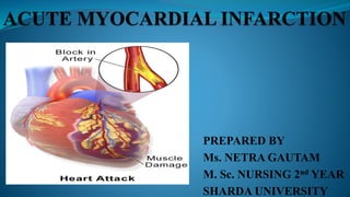
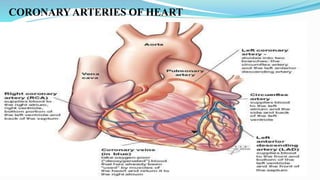
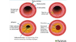
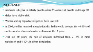
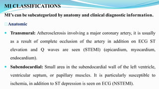
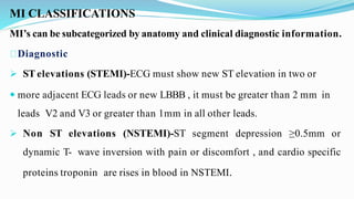
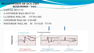
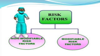
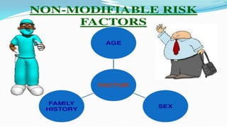
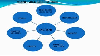
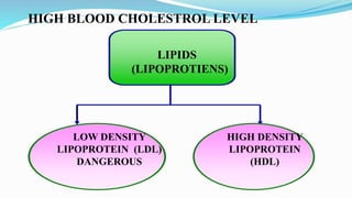
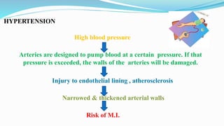
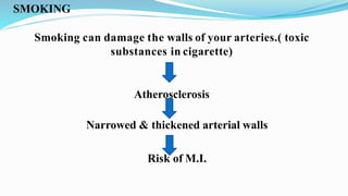
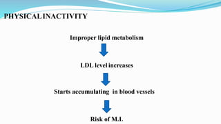
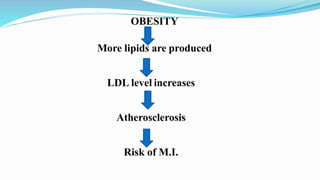
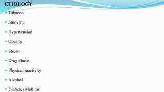
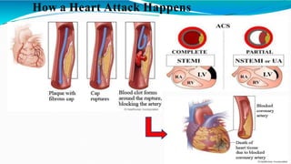
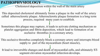
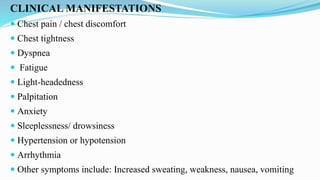
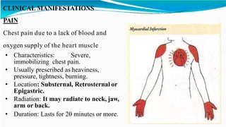
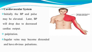
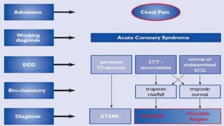
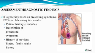
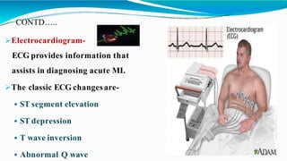
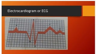
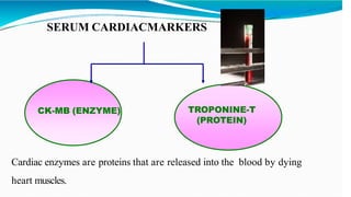
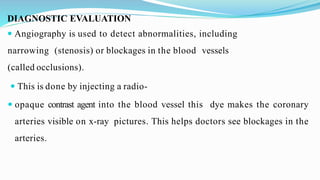
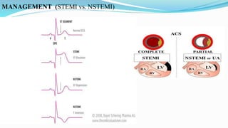
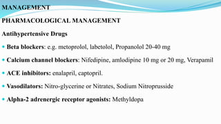
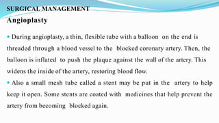
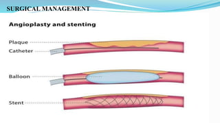
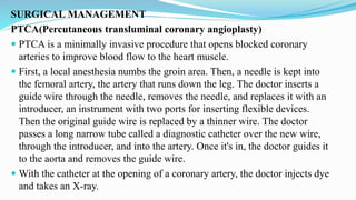
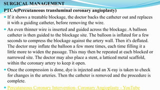
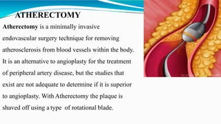
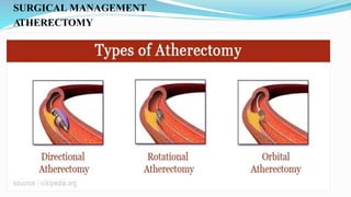
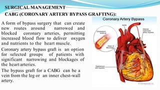
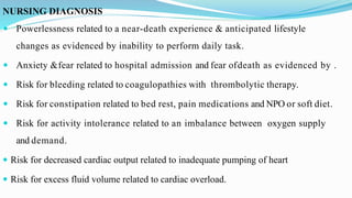
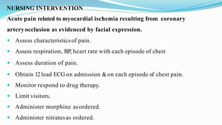
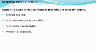
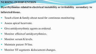
PREPARED BY
Ms. NETRA GAUTAM
M. Sc. NURSING 2nd YEAR
SHARDA UNIVERSITY
OBJECTIVES
General Objectives: At the end of the class, students will be able to explain about acute myocardial infarction.
Specific Objective: At the end of this class, students will be able:
 To introduce acute myocardial infarction.
 To define the acute myocardial infarction.
 To enumerate the incidence of acute myocardial infarction.
 To explain the classifications of acute myocardial infarction.
 To enlist the risk factors of acute myocardial infarction.
 To state the etiological factors of acute myocardial infarction.
 To explain pathophysiology of acute myocardial infarction
 To enumerate the clinical features of acute myocardial infarction.
 To state the diagnostic evaluation of acute myocardial infarction.
 To describe the management of acute myocardial infarction.
 To state the complications of acute myocardial infarction.
 To explain nursing management of acute myocardial infarction
 To state the preventive measures for patient with acute myocardial infarction.
 To enlist health education for patient with acute myocardial infarction
 To summarize
CORONARYARTERIES OF HEART
Tunica Intima
Tunica media
T.Adventitia
plaques
Thrombus
Atherosclerosis –is a narrowing of the
arteries caused by a buildup of plaque
INTRODUCTION
 Acute Myocardial infarction (MI) is also known as heart attack. A heart
attack occurs when one of the heart's coronary arteries is blocked due to
the formation of a blood clot (thrombus) suddenly or has extremely slow
blood flow. When blood flow decreases or stops to a part of the heart that
cause damage to the heart muscle.
 Myocardial infarction is the irreversible death (necrosis) of heart muscle
secondary to prolonged lack of oxygen supply (ischemia).
 MI can be fatal, but treatment has improved dramatically over the years.
DEFINITION
 Myocardial infarction (MI) is defined as a disease condition which is
caused by reduced blood flow in a coronary artery due to atherosclerosis &
occlusion of an artery by an embolus or thrombus.
 A heart attack occurs when the flow of blood to the heart is blocked. The
blockage is most often a buildup of fat, cholesterol and other substances,
which form a plaque in the arteries that feed the heart (coronary arteries).
The plaque eventually breaks away and forms a clot. The interrupted blood
flow can damage or destroy part of the heart muscle.
INCIDENCE
 Incidence is higher in elderly people, about 5% occurs at people under age 40.
 Males have higher risk.
 Women during reproductive period have low risk.
 In 2006, studies revealed a prediction that India would account for 40-60% of
cardiovascular diseases burden within next 10-15 years.
 Over last 30 years, the rate of diseases increased from 2- 6% in rural
population and 4-12% in urban population.
MI CLASSIFICATIONS
MI’s can be subcategorized by anatomy and clinical diagnostic information.
Anatomic
 Transmural: Atherosclerosis involving a major coronary artery, it is usually
as a result of complete occlusion of the artery in addition on ECG ST
elevation and Q waves are seen (STEMI) (epicardium, myocardium,
endocardium).
 Subendocardial: Small area in the subendocardial wall of the left ventricle,
ventricular septum, or papillary muscles. It is particularly susceptible to
ischemia, in addition to ST depression is seen on ECG (NSTEMI).
MI CLASSIFICATIONS
MI’s can be subcategorized by anatomy and clinical diagnostic information.
Diagnostic
 ST elevations (STEMI)-ECG must show new ST elevation in two or
 more adjacent ECG leads or new LBBB , it must be greater than 2 mm in
leads V2 and V3 or greater than 1mm in all other leads.
 Non ST elevations (NSTEMI)-ST segment depression ≥0.5mm or
dynamic T- wave inversion with pain or discomfort , and cardio specific
proteins troponin are rises in blood in NSTEMI.
RECOGNITION OF ECG TIPS
REMEMEBER - SALI
• S-SEPTALMI-V1 V2
• A-ANTERIOR WALLMI-V3 V4
• L-LATERALWALL MI -V5 V6-1-AVL
• I-INFERIOR WALLMI- 11-111-A
VF
• POSTERIOR WALL MI- ST V1-V3,ST V7-V9
MODIFIABLE RISK FACTORS
FACTOR
HIGH BLOOD
LIPIDS LEVEL
HYPERTENSION
SMOKING
PHYSICAL
INACTIVITY
OBESITY
DIABETES
MELLITUS
STRESS
LIPIDS
(LIPOPROTIENS)
LOW DENSITY
LIPOPROTEIN (LDL)
DANGEROUS
HIGH DENSITY
LIPOPROTEIN
(HDL)
HIGH BLOOD CHOLESTROL LEVEL
HYPERTENSION
High blood pressure
Arteries are designed to pump blood at a certain pressure. If that
pressure is exceeded, the walls of the arteries will be damaged.
Injury to endothelial lining , atherosclerosis
Narrowed & thickened arterial walls
Risk of M.I.
SMOKING
Smoking can damage the walls of your arteries.( toxic
substances in cigarette)
Atherosclerosis
Narrowed & thickened arterial walls
Risk of M.I.
PHYSICALINACTIVITY
Improper lipid metabolism
LDL level increases
Starts accumulating in blood vessels
Risk of M.I.
OBESITY
More lipids are produced
LDL level increases
Atherosclerosis
Risk of M.I.
DIABETESMELLITUS
Diabetes increases the risk of MI because it increases the rate of
atherosclerotic progression and adversely affects the lipid profile.
Risk of having M.I.
STRESS
Release stress hormones like adrenaline, noradrenaline, and cortisol
Increase in heart rate, and elevated blood pressure
It’s causing damage over time to all your blood vessel
That damage increases the risk of plaque buildup in coronary arteries or can
even cause a rupture of plaque
MI
The way we handle stress also matters. If person respond to it in unhealthy
ways; such as smoking, over eating, or not exercising that makes matters
worse.
ETIOLOGY
 Tobacco
 Smoking
 Hypertension
 Obesity
 Stress
 Drug abuse
 Physical inactivity
 Alcohol
 Diabetes Mellitus
How a Heart Attack Happens
PATHOPHYSIOLOGY
Cholesterol deposition within the wall of the main artery
This deposited cholesterol ultimately forms a plaque in the wall of the artery
called atherosclerotic plaque. Atherosclerotic plaque formation is a long term
process, required many years to establish.
Sometimes this plaque may rupture, it leads to activate clotting mechanism so
platelet aggregation and fibrin deposition, which lead to formation of an
occlusive thrombus in a coronary artery.
This occlusive thrombus completely block a coronary artery and interrupts blood
supply to part of the myocardium (heart muscle),
It lead to irreversible changes and death of myocardial cells, and ultimately ST-
segment elevation myocardial infarction develops.
CLINICAL MANIFESTATIONS
 Chest pain / chest discomfort
 Chest tightness
 Dyspnea
 Fatigue
 Light-headedness
 Palpitation
 Anxiety
 Sleeplessness/ drowsiness
 Hypertension or hypotension
 Arrhythmia
 Other symptoms include: Increased sweating, weakness, nausea, vomiting
CLINICAL MANIFESTATIONS
PAIN
Chest pain due to a lack of blood and
oxygen supply of the heart muscle
• Characteristics: Severe,
immobilizing chest pain.
• Usually prescribed as heaviness,
pressure, tightness, burning.
• Location: Substernal, Retrosternal or
Epigastric.
• Radiation: It may radiate to neck, jaw,
arm or back.
• Duration: Lasts for 20 minutes or more.
Cardiovascular System
Initially the BP and pulse
may be elevated. Later, BP
will drop due to decreased
cardiac output.
 palpitation.
Jugular veins may become distended
and have obvious pulsations.
Respiratory system
Respiratory symptoms occur when the damaged the
heart muscle limits the pumping action of the left
ventricle, causing acute left heart failure and
consequent lung congestion.
 Shortness of breath
 Dyspnea/Tachypnea
 Crackles
 Pulmonary edema
Gastrointestinal system
 Nausea
 Vomiting
Stimulation of vomiting center by severe pain causes
nausea & vomiting
FEVER
It is due to inflammatory process caused by
Myocardial cell death.
Integumentary system (Skin)
• Cool and clammy skin/ Coolness of extremities
• Diaphoresis
• Pallor/ Cyanosis
In response to pain and the blood flow abnormalities that result from
dysfunction of the heart muscle
Sympathetic nervous system stimulation
Increased catecholamine releases.(Adrenal medulla)
Diaphoresis (perfuse sweating
Cold & clammy skin (“cold sweat”).
GENITOURINARY-SYSTEM:
Myocardial damage
Failure of the pump action of the heart, resulting in reduced
cardiac output
Hypoperfusion to the kidneys leads to decrease renal perfusion pressure which
is required to maintain
glomerular filteration rate in the kidney
Decrease GFR leads to decrease urinary output
Urine output (Oliguria): 30ml/HR or <400ml/day.
 NEUROGENIC SYSTEM: due to inadequate blood flow to the brain
 Light- headedness
 Headache
 Visual Disturbances
 Altered speech
 Altered motor functions
 Altered level of consciousness
PSYCHOSOCIAL
• Fear feeling
• Patient may deny that anything is wrong
DIAGNOSTIC EVALUATION
 History collection
 Physical examination
 Serum cardiac markers
 Blood tests
 ECG
 Chest X-ray
 Ct scan
 MRI
 Angiography
 PET scan
 A stress test
 Coronary catheterization
 An echocardiogram
ASSESSMENT/DIAGNOSTIC FINDINGS
It is generally based on presenting symptoms,
ECG and laboratory test results.
 Patient history-it includes
• Description of
presenting
symptoms
• History of previous
illness, family health
history
CONTD…..
Electrocardiogram-
ECG provides information that
assists in diagnosing acute MI.
The classic ECG changesare-
 ST segment elevation
 ST depression
 T wave inversion
 Abnormal Q wave
RECOGNITION OF ECG TIPS
REMEMEBER - SALI
• S-SEPTALMI-V1 V2
• A-ANTERIOR WALLMI-V3 V4
• L-LATERALWALL MI -V5 V6-1-AVL
• I-INFERIOR WALLMI- 11-111-A
VF
• POSTERIOR WALL MI- ST V1-V3,ST V7-V9
SERUM CARDIACMARKERS
CK-MB (ENZYME) TROPONINE-T
(PROTEIN)
Cardiac enzymes are proteins that are released into the blood by dying
heart muscles.
DIAGNOSTIC EVALUATION
 Angiography is used to detect abnormalities, including
narrowing (stenosis) or blockages in the blood vessels
(called occlusions).
 This is done by injecting a radio-
 opaque contrast agent into the blood vessel this dye makes the coronary
arteries visible on x-ray pictures. This helps doctors see blockages in the
arteries.
MANAGEMENT
GOALS
• Restoration of the balance between the oxygen supply and
demand to prevent further ischemia
• Pain relief
• Prevention and treatment of any complications that may
arise
Chest pain suggestive of ischemia
Vital signs
12lead ECG
Obtain initial
cardiac enzymes
electrolytes, cbc
lipids, bun/cr,
glucose, coags
CXR
Immediate assessment within 10
Minutes
Initial labs
and tests
History &
Physical
Emergent
care
MANAGEMENT (STEMI vs. NSTEMI)
MANAGEMENT
PHARMACOLOGICAL MANAGEMENT
Antihypertensive Drugs
 Beta blockers: e.g. metoprolol, labetolol, Propanolol 20-40 mg
 Calcium channel blockers: Nifedipine, amlodipine 10 mg or 20 mg, Verapamil
 ACE inhibitors: enalapril, captopril.
 Vasodilators: Nitro-glycerine or Nitrates, Sodium Nitroprusside
 Alpha-2 adrenergic receptor agonists: Methyldopa
MANAGEMENT
PHARMACOLOGICAL MANAGEMENT
 Statins: Atorvastatin, Lovastatin
 Antiplatelet drug:
Aspirin with a rapid effect, which reduces mortality by 20%. Aspirin, 150-300
mg, should be swallowed as early as possible.
Clopidorgel 300mg
 Thrombolytic drugs: Streptokinase, Urokinase
 Opiate Analgesics: Morphine Sulphate
 Anticoagulants: Heparin
SURGICAL MANAGEMENT
Angioplasty
 During angioplasty, a thin, flexible tube with a balloon on the end is
threaded through a blood vessel to the blocked coronary artery. Then, the
balloon is inflated to push the plaque against the wall of the artery. This
widens the inside of the artery, restoring blood flow.
 Also a small mesh tube called a stent may be put in the artery to help
keep it open. Some stents are coated with medicines that help prevent the
artery from becoming blocked again.
SURGICAL MANAGEMENT
SURGICAL MANAGEMENT
PTCA(Percutaneous transluminal coronary angioplasty)
 PTCA is a minimally invasive procedure that opens blocked coronary
arteries to improve blood flow to the heart muscle.
 First, a local anesthesia numbs the groin area. Then, a needle is kept into
the femoral artery, the artery that runs down the leg. The doctor inserts a
guide wire through the needle, removes the needle, and replaces it with an
introducer, an instrument with two ports for inserting flexible devices.
Then the original guide wire is replaced by a thinner wire. The doctor
passes a long narrow tube called a diagnostic catheter over the new wire,
through the introducer, and into the artery. Once it's in, the doctor guides it
to the aorta and removes the guide wire.
 With the catheter at the opening of a coronary artery, the doctor injects dye
and takes an X-ray.
SURGICAL MANAGEMENT
PTCA(Percutaneous transluminal coronary angioplasty)
 If it shows a treatable blockage, the doctor backs the catheter out and replaces
it with a guiding catheter, before removing the wire.
 An even thinner wire is inserted and guided across the blockage. A balloon
catheter is then guided to the blockage site. The balloon is inflated for a few
seconds to compress the blockage against the artery wall. Then it's deflated.
The doctor may inflate the balloon a few more times, each time filling it a
little more to widen the passage. This may then be repeated at each blocked or
narrowed site. The doctor may also place a stent, a latticed metal scaffold,
within the coronary artery to keep it open.
 Once the compression is done, dye is injected and an X-ray is taken to check
for changes in the arteries. Then the catheter is removed and the procedure is
complete.
 Percutaneous Coronary Intervention- Coronary Angioplasty - YouTube
ATHERECTOMY
Atherectomy is a minimally invasive
endovascular surgery technique for removing
atherosclerosis from blood vessels within the body.
It is an alternative to angioplasty for the treatment
of peripheral artery disease, but the studies that
exist are not adequate to determine if it is superior
to angioplasty. With Atherectomy the plaque is
shaved off using a type of rotational blade.
SURGICAL MANAGEMENT
ATHERECTOMY
SURGICAL MANAGEMENT
CABG (CORONARY ARTERY BYPASS GRAFTING):
A form of bypass surgery that can create
new routes around narrowed and
blocked coronary arteries, permitting
increased blood flow to deliver oxygen
and nutrients to the heart muscle.
Coronary artery bypass graft is an option
for selected groups of patients with
significant narrowing and blockages of
the heart arteries.
The bypass graft for a CABG can be a
vein from the leg or an inner chest-wall
artery.
SURGICAL MANAGEMENT
CABG (CORONARY ARTERY BYPASS GRAFTING):
 Coronary artery bypass surgery is performed as a emergency bypass
surgery at the time of a heart attack. After bypass surgery heart takes about
three to seven days to recover from heart attack. Bypass surgery involves
sewing veins or arteries in place beyond a blocked or narrowed coronary
artery, allowing blood flow to the heart to bypass the narrowed section.
Once blood flow to heart is restored and condition is stable, patient is likely
to remain in the hospital for several days.
 https://www.youtube.com/watch?v=e1PUec4i0G0
COMPLICATIONS
Dysrrythmias
Heart failure
Acute pulmonary edema
Cardiogenic shock
Embolism
Papillary muscle dysfunction
Sudden cardiac arrest
Mechanical complications: It includes: Severemitral regurgitation, Cardiac tamponade,
Pericarditis and Right heart failure.
Late complications: It include Dressler’ssyndrome
NURSING MANAGEMENT
ASSESSMENT
One of the most important aspects of care of the patient with MI is the
assessment.
 Assess for chest pain not relieved by rest or medications.
 Monitor vital signs, especially the blood pressure and pulse rate.
 Assess for presence of shortness of breath, dyspnea, tachypnea, and crackles.
 Assess for nausea and vomiting.
 Assess for decreased urinary output.
 Assess for the history of illnesses.
 Perform a precise and complete physical assessment to detect complications
and changes in the patient’s status.
 Assess IV sites frequently.
NURSING DIAGNOSIS
 Acute pain related to ischemia of heart muscles from coronary artery occlusion as evidenced
by facial expression.
 Ineffective cardiac tissue perfusion related to reduced coronary blood flow from coronary
thrombus and atherosclerotic plaque as evidenced by less Spo2 .
 Dysrrhythmias related to electrical instability or irritability secondary to infarctedtissue.
 Ineffective gas exchange related to ineffective breathing pattern as evidenced by increased
breath rate.
 Ineffective health maintenance related to MI &implications for lifestylechanges.
 Activity intolerance related to fatigue as evidenced by visualization.
 Deficient Knowledge related to post MI and self care as evidenced by poor knowledge on care.
NURSING DIAGNOSIS
 Powerlessness related to a near-death experience & anticipated lifestyle
changes as evidenced by inability to perform daily task.
 Anxiety &fear related to hospital admission and fear ofdeath as evidenced by .
 Risk for bleeding related to coagulopathies with thrombolytic therapy.
 Risk for constipation related to bed rest, pain medications and NPO or soft diet.
 Risk for activity intolerance related to an imbalance between oxygen supply
and demand.
 Risk for decreased cardiac output related to inadequate pumping of heart
 Risk for excess fluid volume related to cardiac overload.
NURSING INTERVENTION
Acute pain related to myocardial ischemia resulting from coronary
artery occlusion as evidenced by facial expression.
 Assess characteristics of pain.
 Assess respiration, BP, heart rate with each episode of chest
 Assess duration of pain.
 Obtain 12lead ECG on admission &on each episode of chest pain.
 Monitor respond to drug therapy.
 Limit visitors.
 Administer morphine asordered.
 Administer nitratesas ordered.
NURSING INTERVENTION
Ineffective tissue perfusion related to thrombus in coronary artery.
 Provide bed rest.
 Administeroxygen as prescribed.
 Administer thrombolytics.
 Monitor STsegments.
NURSING INTERVENTION
Dysrrhythmias related to electrical instability or irritability secondary to
infarcted tissue.
 Teach client &family about need for continous monitoring.
 Assess apical heart rate.
 Give antidysrrythmic agents as ordered.
 Monitor effectsof antidysrrythmics.
 Monitor serum Klevels.
 Maintain patent IVline.
 Monitor ST segments &document changes.
NURSING INTERVENTION
 Closely watch the vital signs and check for consciousness.
 Monitor ECG waves
 Monitor laboratory values
 Administer oxygen as per need or as per prescription.
 Assess the level of pain.
 Educate the patient to avoid heavy lifting and to take proper rest.
 Encourage patient to take nutritious diet.
 Educate patient to avoid smoking, alcohol, consumption of junk food.
PREVENTION
 Medications
 Lifestyle factors
 Avoid smoke
 Control your blood pressure and cholesterol levels
 Get regular medical checkups
 Exercise
 Maintain a healthy weight
 Eat a heart-healthy diet
 Control stress
 Avoid drinking alcohol
HEALTH EDUCATION
Proper medication compliance (right dose and right time)
Perform exercise
Do not smoke
Follow thediet plan
Maintain a healthy weight
Manage your stress
Signs and symptoms to be reported to physician
SUMMARY
We discussed on definition, incidence, etiology, risk factors, classification,
clinical features, diagnostic studies, management, complications, nursing
management and preventive measures of acute myocardial infarction..
CONCLUSION
Myocardial infarction (MI) is a life threatening disease caused by many
factors. MI is a major public health problem and the leading cause of mortality
in both developed and developing countries. Patients with severe heart failure
require invasive treatment such as mechanical circulatory support, continuous
inotropic infusion, or cardiac transplantation. Health education must be
given to the patients with predisposing or risk factors to prevent it. Early
diagnosis is also very important for saving the life of the patient.

Acute myocardial infraction

  • 1.
    PREPARED BY Ms. NETRAGAUTAM M. Sc. NURSING 2nd YEAR SHARDA UNIVERSITY
  • 2.
    OBJECTIVES General Objectives: Atthe end of the class, students will be able to explain about acute myocardial infarction. Specific Objective: At the end of this class, students will be able:  To introduce acute myocardial infarction.  To define the acute myocardial infarction.  To enumerate the incidence of acute myocardial infarction.  To explain the classifications of acute myocardial infarction.  To enlist the risk factors of acute myocardial infarction.  To state the etiological factors of acute myocardial infarction.  To explain pathophysiology of acute myocardial infarction  To enumerate the clinical features of acute myocardial infarction.  To state the diagnostic evaluation of acute myocardial infarction.  To describe the management of acute myocardial infarction.  To state the complications of acute myocardial infarction.  To explain nursing management of acute myocardial infarction  To state the preventive measures for patient with acute myocardial infarction.  To enlist health education for patient with acute myocardial infarction  To summarize
  • 3.
  • 4.
    Tunica Intima Tunica media T.Adventitia plaques Thrombus Atherosclerosis–is a narrowing of the arteries caused by a buildup of plaque
  • 5.
    INTRODUCTION  Acute Myocardialinfarction (MI) is also known as heart attack. A heart attack occurs when one of the heart's coronary arteries is blocked due to the formation of a blood clot (thrombus) suddenly or has extremely slow blood flow. When blood flow decreases or stops to a part of the heart that cause damage to the heart muscle.  Myocardial infarction is the irreversible death (necrosis) of heart muscle secondary to prolonged lack of oxygen supply (ischemia).  MI can be fatal, but treatment has improved dramatically over the years.
  • 7.
    DEFINITION  Myocardial infarction(MI) is defined as a disease condition which is caused by reduced blood flow in a coronary artery due to atherosclerosis & occlusion of an artery by an embolus or thrombus.  A heart attack occurs when the flow of blood to the heart is blocked. The blockage is most often a buildup of fat, cholesterol and other substances, which form a plaque in the arteries that feed the heart (coronary arteries). The plaque eventually breaks away and forms a clot. The interrupted blood flow can damage or destroy part of the heart muscle.
  • 9.
    INCIDENCE  Incidence ishigher in elderly people, about 5% occurs at people under age 40.  Males have higher risk.  Women during reproductive period have low risk.  In 2006, studies revealed a prediction that India would account for 40-60% of cardiovascular diseases burden within next 10-15 years.  Over last 30 years, the rate of diseases increased from 2- 6% in rural population and 4-12% in urban population.
  • 10.
    MI CLASSIFICATIONS MI’s canbe subcategorized by anatomy and clinical diagnostic information. Anatomic  Transmural: Atherosclerosis involving a major coronary artery, it is usually as a result of complete occlusion of the artery in addition on ECG ST elevation and Q waves are seen (STEMI) (epicardium, myocardium, endocardium).  Subendocardial: Small area in the subendocardial wall of the left ventricle, ventricular septum, or papillary muscles. It is particularly susceptible to ischemia, in addition to ST depression is seen on ECG (NSTEMI).
  • 11.
    MI CLASSIFICATIONS MI’s canbe subcategorized by anatomy and clinical diagnostic information. Diagnostic  ST elevations (STEMI)-ECG must show new ST elevation in two or  more adjacent ECG leads or new LBBB , it must be greater than 2 mm in leads V2 and V3 or greater than 1mm in all other leads.  Non ST elevations (NSTEMI)-ST segment depression ≥0.5mm or dynamic T- wave inversion with pain or discomfort , and cardio specific proteins troponin are rises in blood in NSTEMI.
  • 12.
    RECOGNITION OF ECGTIPS REMEMEBER - SALI • S-SEPTALMI-V1 V2 • A-ANTERIOR WALLMI-V3 V4 • L-LATERALWALL MI -V5 V6-1-AVL • I-INFERIOR WALLMI- 11-111-A VF • POSTERIOR WALL MI- ST V1-V3,ST V7-V9
  • 15.
    MODIFIABLE RISK FACTORS FACTOR HIGHBLOOD LIPIDS LEVEL HYPERTENSION SMOKING PHYSICAL INACTIVITY OBESITY DIABETES MELLITUS STRESS
  • 16.
    LIPIDS (LIPOPROTIENS) LOW DENSITY LIPOPROTEIN (LDL) DANGEROUS HIGHDENSITY LIPOPROTEIN (HDL) HIGH BLOOD CHOLESTROL LEVEL
  • 17.
    HYPERTENSION High blood pressure Arteriesare designed to pump blood at a certain pressure. If that pressure is exceeded, the walls of the arteries will be damaged. Injury to endothelial lining , atherosclerosis Narrowed & thickened arterial walls Risk of M.I.
  • 18.
    SMOKING Smoking can damagethe walls of your arteries.( toxic substances in cigarette) Atherosclerosis Narrowed & thickened arterial walls Risk of M.I.
  • 19.
    PHYSICALINACTIVITY Improper lipid metabolism LDLlevel increases Starts accumulating in blood vessels Risk of M.I.
  • 20.
    OBESITY More lipids areproduced LDL level increases Atherosclerosis Risk of M.I.
  • 21.
    DIABETESMELLITUS Diabetes increases therisk of MI because it increases the rate of atherosclerotic progression and adversely affects the lipid profile. Risk of having M.I.
  • 22.
    STRESS Release stress hormoneslike adrenaline, noradrenaline, and cortisol Increase in heart rate, and elevated blood pressure It’s causing damage over time to all your blood vessel That damage increases the risk of plaque buildup in coronary arteries or can even cause a rupture of plaque MI The way we handle stress also matters. If person respond to it in unhealthy ways; such as smoking, over eating, or not exercising that makes matters worse.
  • 23.
    ETIOLOGY  Tobacco  Smoking Hypertension  Obesity  Stress  Drug abuse  Physical inactivity  Alcohol  Diabetes Mellitus
  • 24.
    How a HeartAttack Happens
  • 25.
    PATHOPHYSIOLOGY Cholesterol deposition withinthe wall of the main artery This deposited cholesterol ultimately forms a plaque in the wall of the artery called atherosclerotic plaque. Atherosclerotic plaque formation is a long term process, required many years to establish. Sometimes this plaque may rupture, it leads to activate clotting mechanism so platelet aggregation and fibrin deposition, which lead to formation of an occlusive thrombus in a coronary artery. This occlusive thrombus completely block a coronary artery and interrupts blood supply to part of the myocardium (heart muscle), It lead to irreversible changes and death of myocardial cells, and ultimately ST- segment elevation myocardial infarction develops.
  • 26.
    CLINICAL MANIFESTATIONS  Chestpain / chest discomfort  Chest tightness  Dyspnea  Fatigue  Light-headedness  Palpitation  Anxiety  Sleeplessness/ drowsiness  Hypertension or hypotension  Arrhythmia  Other symptoms include: Increased sweating, weakness, nausea, vomiting
  • 27.
    CLINICAL MANIFESTATIONS PAIN Chest paindue to a lack of blood and oxygen supply of the heart muscle • Characteristics: Severe, immobilizing chest pain. • Usually prescribed as heaviness, pressure, tightness, burning. • Location: Substernal, Retrosternal or Epigastric. • Radiation: It may radiate to neck, jaw, arm or back. • Duration: Lasts for 20 minutes or more.
  • 28.
    Cardiovascular System Initially theBP and pulse may be elevated. Later, BP will drop due to decreased cardiac output.  palpitation. Jugular veins may become distended and have obvious pulsations.
  • 29.
    Respiratory system Respiratory symptomsoccur when the damaged the heart muscle limits the pumping action of the left ventricle, causing acute left heart failure and consequent lung congestion.  Shortness of breath  Dyspnea/Tachypnea  Crackles  Pulmonary edema
  • 30.
    Gastrointestinal system  Nausea Vomiting Stimulation of vomiting center by severe pain causes nausea & vomiting FEVER It is due to inflammatory process caused by Myocardial cell death.
  • 31.
    Integumentary system (Skin) •Cool and clammy skin/ Coolness of extremities • Diaphoresis • Pallor/ Cyanosis In response to pain and the blood flow abnormalities that result from dysfunction of the heart muscle Sympathetic nervous system stimulation Increased catecholamine releases.(Adrenal medulla) Diaphoresis (perfuse sweating Cold & clammy skin (“cold sweat”).
  • 32.
    GENITOURINARY-SYSTEM: Myocardial damage Failure ofthe pump action of the heart, resulting in reduced cardiac output Hypoperfusion to the kidneys leads to decrease renal perfusion pressure which is required to maintain glomerular filteration rate in the kidney Decrease GFR leads to decrease urinary output Urine output (Oliguria): 30ml/HR or <400ml/day.
  • 33.
     NEUROGENIC SYSTEM:due to inadequate blood flow to the brain  Light- headedness  Headache  Visual Disturbances  Altered speech  Altered motor functions  Altered level of consciousness PSYCHOSOCIAL • Fear feeling • Patient may deny that anything is wrong
  • 34.
    DIAGNOSTIC EVALUATION  Historycollection  Physical examination  Serum cardiac markers  Blood tests  ECG  Chest X-ray  Ct scan  MRI  Angiography  PET scan  A stress test  Coronary catheterization  An echocardiogram
  • 36.
    ASSESSMENT/DIAGNOSTIC FINDINGS It isgenerally based on presenting symptoms, ECG and laboratory test results.  Patient history-it includes • Description of presenting symptoms • History of previous illness, family health history
  • 37.
    CONTD….. Electrocardiogram- ECG provides informationthat assists in diagnosing acute MI. The classic ECG changesare-  ST segment elevation  ST depression  T wave inversion  Abnormal Q wave
  • 39.
    RECOGNITION OF ECGTIPS REMEMEBER - SALI • S-SEPTALMI-V1 V2 • A-ANTERIOR WALLMI-V3 V4 • L-LATERALWALL MI -V5 V6-1-AVL • I-INFERIOR WALLMI- 11-111-A VF • POSTERIOR WALL MI- ST V1-V3,ST V7-V9
  • 40.
    SERUM CARDIACMARKERS CK-MB (ENZYME)TROPONINE-T (PROTEIN) Cardiac enzymes are proteins that are released into the blood by dying heart muscles.
  • 41.
    DIAGNOSTIC EVALUATION  Angiographyis used to detect abnormalities, including narrowing (stenosis) or blockages in the blood vessels (called occlusions).  This is done by injecting a radio-  opaque contrast agent into the blood vessel this dye makes the coronary arteries visible on x-ray pictures. This helps doctors see blockages in the arteries.
  • 42.
    MANAGEMENT GOALS • Restoration ofthe balance between the oxygen supply and demand to prevent further ischemia • Pain relief • Prevention and treatment of any complications that may arise
  • 43.
    Chest pain suggestiveof ischemia Vital signs 12lead ECG Obtain initial cardiac enzymes electrolytes, cbc lipids, bun/cr, glucose, coags CXR Immediate assessment within 10 Minutes Initial labs and tests History & Physical Emergent care
  • 44.
  • 45.
    MANAGEMENT PHARMACOLOGICAL MANAGEMENT Antihypertensive Drugs Beta blockers: e.g. metoprolol, labetolol, Propanolol 20-40 mg  Calcium channel blockers: Nifedipine, amlodipine 10 mg or 20 mg, Verapamil  ACE inhibitors: enalapril, captopril.  Vasodilators: Nitro-glycerine or Nitrates, Sodium Nitroprusside  Alpha-2 adrenergic receptor agonists: Methyldopa
  • 46.
    MANAGEMENT PHARMACOLOGICAL MANAGEMENT  Statins:Atorvastatin, Lovastatin  Antiplatelet drug: Aspirin with a rapid effect, which reduces mortality by 20%. Aspirin, 150-300 mg, should be swallowed as early as possible. Clopidorgel 300mg  Thrombolytic drugs: Streptokinase, Urokinase  Opiate Analgesics: Morphine Sulphate  Anticoagulants: Heparin
  • 47.
    SURGICAL MANAGEMENT Angioplasty  Duringangioplasty, a thin, flexible tube with a balloon on the end is threaded through a blood vessel to the blocked coronary artery. Then, the balloon is inflated to push the plaque against the wall of the artery. This widens the inside of the artery, restoring blood flow.  Also a small mesh tube called a stent may be put in the artery to help keep it open. Some stents are coated with medicines that help prevent the artery from becoming blocked again.
  • 48.
  • 49.
    SURGICAL MANAGEMENT PTCA(Percutaneous transluminalcoronary angioplasty)  PTCA is a minimally invasive procedure that opens blocked coronary arteries to improve blood flow to the heart muscle.  First, a local anesthesia numbs the groin area. Then, a needle is kept into the femoral artery, the artery that runs down the leg. The doctor inserts a guide wire through the needle, removes the needle, and replaces it with an introducer, an instrument with two ports for inserting flexible devices. Then the original guide wire is replaced by a thinner wire. The doctor passes a long narrow tube called a diagnostic catheter over the new wire, through the introducer, and into the artery. Once it's in, the doctor guides it to the aorta and removes the guide wire.  With the catheter at the opening of a coronary artery, the doctor injects dye and takes an X-ray.
  • 50.
    SURGICAL MANAGEMENT PTCA(Percutaneous transluminalcoronary angioplasty)  If it shows a treatable blockage, the doctor backs the catheter out and replaces it with a guiding catheter, before removing the wire.  An even thinner wire is inserted and guided across the blockage. A balloon catheter is then guided to the blockage site. The balloon is inflated for a few seconds to compress the blockage against the artery wall. Then it's deflated. The doctor may inflate the balloon a few more times, each time filling it a little more to widen the passage. This may then be repeated at each blocked or narrowed site. The doctor may also place a stent, a latticed metal scaffold, within the coronary artery to keep it open.  Once the compression is done, dye is injected and an X-ray is taken to check for changes in the arteries. Then the catheter is removed and the procedure is complete.  Percutaneous Coronary Intervention- Coronary Angioplasty - YouTube
  • 51.
    ATHERECTOMY Atherectomy is aminimally invasive endovascular surgery technique for removing atherosclerosis from blood vessels within the body. It is an alternative to angioplasty for the treatment of peripheral artery disease, but the studies that exist are not adequate to determine if it is superior to angioplasty. With Atherectomy the plaque is shaved off using a type of rotational blade.
  • 52.
  • 53.
    SURGICAL MANAGEMENT CABG (CORONARYARTERY BYPASS GRAFTING): A form of bypass surgery that can create new routes around narrowed and blocked coronary arteries, permitting increased blood flow to deliver oxygen and nutrients to the heart muscle. Coronary artery bypass graft is an option for selected groups of patients with significant narrowing and blockages of the heart arteries. The bypass graft for a CABG can be a vein from the leg or an inner chest-wall artery.
  • 54.
    SURGICAL MANAGEMENT CABG (CORONARYARTERY BYPASS GRAFTING):  Coronary artery bypass surgery is performed as a emergency bypass surgery at the time of a heart attack. After bypass surgery heart takes about three to seven days to recover from heart attack. Bypass surgery involves sewing veins or arteries in place beyond a blocked or narrowed coronary artery, allowing blood flow to the heart to bypass the narrowed section. Once blood flow to heart is restored and condition is stable, patient is likely to remain in the hospital for several days.  https://www.youtube.com/watch?v=e1PUec4i0G0
  • 55.
    COMPLICATIONS Dysrrythmias Heart failure Acute pulmonaryedema Cardiogenic shock Embolism Papillary muscle dysfunction Sudden cardiac arrest Mechanical complications: It includes: Severemitral regurgitation, Cardiac tamponade, Pericarditis and Right heart failure. Late complications: It include Dressler’ssyndrome
  • 56.
    NURSING MANAGEMENT ASSESSMENT One ofthe most important aspects of care of the patient with MI is the assessment.  Assess for chest pain not relieved by rest or medications.  Monitor vital signs, especially the blood pressure and pulse rate.  Assess for presence of shortness of breath, dyspnea, tachypnea, and crackles.  Assess for nausea and vomiting.  Assess for decreased urinary output.  Assess for the history of illnesses.  Perform a precise and complete physical assessment to detect complications and changes in the patient’s status.  Assess IV sites frequently.
  • 57.
    NURSING DIAGNOSIS  Acutepain related to ischemia of heart muscles from coronary artery occlusion as evidenced by facial expression.  Ineffective cardiac tissue perfusion related to reduced coronary blood flow from coronary thrombus and atherosclerotic plaque as evidenced by less Spo2 .  Dysrrhythmias related to electrical instability or irritability secondary to infarctedtissue.  Ineffective gas exchange related to ineffective breathing pattern as evidenced by increased breath rate.  Ineffective health maintenance related to MI &implications for lifestylechanges.  Activity intolerance related to fatigue as evidenced by visualization.  Deficient Knowledge related to post MI and self care as evidenced by poor knowledge on care.
  • 58.
    NURSING DIAGNOSIS  Powerlessnessrelated to a near-death experience & anticipated lifestyle changes as evidenced by inability to perform daily task.  Anxiety &fear related to hospital admission and fear ofdeath as evidenced by .  Risk for bleeding related to coagulopathies with thrombolytic therapy.  Risk for constipation related to bed rest, pain medications and NPO or soft diet.  Risk for activity intolerance related to an imbalance between oxygen supply and demand.  Risk for decreased cardiac output related to inadequate pumping of heart  Risk for excess fluid volume related to cardiac overload.
  • 59.
    NURSING INTERVENTION Acute painrelated to myocardial ischemia resulting from coronary artery occlusion as evidenced by facial expression.  Assess characteristics of pain.  Assess respiration, BP, heart rate with each episode of chest  Assess duration of pain.  Obtain 12lead ECG on admission &on each episode of chest pain.  Monitor respond to drug therapy.  Limit visitors.  Administer morphine asordered.  Administer nitratesas ordered.
  • 60.
    NURSING INTERVENTION Ineffective tissueperfusion related to thrombus in coronary artery.  Provide bed rest.  Administeroxygen as prescribed.  Administer thrombolytics.  Monitor STsegments.
  • 61.
    NURSING INTERVENTION Dysrrhythmias relatedto electrical instability or irritability secondary to infarcted tissue.  Teach client &family about need for continous monitoring.  Assess apical heart rate.  Give antidysrrythmic agents as ordered.  Monitor effectsof antidysrrythmics.  Monitor serum Klevels.  Maintain patent IVline.  Monitor ST segments &document changes.
  • 62.
    NURSING INTERVENTION  Closelywatch the vital signs and check for consciousness.  Monitor ECG waves  Monitor laboratory values  Administer oxygen as per need or as per prescription.  Assess the level of pain.  Educate the patient to avoid heavy lifting and to take proper rest.  Encourage patient to take nutritious diet.  Educate patient to avoid smoking, alcohol, consumption of junk food.
  • 63.
    PREVENTION  Medications  Lifestylefactors  Avoid smoke  Control your blood pressure and cholesterol levels  Get regular medical checkups  Exercise  Maintain a healthy weight  Eat a heart-healthy diet  Control stress  Avoid drinking alcohol
  • 64.
    HEALTH EDUCATION Proper medicationcompliance (right dose and right time) Perform exercise Do not smoke Follow thediet plan Maintain a healthy weight Manage your stress Signs and symptoms to be reported to physician
  • 65.
    SUMMARY We discussed ondefinition, incidence, etiology, risk factors, classification, clinical features, diagnostic studies, management, complications, nursing management and preventive measures of acute myocardial infarction..
  • 66.
    CONCLUSION Myocardial infarction (MI)is a life threatening disease caused by many factors. MI is a major public health problem and the leading cause of mortality in both developed and developing countries. Patients with severe heart failure require invasive treatment such as mechanical circulatory support, continuous inotropic infusion, or cardiac transplantation. Health education must be given to the patients with predisposing or risk factors to prevent it. Early diagnosis is also very important for saving the life of the patient.