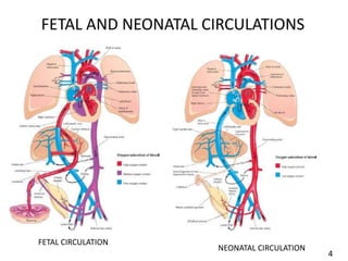
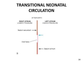
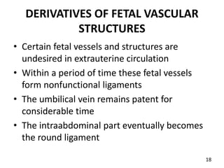
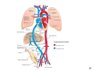
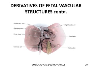
FETAL AND NEONATAL
CIRCULATION
DR KELECHI DURUH
DEPARTMENT OF HUMAN ANATOMY
FBMS COLLEGE OF MEDICINE
UNEC
27/7/23
BY
OUTLINE
• INTRODUCTION
• FETAL CIRCULATION
• TRANSITIONAL CHANGES/TRANSITIONAL
NEONATAL CIRCULATION
• DERIVATIVES FROM THE TRANSITIONAL
CHANGES
• CLINICAL CORRELATES
1
INTRODUCTION
• In fetal circulation, the placenta supplies oxygen,
nutrition and aids in detoxification.
• The lungs are non-functional
• Shunts exist bypassing the lungs and liver
• Though the partial Oxygen pressure is relatively
low, tissues are adequately supplied
• Fetal circulatory system provides prenatal and
perinatal needs and allows for modification at
birth
2
INTRODUCTION contd.
• Modifications establish neonetal circulatory
pattern
• In neonatal circulation, the lungs are aerated
• The severance of placenta from the fetus has
consequential changes
• Some circulatory components undergo functional
and anatomical changes
• Changes bring about directional and dynamic
alterations in blood flow
3
FETAL AND NEONATAL CIRCULATIONS
FETAL CIRCULATION
NEONATAL CIRCULATION
4
FETAL CIRCULATION
• Oxygenated, nutrient rich blood from placenta
passes through the umbilical vein
• At the area of the liver, 50% pass through the
ductus venosus(DV) into the IVC
• The other 50% pass through the portal sinus , the
portal vein, liver sinusoids, hepatic veins into the
IVC
• A sphincter in the DV regulates blood flow
• Contraction of sphincter reduces flow through
the DV increasing flow through liver sinusoids
5
FETAL CIRCULATION contd
• The IVC drains the DV, hepatic veins and the
abdominal vein
• After a short course the IVC, empties into the
right atrium
• Most blood arriving right atrium from IVC are
directed through the foramen ovale into the left
atrium
• In the left atrium ,it mixes with a small quantity
of poorly oxygenated blood returning from the
pulmonary veins
• Left atrium empties into the left ventricle
6
FETAL CIRCULATION contd.
FORAMEN OVALE
7
FETAL CIRCULATION contd
• Left ventricle transmits blood through the
ascending aorta , the arch, the descending aorta,
to the arteries of the trunk and lower body
• The ascending aorta supplies the heart
• The arch supplies the head ,neck and upper limbs
• The residual volume of blood in the right atrium
mixes with poorly oxygenated blood from SVC
and coronary sinus and enters the right ventricle
• The right ventricle ejects blood through the
pulmonary trunk
8
FETAL CIRCULATION contd
• Pulmonary vascular resistance is higher than
that of the systemic
• 90% of pulmonary trunk blood pass through
the ductus arteriosus(DA) into the descending
aorta
• 10% goes to nourish the lungs
• DA protects the lungs from circulatory
overload also aids the right ventricle in
adjusting to postnatal functioning
9
FETAL CIRCULATION contd
• 65% of blood in the descending aorta passes
into the umbilical arteries into the placenta
• 35% nourishes the viscera and inferior part of
the body then returns to the heart through
IVC and SVC
• 90% of left ventricular ejection nourishes the
head, neck and upper limb
• 10% pass down to the descending aorta
10
TRANSITIONAL NEONATAL
CIRCULATION
• Change from fetal to neonatal circulation is not
sudden occurrence
• Some occur with first breath others over hours or
days
• The closure of fetal vessels and oval foramen is
initially a functional change then anatomical
• At cessation of placental flow, changes occur to
exclude placenta in the circulation
• Shunt points are shut down
11
TRANSITIONAL NEONATAL
CIRCULATION
• The foramen ovale, DA, DV and umbilical vessels
become needless
• DV sphincter constricts , diverting all blood to
portal vein
• Pressure in IVC and right atrium consequently
reduce
• Lung expansion and aeration causes decrease in
pulmonary vascular resistance and increase in
pulmonary flow
• Stretching of the lungs causes thinning of
pulmonary artery walls
12
TRANSITIONAL NEONATAL
CIRCULATION
13
TRANSITIONAL NEONATAL
CIRCULATION
14
TRANSITIONAL NEONATAL
CIRCULATION contd.
• Increased pulmonary flow and cessation of
umbilical flow reduces pressure in the right
heart
• Increased pulmonary venous return builds left
atrial pressure
• Higher pressure in the left atrium relative to
right functionally closes the foramen ovale
• DA constricts at birth
15
TRANSITIONAL NEONATAL
CIRCULATION contd.
• Small volume flow from descending aorta into
pulmonary trunk may linger for about 72hrs
• By 96hrs 100% of DA should be functionally
closed
• Oxygen is the main enhancing factor
• Closure is mediated by prostaglandin E2 and
bradykinin
• Factors that may impede normal closure
include prematurity, hypoxia etc
16
TRANSITIONAL NEONATAL
CIRCULATION contd.
• DA constricts when PaO2 of blood in the aorta
approximates 50mmHg
• During fetal life ,patency of DA is maintained
by low oxygen and some endogenous
prostaglandins
• Umbilical arteries constricts at birth
• Blood flow in the vein continues for 1min or
more prior to tying of cord
17
DERIVATIVES OF FETAL VASCULAR
STRUCTURES
• Certain fetal vessels and structures are
undesired in extrauterine circulation
• Within a period of time these fetal vessels
form nonfunctional ligaments
• The umbilical vein remains patent for
considerable time
• The intraabdominal part eventually becomes
the round ligament
18
19
DERIVATIVES OF FETAL VASCULAR
STRUCTURES contd.
UMBILICAL VEIN, DUCTUS VENOSUS 20
DERIVATIVES OF FETAL VASCULAR
STRUCTURES contd.
• DV becomes ligamentum venosum
• Distal part of umbilical arteries become the
medial umbilical ligament
• The proximal part persist as the superior
vesical arteries
• Foramen ovale closes functionally at birth
then anatomically by 3rd month
21
DERIVATIVES OF FETAL VASCULAR
STRUCTURES contd.
• The septum primum forms the floor of the
oval fossa
• The inferior edge of the septum secundum
forms the rounded fold
• DA closes functionally within first four days of
birth
• Anatomical closure and formation of
ligamentum arteriosum occurs at 12th
postnatal week
22
CLINICAL CORRELATES
• Patent ductus arteriosus
• Coartation of the aorta
• Double IVC, non formation of IVC
• Patent foramen ovale
• Pulmonary stenosis
• Double arch of the aorta
• Abnormal origin rt.subclavian
23
CLINICAL CORRELATES
PATENT DUCTUS ARTERIOSUS 24
CLINICAL CORRELATES
PRE AND POST-DUCTAL COARTATIONS OF THE AORTA
25
CLINICAL CORRELATES
PATENT FORAMEN OVALE
26
GOD BLESS YOU
27

FETAL AND NEONATAL CIRCULATION lecture.pptx

  • 1.
    FETAL AND NEONATAL CIRCULATION DRKELECHI DURUH DEPARTMENT OF HUMAN ANATOMY FBMS COLLEGE OF MEDICINE UNEC 27/7/23 BY
  • 2.
    OUTLINE • INTRODUCTION • FETALCIRCULATION • TRANSITIONAL CHANGES/TRANSITIONAL NEONATAL CIRCULATION • DERIVATIVES FROM THE TRANSITIONAL CHANGES • CLINICAL CORRELATES 1
  • 3.
    INTRODUCTION • In fetalcirculation, the placenta supplies oxygen, nutrition and aids in detoxification. • The lungs are non-functional • Shunts exist bypassing the lungs and liver • Though the partial Oxygen pressure is relatively low, tissues are adequately supplied • Fetal circulatory system provides prenatal and perinatal needs and allows for modification at birth 2
  • 4.
    INTRODUCTION contd. • Modificationsestablish neonetal circulatory pattern • In neonatal circulation, the lungs are aerated • The severance of placenta from the fetus has consequential changes • Some circulatory components undergo functional and anatomical changes • Changes bring about directional and dynamic alterations in blood flow 3
  • 5.
    FETAL AND NEONATALCIRCULATIONS FETAL CIRCULATION NEONATAL CIRCULATION 4
  • 6.
    FETAL CIRCULATION • Oxygenated,nutrient rich blood from placenta passes through the umbilical vein • At the area of the liver, 50% pass through the ductus venosus(DV) into the IVC • The other 50% pass through the portal sinus , the portal vein, liver sinusoids, hepatic veins into the IVC • A sphincter in the DV regulates blood flow • Contraction of sphincter reduces flow through the DV increasing flow through liver sinusoids 5
  • 7.
    FETAL CIRCULATION contd •The IVC drains the DV, hepatic veins and the abdominal vein • After a short course the IVC, empties into the right atrium • Most blood arriving right atrium from IVC are directed through the foramen ovale into the left atrium • In the left atrium ,it mixes with a small quantity of poorly oxygenated blood returning from the pulmonary veins • Left atrium empties into the left ventricle 6
  • 8.
  • 9.
    FETAL CIRCULATION contd •Left ventricle transmits blood through the ascending aorta , the arch, the descending aorta, to the arteries of the trunk and lower body • The ascending aorta supplies the heart • The arch supplies the head ,neck and upper limbs • The residual volume of blood in the right atrium mixes with poorly oxygenated blood from SVC and coronary sinus and enters the right ventricle • The right ventricle ejects blood through the pulmonary trunk 8
  • 10.
    FETAL CIRCULATION contd •Pulmonary vascular resistance is higher than that of the systemic • 90% of pulmonary trunk blood pass through the ductus arteriosus(DA) into the descending aorta • 10% goes to nourish the lungs • DA protects the lungs from circulatory overload also aids the right ventricle in adjusting to postnatal functioning 9
  • 11.
    FETAL CIRCULATION contd •65% of blood in the descending aorta passes into the umbilical arteries into the placenta • 35% nourishes the viscera and inferior part of the body then returns to the heart through IVC and SVC • 90% of left ventricular ejection nourishes the head, neck and upper limb • 10% pass down to the descending aorta 10
  • 12.
    TRANSITIONAL NEONATAL CIRCULATION • Changefrom fetal to neonatal circulation is not sudden occurrence • Some occur with first breath others over hours or days • The closure of fetal vessels and oval foramen is initially a functional change then anatomical • At cessation of placental flow, changes occur to exclude placenta in the circulation • Shunt points are shut down 11
  • 13.
    TRANSITIONAL NEONATAL CIRCULATION • Theforamen ovale, DA, DV and umbilical vessels become needless • DV sphincter constricts , diverting all blood to portal vein • Pressure in IVC and right atrium consequently reduce • Lung expansion and aeration causes decrease in pulmonary vascular resistance and increase in pulmonary flow • Stretching of the lungs causes thinning of pulmonary artery walls 12
  • 14.
  • 15.
  • 16.
    TRANSITIONAL NEONATAL CIRCULATION contd. •Increased pulmonary flow and cessation of umbilical flow reduces pressure in the right heart • Increased pulmonary venous return builds left atrial pressure • Higher pressure in the left atrium relative to right functionally closes the foramen ovale • DA constricts at birth 15
  • 17.
    TRANSITIONAL NEONATAL CIRCULATION contd. •Small volume flow from descending aorta into pulmonary trunk may linger for about 72hrs • By 96hrs 100% of DA should be functionally closed • Oxygen is the main enhancing factor • Closure is mediated by prostaglandin E2 and bradykinin • Factors that may impede normal closure include prematurity, hypoxia etc 16
  • 18.
    TRANSITIONAL NEONATAL CIRCULATION contd. •DA constricts when PaO2 of blood in the aorta approximates 50mmHg • During fetal life ,patency of DA is maintained by low oxygen and some endogenous prostaglandins • Umbilical arteries constricts at birth • Blood flow in the vein continues for 1min or more prior to tying of cord 17
  • 19.
    DERIVATIVES OF FETALVASCULAR STRUCTURES • Certain fetal vessels and structures are undesired in extrauterine circulation • Within a period of time these fetal vessels form nonfunctional ligaments • The umbilical vein remains patent for considerable time • The intraabdominal part eventually becomes the round ligament 18
  • 20.
  • 21.
    DERIVATIVES OF FETALVASCULAR STRUCTURES contd. UMBILICAL VEIN, DUCTUS VENOSUS 20
  • 22.
    DERIVATIVES OF FETALVASCULAR STRUCTURES contd. • DV becomes ligamentum venosum • Distal part of umbilical arteries become the medial umbilical ligament • The proximal part persist as the superior vesical arteries • Foramen ovale closes functionally at birth then anatomically by 3rd month 21
  • 23.
    DERIVATIVES OF FETALVASCULAR STRUCTURES contd. • The septum primum forms the floor of the oval fossa • The inferior edge of the septum secundum forms the rounded fold • DA closes functionally within first four days of birth • Anatomical closure and formation of ligamentum arteriosum occurs at 12th postnatal week 22
  • 24.
    CLINICAL CORRELATES • Patentductus arteriosus • Coartation of the aorta • Double IVC, non formation of IVC • Patent foramen ovale • Pulmonary stenosis • Double arch of the aorta • Abnormal origin rt.subclavian 23
  • 25.
  • 26.
    CLINICAL CORRELATES PRE ANDPOST-DUCTAL COARTATIONS OF THE AORTA 25
  • 27.
  • 28.