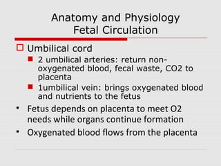
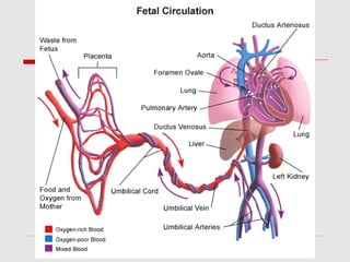
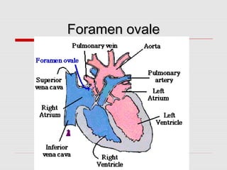
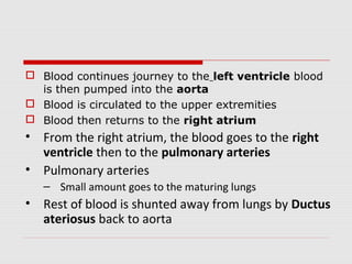
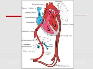
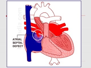
FETAL HEART CIRCULATION
VD. MANOJ JAGTAP
LECTURER, SHARIR RACHANA VIBHAG
Objectives
• Discuss anatomy and physiology of
fetal circulation
• Define specialized structures of fetal
circulation
Anatomy and Physiology
Fetal Circulation
 Umbilical cord
 2 umbilical arteries: return non-
oxygenated blood, fecal waste, CO2 to
placenta
 1umbilical vein: brings oxygenated blood
and nutrients to the fetus
• Fetus depends on placenta to meet O2
needs while organs continue formation
• Oxygenated blood flows from the placenta
• To the fetus via the umbilical vein
• After reaching fetus the blood flows through
the inferior vena cava
• Blood continues to travel from the inferior
vena cava to the ductus venosis
• Ductus Venosis
– Small amount of blood routed to growing liver
– Increased blood flow leads to large liver in
newborns
• Blood continues to travel up the inferior vena cava
• Empties into the right atrium of the heart
• The blood then passes to the left atrium through the
foramen ovale
• Foramen ovale
– Small opening in the septum of the heart
– Completely bypasses the non-functioning lungs
Foramen ovaleForamen ovale
 Blood continues journey to the left ventricle blood
is then pumped into the aorta
 Blood is circulated to the upper extremities
 Blood then returns to the right atrium
• From the right atrium, the blood goes to the right
ventricle then to the pulmonary arteries
• Pulmonary arteries
– Small amount goes to the maturing lungs
• Rest of blood is shunted away from lungs by Ductus
ateriosus back to aorta
• Blood travels back from aorta to the two
umbilical arteries to the placenta
• The placenta will re-supply the blood with
oxygen
What happens at birth?
Foramen ovale Closes shortly after birth,
fuses completely in first
year.
Ductus arteriousus Closes soon after birth,
becomes ligamentum
arteriousum in about 3
months.
Ductus venosus Ligamentum venosum
Umbilical arteries Medial umbilical ligaments
Umbilical vein Ligamentum teres
Problem with persistence
of fetal circulation
 Patent (open) Ductus arteriousus and patent
foramen ovale each characterize about 8% of
congenital heart defects.
 Both cause a mixing of oxygen-rich and oxygen-
poor blood; blood reaching tissues not fully
oxygenated. Can cause cyanosis.
 Surgical correction now available, ideally
completed around age two.
 Many of these defects go undetected until child
is at least school age.
Fetal heart circulation

Fetal heart circulation

  • 1.
    FETAL HEART CIRCULATION VD.MANOJ JAGTAP LECTURER, SHARIR RACHANA VIBHAG
  • 2.
    Objectives • Discuss anatomyand physiology of fetal circulation • Define specialized structures of fetal circulation
  • 3.
    Anatomy and Physiology FetalCirculation  Umbilical cord  2 umbilical arteries: return non- oxygenated blood, fecal waste, CO2 to placenta  1umbilical vein: brings oxygenated blood and nutrients to the fetus • Fetus depends on placenta to meet O2 needs while organs continue formation • Oxygenated blood flows from the placenta
  • 6.
    • To thefetus via the umbilical vein • After reaching fetus the blood flows through the inferior vena cava • Blood continues to travel from the inferior vena cava to the ductus venosis • Ductus Venosis – Small amount of blood routed to growing liver – Increased blood flow leads to large liver in newborns
  • 7.
    • Blood continuesto travel up the inferior vena cava • Empties into the right atrium of the heart • The blood then passes to the left atrium through the foramen ovale • Foramen ovale – Small opening in the septum of the heart – Completely bypasses the non-functioning lungs
  • 8.
  • 9.
     Blood continuesjourney to the left ventricle blood is then pumped into the aorta  Blood is circulated to the upper extremities  Blood then returns to the right atrium • From the right atrium, the blood goes to the right ventricle then to the pulmonary arteries • Pulmonary arteries – Small amount goes to the maturing lungs • Rest of blood is shunted away from lungs by Ductus ateriosus back to aorta
  • 11.
    • Blood travelsback from aorta to the two umbilical arteries to the placenta • The placenta will re-supply the blood with oxygen
  • 12.
    What happens atbirth? Foramen ovale Closes shortly after birth, fuses completely in first year. Ductus arteriousus Closes soon after birth, becomes ligamentum arteriousum in about 3 months. Ductus venosus Ligamentum venosum Umbilical arteries Medial umbilical ligaments Umbilical vein Ligamentum teres
  • 13.
    Problem with persistence offetal circulation  Patent (open) Ductus arteriousus and patent foramen ovale each characterize about 8% of congenital heart defects.  Both cause a mixing of oxygen-rich and oxygen- poor blood; blood reaching tissues not fully oxygenated. Can cause cyanosis.  Surgical correction now available, ideally completed around age two.  Many of these defects go undetected until child is at least school age.