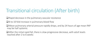
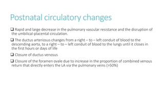
Fetal circulation
How is it different?
Shunts
• Venous system ( Ductus venosus )
• Heart ( Foramen ovale )
• Arterial system ( Ductus arteriosus )
Preferential blood flow pattern
Purpose
1. To deliver highly oxygenated to the metabolically active tissues ( Heart and brain )
2. To deliver less oxygenated blood to placenta
The RV supplies most of its blood via the ductus arteriosus and descending aorta to
the placenta for oxygenation.
The LV supplies most of its blood via the ascending aorta to the heart and brain for
oxygen delivery
5 components of the central venous
circulation
Venous return from
1. The upper body
2. The myocardium
3. The lungs
4. The lower body
5. The placenta
Among these, the least saturated blood is from the upper body and the myocardium
Preferential blood flow pattern
 The course of the IVC ( Eustacian ) valve is positioned in such a way that >95% of
the blood entering the RA from the SVC is directed towards the tricuspid valve to the
RV preventing it from going through the Foramen ovale
 The location coronary sinus cauded to the Foramen ovale assists venous blood
coming from the myocardium to flow through the tricuspid valve to the RV
 The relatively desaturated blood from the lower body and the placenta ascends the
distal IVC and enters the lateral margin of the RA and is directed primarily through the
tricuspid valve
 Exception in the central venous blood flow pattern is that the pulmonary veins from
the lungs doesn’t drain into the RA. It flows into the LA like it normally does in
adults.But it doesn’t matter much because
- it has only an intermediate saturation
- it amounts to only about 5 % of the combined ventricular output
(maximum 25%)
Therefore it doesn’t have a significant effect on the oxygen delivery
Placental (umbilical venous) and liver
venous return
 Majority (≈55%) of the umbilical venous blood passes through the ductus venosus
to reach the RA
 ≈45% enters the left lobe of liver – joins left hepatic vein – which joins the ductus
venosus near the IVC and eventually pours into the RA
 Well oxygenated blood from these two sources on entering the RA preferentially
crosses the Foramen ovale to enter the LA thereby supplying the systemic circulation.
 The remainder of the umbilical venous blood along with >95% of the poorly
saturated portal venous blood enters the right lobe of liver and from there through
the right hepatic vein joins the distal IVC and empties into the RV finally
Other dynamic changes in fetal
circulation
 Pulmonary blood flow is low despite the dominance of the RV which in fetus ejects
55% to 60% of the total cardiac output.
 Most of the RV output is diverted away from the lungs through the widely patent
ductus arteriosus.
 Fetal pulmonary arterial mean pressure increases progressively with gestation and
at at term it is about 50 mmHg exceeding the mean aortic blood pressure by 1 to 2
mmHg
Transitional circulation (After birth)
 Rapid decrease in the pulmonary vascular resistance
 8 to 10 fold increase in pulmonary blood flow
 Mean pulmonary arterial pressure rapidly drops, and by 24 hours of age mean PAP
may be half systemic
 After the initial rapid fall, there is slow progressive decrease, with adult levels
reached after 2 to 6 weeks
Postnatal circulatory changes
 Rapid and large decrease in the pulmonary vascular resistance and the disruption of
the umbilical-placental circulation.
 The ductus arteriosus changes from a right – to – left conduit of blood to the
descending aorta, to a right – to – left conduit of blood to the lungs until it closes in
the first hours or days of life
 Closure of ductus venosus
 Closure of the foramen ovale due to increase in the proportion of combined venous
return that directly enters the LA via the pulmonary veins (>50%)
Physiologic regulation of pulmonary
vascular resistance
• Pulmonary vessels produce and actively metabolise many vasoactive substances
• The three main factors influencing the regulation of the fetal pulmonary circulation
are
mechanical effects
state of oxygenation and
production of vaso active substances
The most prominent among these is the normally low blood and alveolar oxygen tension that is
responsible for the high fetal pulmonary vascular resistance
Other factors that increase pulmonary
vascular resistance
Apart from the low oxygen tension, the other factors responsible for constriction in pulmonary fetal
circulation are
alpha agonists
thromboxane
leuokotrienes ( present in fetal tracheal fluid )
EDRF – Endothelium Derived Relaxing
Factor
• NO – nitric oxide / Endothelium Derived NO (EDNO)
• Produced by endothelial cells in response to various stimuli , generally involving
specific receptors and the activation of endothelial NO synthase (eNOS)
• Subsequent smooth muscle relaxation is produced by a NO/guanylyl cyclase/cGMP
mediated mechanism
Effect of EDNO on fetal circulation
• After certain stimuli such as shear stress or receptor binding of specific vasodilator
NO is synthesized by the activation of NO synthase
• NO released from endothelial cell
• NO diffuses into smooth muscle cell
• Activates guanylate cyclase
• Increase concentration of cGMP
•This causes smooth muscle relaxation
• Inhibition of EDNO synthesis produces marked increase in resting fetal pulmonary
vascular resistance
• Studies of intra pulmonary arterial and isolated lung preparation of sheep show
maturative increase in NO mediated relaxation during the late fetal and early
postnatal period
• Therefore basal EDNO production is an important mediator of both normal fetal
Pulmonary vascular tone and dramatic decrease in resistance that occurs with O2
ventilation at birth
Prostacyclins
•Prostaglandins ( arachidonic acid metabolite ) are not the central factor in the
regulation of resting fetal pulmonary circulation
• But they ( PGI2) act in modulating the tone and there by maintaining the pulmonary
vascular resistance relatively constant
•PC – primarily synthesized in vascular endothelial cells
• Produce vasodilation by activating adenylate cyclase and producing increased
concentration of cAMP
• Causes smooth muscle relaxation
Endothelin
• ET1 – endothelially derived vasoactive factor
• Has both vasoconstrictive and vasodilatory effect
• Acts on two receptors – ETa , ETb
• ETa – vascular smooth muscle – Vasoconstriction
• ETb – endothelial cell - Vasodilatation
• Predominant action of ET1 is VD by acting on ETb and EDNO release
Role of oxygen in pulmonary
vasodilation
• Increase in alveolar or arterial O2 tension may decrease the pulmonary vascular
resistance either directly via K+ channel activation or indirecty by stimulating the
production of vasodilator substances like PGI2, bradykynin, adenosine, adenosine 5’ –
triphosphate, or EDNO
• EDNO is the most important mediator for casusing decreasing in PVR at birth
associated with increased oxygenation
Exogenous prostaglandins
• Particularly PGI2, lowers the PVR in the fetus
• Indomethacin acts by inhibiting the PG production when given before the fetal lung
starts ventilating – thereby attenuating the subsequent decrease in the pulmonary
vascular resistance that occurs after the initial rapid decrease in resistance
Other vasoactive substances
Adenosine
Adenosine triphosphate
Bradykynin
Immediate and late vasodilatory effects
after birth
• Atleast two components appear to determine the decrease in PVR with the initiation
of ventilation and oxygenation
• First ( independent of fetal oxygenation ) – physical expansion and production of
prostaglandins ( PGI2 and PGD2)
• Second – EDNO ( associated with the fetal oxygenation )
• Both components are necessary for the successful extrauterine life.
PULMONARY VASOCONSTRICTORS PULMONARY VASODILATORS
1. Leukotrienes 1. EDNO
2. Low oxygen 2. ET1 – acting through ETb
3. ET1 – acting through ETa 3. Bradykynin
4. Prostaglandins
Fetal circulation

Fetal circulation

  • 1.
  • 3.
    How is itdifferent? Shunts • Venous system ( Ductus venosus ) • Heart ( Foramen ovale ) • Arterial system ( Ductus arteriosus ) Preferential blood flow pattern
  • 4.
    Purpose 1. To deliverhighly oxygenated to the metabolically active tissues ( Heart and brain ) 2. To deliver less oxygenated blood to placenta
  • 5.
    The RV suppliesmost of its blood via the ductus arteriosus and descending aorta to the placenta for oxygenation. The LV supplies most of its blood via the ascending aorta to the heart and brain for oxygen delivery
  • 6.
    5 components ofthe central venous circulation Venous return from 1. The upper body 2. The myocardium 3. The lungs 4. The lower body 5. The placenta Among these, the least saturated blood is from the upper body and the myocardium
  • 7.
    Preferential blood flowpattern  The course of the IVC ( Eustacian ) valve is positioned in such a way that >95% of the blood entering the RA from the SVC is directed towards the tricuspid valve to the RV preventing it from going through the Foramen ovale
  • 9.
     The locationcoronary sinus cauded to the Foramen ovale assists venous blood coming from the myocardium to flow through the tricuspid valve to the RV
  • 10.
     The relativelydesaturated blood from the lower body and the placenta ascends the distal IVC and enters the lateral margin of the RA and is directed primarily through the tricuspid valve  Exception in the central venous blood flow pattern is that the pulmonary veins from the lungs doesn’t drain into the RA. It flows into the LA like it normally does in adults.But it doesn’t matter much because - it has only an intermediate saturation - it amounts to only about 5 % of the combined ventricular output (maximum 25%) Therefore it doesn’t have a significant effect on the oxygen delivery
  • 11.
    Placental (umbilical venous)and liver venous return  Majority (≈55%) of the umbilical venous blood passes through the ductus venosus to reach the RA  ≈45% enters the left lobe of liver – joins left hepatic vein – which joins the ductus venosus near the IVC and eventually pours into the RA  Well oxygenated blood from these two sources on entering the RA preferentially crosses the Foramen ovale to enter the LA thereby supplying the systemic circulation.  The remainder of the umbilical venous blood along with >95% of the poorly saturated portal venous blood enters the right lobe of liver and from there through the right hepatic vein joins the distal IVC and empties into the RV finally
  • 13.
    Other dynamic changesin fetal circulation  Pulmonary blood flow is low despite the dominance of the RV which in fetus ejects 55% to 60% of the total cardiac output.  Most of the RV output is diverted away from the lungs through the widely patent ductus arteriosus.  Fetal pulmonary arterial mean pressure increases progressively with gestation and at at term it is about 50 mmHg exceeding the mean aortic blood pressure by 1 to 2 mmHg
  • 14.
    Transitional circulation (Afterbirth)  Rapid decrease in the pulmonary vascular resistance  8 to 10 fold increase in pulmonary blood flow  Mean pulmonary arterial pressure rapidly drops, and by 24 hours of age mean PAP may be half systemic  After the initial rapid fall, there is slow progressive decrease, with adult levels reached after 2 to 6 weeks
  • 15.
    Postnatal circulatory changes Rapid and large decrease in the pulmonary vascular resistance and the disruption of the umbilical-placental circulation.  The ductus arteriosus changes from a right – to – left conduit of blood to the descending aorta, to a right – to – left conduit of blood to the lungs until it closes in the first hours or days of life  Closure of ductus venosus  Closure of the foramen ovale due to increase in the proportion of combined venous return that directly enters the LA via the pulmonary veins (>50%)
  • 16.
    Physiologic regulation ofpulmonary vascular resistance • Pulmonary vessels produce and actively metabolise many vasoactive substances • The three main factors influencing the regulation of the fetal pulmonary circulation are mechanical effects state of oxygenation and production of vaso active substances The most prominent among these is the normally low blood and alveolar oxygen tension that is responsible for the high fetal pulmonary vascular resistance
  • 17.
    Other factors thatincrease pulmonary vascular resistance Apart from the low oxygen tension, the other factors responsible for constriction in pulmonary fetal circulation are alpha agonists thromboxane leuokotrienes ( present in fetal tracheal fluid )
  • 18.
    EDRF – EndotheliumDerived Relaxing Factor • NO – nitric oxide / Endothelium Derived NO (EDNO) • Produced by endothelial cells in response to various stimuli , generally involving specific receptors and the activation of endothelial NO synthase (eNOS) • Subsequent smooth muscle relaxation is produced by a NO/guanylyl cyclase/cGMP mediated mechanism
  • 19.
    Effect of EDNOon fetal circulation • After certain stimuli such as shear stress or receptor binding of specific vasodilator NO is synthesized by the activation of NO synthase • NO released from endothelial cell • NO diffuses into smooth muscle cell • Activates guanylate cyclase • Increase concentration of cGMP •This causes smooth muscle relaxation
  • 21.
    • Inhibition ofEDNO synthesis produces marked increase in resting fetal pulmonary vascular resistance • Studies of intra pulmonary arterial and isolated lung preparation of sheep show maturative increase in NO mediated relaxation during the late fetal and early postnatal period • Therefore basal EDNO production is an important mediator of both normal fetal Pulmonary vascular tone and dramatic decrease in resistance that occurs with O2 ventilation at birth
  • 22.
    Prostacyclins •Prostaglandins ( arachidonicacid metabolite ) are not the central factor in the regulation of resting fetal pulmonary circulation • But they ( PGI2) act in modulating the tone and there by maintaining the pulmonary vascular resistance relatively constant
  • 23.
    •PC – primarilysynthesized in vascular endothelial cells • Produce vasodilation by activating adenylate cyclase and producing increased concentration of cAMP • Causes smooth muscle relaxation
  • 24.
    Endothelin • ET1 –endothelially derived vasoactive factor • Has both vasoconstrictive and vasodilatory effect • Acts on two receptors – ETa , ETb • ETa – vascular smooth muscle – Vasoconstriction • ETb – endothelial cell - Vasodilatation • Predominant action of ET1 is VD by acting on ETb and EDNO release
  • 25.
    Role of oxygenin pulmonary vasodilation • Increase in alveolar or arterial O2 tension may decrease the pulmonary vascular resistance either directly via K+ channel activation or indirecty by stimulating the production of vasodilator substances like PGI2, bradykynin, adenosine, adenosine 5’ – triphosphate, or EDNO • EDNO is the most important mediator for casusing decreasing in PVR at birth associated with increased oxygenation
  • 26.
    Exogenous prostaglandins • ParticularlyPGI2, lowers the PVR in the fetus • Indomethacin acts by inhibiting the PG production when given before the fetal lung starts ventilating – thereby attenuating the subsequent decrease in the pulmonary vascular resistance that occurs after the initial rapid decrease in resistance
  • 27.
  • 28.
    Immediate and latevasodilatory effects after birth • Atleast two components appear to determine the decrease in PVR with the initiation of ventilation and oxygenation • First ( independent of fetal oxygenation ) – physical expansion and production of prostaglandins ( PGI2 and PGD2) • Second – EDNO ( associated with the fetal oxygenation ) • Both components are necessary for the successful extrauterine life.
  • 29.
    PULMONARY VASOCONSTRICTORS PULMONARYVASODILATORS 1. Leukotrienes 1. EDNO 2. Low oxygen 2. ET1 – acting through ETb 3. ET1 – acting through ETa 3. Bradykynin 4. Prostaglandins