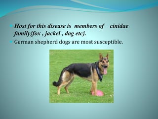
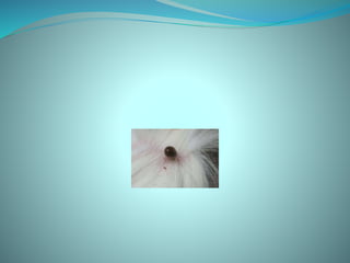
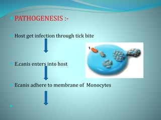
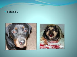
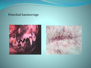
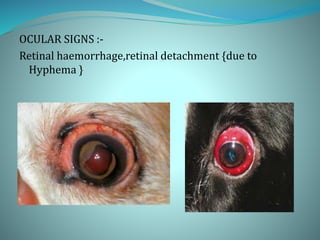
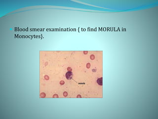
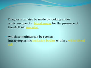
KARNATAKA VETERINARY,
ANIMAL & FISHERIES
SCIENCE UNIVERSITY, BIDAR.
VETERINARY COLLEGE,
SHIMOGA
To :
Dr. Maltesh
Asst professor
Dept of MEDICINE
INTRODUCTION
Canine Ehrlichiosis (Canine monocytic
Ehrlichiosis)
 It is at tick borne disease caused by
Erhlichia species.
 Dr Ehrlichia described this organism
 Hence it was name as Erhlichia sps.
Characteristics of E canis
 They are gram negative.
 pleomorphic coccobacilli rickettia.
 Obligatory intracellular .
 Ecanis usually appears in monocytes as a cluster of
organism called as MORULAE.
 .
 Etiology
 EHRLICHIA CANIS
 They are richkettsial organism.
{ They are intermediate between VIRUSES and BACTERIA.}
 E canis is transmitted by the brown dog
tick, Rhipicephalus sanguineus.
 Experimentally it can also be transmitted by Dermacentor
variabilis.
 Host for this disease is members of cinidae
family{fox , jackel , dog etc}.
 German shepherd dogs are most susceptible.
Epidemiology
 It has world wide distribution including Asia ,Africa
and America.
 Australia is free of E.Canis.
 Transmission:-
 Ticks aquired Ecanis while feeding on infected dog.
 this ticks carrying Ecanis transmit to other Dogs upon
feeding on them.
 PATHOGENESIS :-
 Host get infection through tick bite
 E.canis enters into host
 Ecanis adhere to membrane of Monocytes

 through endocytosis enters into the cell

 Divide by binary fission
 They form morulae

 Release of organism through rupture of morulae
 They spread to adjacent cells through cytoplasmic
projections
 Spread throughout the body
 There includes three phases :-
 Acute phase
 Persistence subclinical infection
 Severe chronic phase
 Once the organism enters the body causes ACUTE
PHASE.
 Which is characterised by :-
 Upto 4 weeks
 Fever,
• Severe Thrombocytopenia
 During this time the platelet count will drop due
an immune-mediated platelet destruction .
 The dog will be listless, off food, and may have
enlarged lymph nodes.
 Most dogs clear the organism if they are treated in this
stage.
 but those that do not receive adequate treatment will
go on to the next phase.
 SUBCLINICAL PHASE:
 In this phase, the dog appears normal.
 The organism has sequestered in the spleen .
 Dogs can stay in this phase for months or even years.
 Intermittent fever ,mild Thrombocytopenia and
anaemia.
 CHRONIC PHASE:
 Charecterised by :-
 Severe pancytopenia { due to bone marrow
Hyperplasia}.
 Fever , wide spread petechia and edema.
 Death due to secondary bacterial infection .
 CLINICAL SIGNS:-
 There are no signs of the subclinical phase.
 Fever , Anorexia , lethargy ,weight loss.
 Epitaxis ,
 Petechial haemorrage
OCULAR SIGNS :-
Retinal haemorrhage,retinal detachment {due to
Hyphema }
 Neurologic effects may also be seen.{due to meningeal
bleeding}
 Glomeruloneprhitis, resulting in serious urinary
protein loss, can also result.
 Increased globulin levels are also seen.
 POST MORTAM LESIONS :-
 Petechial haemorrhage seen on serosal surface of
organs like lungs ,kidney ,brain, nasal cavity GI tract.
 Generalised Lymphadenomegaly , splenomegaly and
Hepatomegaly
 Postmortem Lesions:
 During the acute or self-limiting phase of E
canis infections, lesions generally are nonspecific,
 but splenomegaly is common.
 Histologically, there is lymphoreticular hyperplasia
and lymphocytic and plasmacytic perivascular cuffing
 . In chronic cases:-
 these lesions may be accompanied by widespread
hemorrhage and
 increased mononuclear cell infiltration in perivascular
regions of many organs.
 DIAGNOSIS:-
 Clinical examination
 Hemotology and Serum biochemistry
 {Thrombocytopenia and Hypo albuminaemia and
hyperglobulinaemia}.
 Dot ELISA kit @field level
 Molecular test –PCR
• Serological test {FAT-flouroscent antibody technique }-
which is widely used.
 Blood smear examination { to find MORULA in
Monocytes}.
• Diagnosis canaiso be made by looking under
a microscope of a blood smear for the presence of
the ehrlichia morulae,
• which sometimes can be seen as
intracytoplasmic inclusion bodies within a white blood
cell
 TREATMENT:-
Doxycycline(5-10mg /kg/day) {PO/IV} for 21-25 days.
 OXYTETRACYCLIN –( 5-10 /Kg /day){I/V} for 21-25 days.
 Supportive therapy ( in anaemia ) – Blood transfusion if Hb
concentration is <4%.
 short term (2-7days)therapy with low immunosuppressive
doses of Glucocorticoides{1-2mg /kg prednisolone,PO}
 This may be beneficial early in the treatment period when
severe Thrombocytopenia is present.
Prevention:-
Tick control is the most effective method of prevention.
No vaccinesn are available.
Chemoprophylaxis by using tetracylin @6.6mg /kg .
Precautions should be taken while tranfusion of blood.
Canin1. ppt
Canin1. ppt

Canin1. ppt

  • 1.
    KARNATAKA VETERINARY, ANIMAL &FISHERIES SCIENCE UNIVERSITY, BIDAR. VETERINARY COLLEGE, SHIMOGA To : Dr. Maltesh Asst professor Dept of MEDICINE
  • 2.
    INTRODUCTION Canine Ehrlichiosis (Caninemonocytic Ehrlichiosis)  It is at tick borne disease caused by Erhlichia species.  Dr Ehrlichia described this organism  Hence it was name as Erhlichia sps.
  • 3.
    Characteristics of Ecanis  They are gram negative.  pleomorphic coccobacilli rickettia.  Obligatory intracellular .  Ecanis usually appears in monocytes as a cluster of organism called as MORULAE.  .
  • 4.
     Etiology  EHRLICHIACANIS  They are richkettsial organism. { They are intermediate between VIRUSES and BACTERIA.}  E canis is transmitted by the brown dog tick, Rhipicephalus sanguineus.
  • 7.
     Experimentally itcan also be transmitted by Dermacentor variabilis.
  • 8.
     Host forthis disease is members of cinidae family{fox , jackel , dog etc}.  German shepherd dogs are most susceptible.
  • 9.
    Epidemiology  It hasworld wide distribution including Asia ,Africa and America.  Australia is free of E.Canis.
  • 10.
     Transmission:-  Ticksaquired Ecanis while feeding on infected dog.  this ticks carrying Ecanis transmit to other Dogs upon feeding on them.
  • 12.
     PATHOGENESIS :- Host get infection through tick bite  E.canis enters into host  Ecanis adhere to membrane of Monocytes 
  • 13.
     through endocytosisenters into the cell   Divide by binary fission  They form morulae
  • 14.
      Release oforganism through rupture of morulae  They spread to adjacent cells through cytoplasmic projections  Spread throughout the body
  • 15.
     There includesthree phases :-  Acute phase  Persistence subclinical infection  Severe chronic phase
  • 16.
     Once theorganism enters the body causes ACUTE PHASE.  Which is characterised by :-  Upto 4 weeks  Fever, • Severe Thrombocytopenia
  • 17.
     During thistime the platelet count will drop due an immune-mediated platelet destruction .  The dog will be listless, off food, and may have enlarged lymph nodes.  Most dogs clear the organism if they are treated in this stage.  but those that do not receive adequate treatment will go on to the next phase.
  • 18.
     SUBCLINICAL PHASE: In this phase, the dog appears normal.  The organism has sequestered in the spleen .  Dogs can stay in this phase for months or even years.  Intermittent fever ,mild Thrombocytopenia and anaemia.
  • 19.
     CHRONIC PHASE: Charecterised by :-  Severe pancytopenia { due to bone marrow Hyperplasia}.  Fever , wide spread petechia and edema.  Death due to secondary bacterial infection .
  • 20.
     CLINICAL SIGNS:- There are no signs of the subclinical phase.  Fever , Anorexia , lethargy ,weight loss.
  • 21.
  • 22.
  • 23.
    OCULAR SIGNS :- Retinalhaemorrhage,retinal detachment {due to Hyphema }
  • 24.
     Neurologic effectsmay also be seen.{due to meningeal bleeding}  Glomeruloneprhitis, resulting in serious urinary protein loss, can also result.  Increased globulin levels are also seen.
  • 25.
     POST MORTAMLESIONS :-  Petechial haemorrhage seen on serosal surface of organs like lungs ,kidney ,brain, nasal cavity GI tract.  Generalised Lymphadenomegaly , splenomegaly and Hepatomegaly
  • 26.
     Postmortem Lesions: During the acute or self-limiting phase of E canis infections, lesions generally are nonspecific,  but splenomegaly is common.  Histologically, there is lymphoreticular hyperplasia and lymphocytic and plasmacytic perivascular cuffing
  • 27.
     . Inchronic cases:-  these lesions may be accompanied by widespread hemorrhage and  increased mononuclear cell infiltration in perivascular regions of many organs.
  • 28.
     DIAGNOSIS:-  Clinicalexamination  Hemotology and Serum biochemistry  {Thrombocytopenia and Hypo albuminaemia and hyperglobulinaemia}.  Dot ELISA kit @field level  Molecular test –PCR • Serological test {FAT-flouroscent antibody technique }- which is widely used.
  • 29.
     Blood smearexamination { to find MORULA in Monocytes}.
  • 30.
    • Diagnosis canaisobe made by looking under a microscope of a blood smear for the presence of the ehrlichia morulae, • which sometimes can be seen as intracytoplasmic inclusion bodies within a white blood cell
  • 31.
     TREATMENT:- Doxycycline(5-10mg /kg/day){PO/IV} for 21-25 days.  OXYTETRACYCLIN –( 5-10 /Kg /day){I/V} for 21-25 days.  Supportive therapy ( in anaemia ) – Blood transfusion if Hb concentration is <4%.  short term (2-7days)therapy with low immunosuppressive doses of Glucocorticoides{1-2mg /kg prednisolone,PO}  This may be beneficial early in the treatment period when severe Thrombocytopenia is present.
  • 32.
    Prevention:- Tick control isthe most effective method of prevention. No vaccinesn are available. Chemoprophylaxis by using tetracylin @6.6mg /kg . Precautions should be taken while tranfusion of blood.