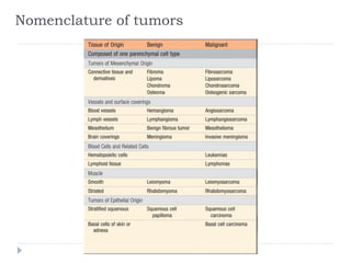
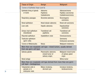
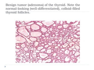
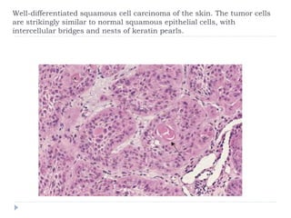
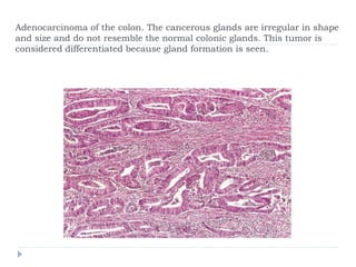
Neoplasia class 1
 Neoplasia means “new growth.”
 Tumor originally applied to the swelling caused by
inflammation, but the non-neoplastic usage of tumor
has almost vanished; the term is now equated with
neoplasm.
 Oncology (Greek oncos = tumor) is the study of
tumors or neoplasms.
Definition
In the pre-molecular era, the eminent British oncologist
Willis defined neoplasia as:
 “A neoplasm is an abnormal mass of tissue, the
growth of which exceeds and is uncoordinated with
that of the normal tissues and persists in the same
excessive manner after cessation of the stimuli
which evoked the change.”
 In the modern era, a neoplasm can be defined as
 “a disorder of cell growth that is triggered by a series
of acquired mutations affecting a single cell and its
clonal progeny”.
 The causative mutations give the neoplastic cells a
survival and growth advantage, resulting in
excessive proliferation that is independent of
physiologic growth signals (autonomous).
 All tumors have two basic components:
(1) neoplastic cells that constitute the tumor
parenchyma and
(2) reactive stroma made up of connective tissue,
blood vessels, and variable numbers of cells of the
adaptive and innate immune system.
 The classification of tumors and their biologic
behavior are based primarily on the parenchymal
component, but their growth and spread are critically
dependent on their stroma.
 In some tumors, connective tissue is scant and so
the neoplasm is soft and fleshy.
 In other cases, the parenchymal cells stimulate the
formation of an abundant collagenous stroma,
referred to as desmoplasia.
 Some demoplastic tumors are stony hard or
scirrhous—for example, some cancers of the female
breast.
Benign Tumors and Malignant Tumors
 A tumor is said to be benign when its gross and
microscopic appearances are considered relatively
innocent, implying that
 it will remain localized,
 will not spread to other sites, and
 is amenable to local surgical removal.
 In general, benign tumors are designated by
attaching the suffix -oma to the name of the cell type
from which the tumor originates.
 Tumors of mesenchymal cells generally follow this
rule.
 For example, a benign tumor arising in fibrous tissue
is called a fibroma, whereas a benign cartilaginous
tumor is a chondroma.
 The nomenclature of benign epithelial tumors is
more complex;
 some are classified based on their cells of origin,
 some on microscopic pattern, and
 others on their macroscopic architecture.
 Adenoma is applied to benign epithelial neoplasms
derived from glands, although they may or may not
form glandular structures.
 On this basis, a benign epithelial neoplasm that
arises from renal tubular cells & grows in the form of
numerous tightly clustered small glands is termed an
adenoma.
 A heterogeneous mass of adrenal cortical cells
growing as a solid sheet is also termed as adenoma.
 Benign epithelial neoplasms producing
microscopically or macroscopically visible fingerlike
or warty projections from epithelial surfaces are
referred to as papillomas.
 Those that form large cystic masses, such as in the
ovary, are referred to as cystadenomas.
 Some tumors produce papillary patterns that
protrude into cystic spaces and are called papillary
cystadenomas.
 When a neoplasm—benign or malignant— produces
a macroscopically visible projection above a mucosal
surface and projects into a lumen, it is termed a
polyp. For example, gastric or colonic polyps.
 If the polyp has glandular tissue, it is called an
adenomatous polyp.
An aedonmatous (glandular) polyp is projecting into the colonic
lumen and is attached to the mucosa by a distinct stalk
 Malignant tumors are collectively referred to as
cancers (Latin word for crab).
 They can invade and destroy adjacent structures
and spread to distant sites (metastasize) to cause
death.
 Not all cancers pursue so deadly a course.
 Some are discovered early enough to be excised
surgically or are treated successfully with
chemotherapy or radiation.
 Malignant tumors arising in solid mesenchymal
tissues are usually called sarcomas (Greek sar =
fleshy); e.g., fibrosarcoma, chondrosarcoma,
liposarcoma, leiomyosarcoma, and
rhabdomyosarcoma.
 Those arising from blood-forming cells are
designated leukemias (literally, white blood) or
lymphomas (tumors of lymphocytes or their
precursors).
 Malignant neoplasms of epithelial cell origin, derived
from any of the three germ layers, are called
carcinomas.
 Thus, cancers arising in the ectodermally derived
epidermis, the mesodermally derived renal tubules,
and the endodermally derived lining of the
gastrointestinal tract are all termed carcinomas.
 Squamous cell carcinoma denotes a cancer in which
the tumor cells resemble stratified squamous
epithelium, and adenocarcinoma denotes a lesion in
which the neoplastic epithelial cells grow in a
glandular pattern.
Nomenclature of tumors
Teratoma
 Neoplasm which contains recognizable mature or
immature cells or tissues belonging to more than one
germ cell layer (and sometimes all three).
 Originates from totipotential germ cells that are
normally present in the ovary and testis and
sometimes also found in abnormal midline
embryonic rests.
 Such cells can differentiate into any of the cell types
found in the adult body and so may give rise to
neoplasms that contain, in a helter-skelter fashion,
bone, epithelium, muscle, fat, nerve, and other
tissues.
 A particularly common pattern is seen in the ovarian
cystic teratoma (dermoid cyst), which differentiates
principally along ectodermal lines to create a cystic
tumor lined by skin replete with hair, sebaceous
glands, and tooth structures.
Benign cystic teratoma of ovary
Mixed Tumors.
 Divergent differentiation of a single neoplastic clone
creates a mixed tumor, such as the mixed tumor of
salivary gland; also called pleomorphic adenoma.
 These tumors contain epithelial components
scattered within a myxoid stroma that may contain
islands of cartilage or bone.
 All of these elements arise from a single clone
capable of producing both epithelial and
myoepithelial cells.
This mixed tumor of the parotid gland contains
epithelial cells forming ducts and myxoid stroma that
resemble cartilage.
 Hamartomas are disorganized but benign masses
composed of cells indigenous to the involved site.
 Choristoma is the term applied to a heterotopic rest
of cells.
 For example, a small nodule of well-developed and
normally organized pancreatic tissue may be found
in the submucosa of the stomach, duodenum, or
small intestine.
 Benign-sounding designations such as lymphoma,
melanoma, mesothelioma, and seminoma are used
for certain malignant neoplasms.
Differentiation and Anaplasia
 Differentiation refers to the extent to which
neoplastic parenchymal cells resemble the
corresponding normal parenchymal cells, both
morphologically and functionally.
 Lack of differentiation is called anaplasia.
 In general, benign tumors are well differentiated.
 In contrast, malignant tumors exhibit a wide range of
parenchymal cell differentiation (well differentiated,
moderately differentiated & poorly differentiated).
Leiomyoma of the uterus. This benign, well-differentiated tumor contains
interlacing bundles of neoplastic smooth muscle cells that are virtually
identical in appearance to normal smooth muscle cells in the
myometrium.
Benign tumor (adenoma) of the thyroid. Note the
normal-looking (well-differentiated), colloid-filled
thyroid follicles.
Well-differentiated squamous cell carcinoma of the skin. The tumor cells
are strikingly similar to normal squamous epithelial cells, with
intercellular bridges and nests of keratin pearls.
Adenocarcinoma of the colon. The cancerous glands are irregular in shape
and size and do not resemble the normal colonic glands. This tumor is
considered differentiated because gland formation is seen.
 Malignant neoplasms that are composed of poorly
differentiated cells are said to be anaplastic.
 Lack of differentiation, or anaplasia, is considered a
hallmark of malignancy.
 Anaplasia is often associated with many other
morphologic changes.
 Pleomorphism.
 Variation in size and shape; cells within the same
tumor are not uniform, but range from small cells
with an undifferentiated appearance, to tumor giant
cells many times larger than their neighbors.
 Some tumor giant cells possess only a single huge
polymorphic nucleus, while others may have two or
more large, hyperchromatic nuclei.
 These giant cells are not to be confused with
inflammatory Langhans or foreign body giant cells,
which are derived from macrophages and contain
many small, normal-appearing nuclei.
Pleomorphic tumor of the skeletal muscle (rhabdomyosarcoma). Note the
marked cellular and nuclear pleomorphism, hyperchromatic nuclei, and
tumor giant cells.
 Abnormal nuclear morphology.
 Characteristically, the nuclei are disproportionately
large for the cell, with a nuclear-to-cytoplasm ratio
that may approach 1: 1 instead of the normal 1: 4 or
1:6.
 The nuclear shape is variable and often irregular,
and the chromatin is often coarsely clumped and
distributed along the nuclear membrane, or more
darkly stained than normal (hyperchromatic).
 Abnormally large nucleoli are also commonly seen.
 Mitoses.
 Mitoses are indicative of rapid cell growth. Hence,
mitosis are often seen in normal tissues exhibiting
rapid turnover, such as the epithelial lining of the gut
and nonneoplastic proliferations such as
hyperplasias.
 More important as a morphologic feature of
malignancy are atypical, bizarre mitotic figures,
sometimes with tripolar, quadripolar, or multipolar
spindles.
Anaplastic tumor showing cellular and nuclear variation in size
and shape. The prominent cell in the center field has an
abnormal tripolar spindle.
 Loss of polarity.
 The orientation of anaplastic cells is markedly disturbed.
 Sheets or large masses of tumor cells grow in an
anarchic, disorganized fashion.
 Other changes.
 Growing tumor cells obviously require a blood supply, but
often the vascular stroma is insufficient, and as a result in
many rapidly growing malignant tumors develop large
central areas of ischemic necrosis.
 The better the differentiation of the transformed cell, the more
completely it retains the functional capabilities of its normal
counterpart.
 Thus, benign neoplasms and well-differentiated carcinomas of
endocrine glands frequently secrete hormones characteristic
of their origin. Increased levels of these hormones in the blood
are used clinically to detect and follow such tumors.
 Well-differentiated squamous cell carcinomas of the epidermis
synthesize keratin, and well-differentiated hepatocellular
carcinomas elaborate bile.
 Highly anaplastic undifferentiated cells, lose their
resemblance to the normal cells from which they have
arisen. In some instances, new and unanticipated
functions emerge.
 Thus, some tumors express fetal proteins that are not
produced by comparable cells in the adult, while others
express proteins that are normally only found in other
types of adult cells.
 For example, bronchogenic carcinomas may produce
corticotropin, parathyroid-like hormone, insulin, glucagon,
and other hormones, giving rise to paraneoplastic
syndromes
Dysplasia
 The term literally means “disordered growth.”
 It is encountered principally in epithelia and is characterized by
a constellation of changes that include
i)a loss in the uniformity of the individual cells as well as
ii)a loss in their architectural orientation.
 Dysplastic cells may exhibit considerable pleomorphism and
often contain large hyperchromatic nuclei with a high nuclear-
to-cytoplasmic ratio.
 The architecture of the tissue may be disorderly.
 In dysplastic squamous epithelium the normal
progressive maturation of tall cells in the basal layer
to flattened squames on the surface may fail in part
or entirely, leading to replacement of the epithelium
by basal-appearing cells with hyperchromatic nuclei.
 In addition, mitotic figures are more abundant than in
the normal tissue and rather than being confined to
the basal layer may be seen at all levels, including
surface cells.
 When dysplastic changes are marked and involve the full
thickness of the epithelium, but the lesion does not
penetrate the basement membrane, it is considered a
pre-invasive neoplasm and is referred to as carcinoma in
situ.
 Once the tumor cells breach the basement membrane,
the tumor is said to be invasive.
 Dysplastic changes are often found adjacent to foci of
invasive carcinoma, and in long-term cigarette smokers
and persons with Barrett esophagus, severe epithelial
dysplasia frequently antedates the appearance of cancer.
 Although dysplasia may be a precursor to malignant
transformation, it does not always progress to cancer.
 With removal of the inciting causes, mild to moderate
dysplasias that do not involve the entire thickness of
epithelium may be completely reversible.
 Even carcinoma in situ may persist for years before it
becomes invasive.
 It should be noted that dysplasia often occurs in
metaplastic epithelium, not all metaplastic epithelium is
dysplastic.
Carcinoma in situ. A low-power view shows that the epithelium is entirely
replaced by atypical dysplastic cells. There is no orderly differentiation of
squamous cells. The basement membrane is intact, and there is no tumor
in the subepithelial stroma.
A high-power view shows failure of normal differentiation, marked nuclear
and cellular pleomorphism, and numerous mitotic figures extending
toward the surface.

Neoplasia class 1.pptx

  • 1.
  • 2.
     Neoplasia means“new growth.”  Tumor originally applied to the swelling caused by inflammation, but the non-neoplastic usage of tumor has almost vanished; the term is now equated with neoplasm.  Oncology (Greek oncos = tumor) is the study of tumors or neoplasms.
  • 3.
    Definition In the pre-molecularera, the eminent British oncologist Willis defined neoplasia as:  “A neoplasm is an abnormal mass of tissue, the growth of which exceeds and is uncoordinated with that of the normal tissues and persists in the same excessive manner after cessation of the stimuli which evoked the change.”
  • 4.
     In themodern era, a neoplasm can be defined as  “a disorder of cell growth that is triggered by a series of acquired mutations affecting a single cell and its clonal progeny”.  The causative mutations give the neoplastic cells a survival and growth advantage, resulting in excessive proliferation that is independent of physiologic growth signals (autonomous).
  • 5.
     All tumorshave two basic components: (1) neoplastic cells that constitute the tumor parenchyma and (2) reactive stroma made up of connective tissue, blood vessels, and variable numbers of cells of the adaptive and innate immune system.  The classification of tumors and their biologic behavior are based primarily on the parenchymal component, but their growth and spread are critically dependent on their stroma.
  • 6.
     In sometumors, connective tissue is scant and so the neoplasm is soft and fleshy.  In other cases, the parenchymal cells stimulate the formation of an abundant collagenous stroma, referred to as desmoplasia.  Some demoplastic tumors are stony hard or scirrhous—for example, some cancers of the female breast.
  • 7.
    Benign Tumors andMalignant Tumors  A tumor is said to be benign when its gross and microscopic appearances are considered relatively innocent, implying that  it will remain localized,  will not spread to other sites, and  is amenable to local surgical removal.
  • 8.
     In general,benign tumors are designated by attaching the suffix -oma to the name of the cell type from which the tumor originates.  Tumors of mesenchymal cells generally follow this rule.  For example, a benign tumor arising in fibrous tissue is called a fibroma, whereas a benign cartilaginous tumor is a chondroma.
  • 9.
     The nomenclatureof benign epithelial tumors is more complex;  some are classified based on their cells of origin,  some on microscopic pattern, and  others on their macroscopic architecture.
  • 10.
     Adenoma isapplied to benign epithelial neoplasms derived from glands, although they may or may not form glandular structures.  On this basis, a benign epithelial neoplasm that arises from renal tubular cells & grows in the form of numerous tightly clustered small glands is termed an adenoma.  A heterogeneous mass of adrenal cortical cells growing as a solid sheet is also termed as adenoma.
  • 11.
     Benign epithelialneoplasms producing microscopically or macroscopically visible fingerlike or warty projections from epithelial surfaces are referred to as papillomas.  Those that form large cystic masses, such as in the ovary, are referred to as cystadenomas.  Some tumors produce papillary patterns that protrude into cystic spaces and are called papillary cystadenomas.
  • 14.
     When aneoplasm—benign or malignant— produces a macroscopically visible projection above a mucosal surface and projects into a lumen, it is termed a polyp. For example, gastric or colonic polyps.  If the polyp has glandular tissue, it is called an adenomatous polyp.
  • 15.
    An aedonmatous (glandular)polyp is projecting into the colonic lumen and is attached to the mucosa by a distinct stalk
  • 16.
     Malignant tumorsare collectively referred to as cancers (Latin word for crab).  They can invade and destroy adjacent structures and spread to distant sites (metastasize) to cause death.  Not all cancers pursue so deadly a course.  Some are discovered early enough to be excised surgically or are treated successfully with chemotherapy or radiation.
  • 17.
     Malignant tumorsarising in solid mesenchymal tissues are usually called sarcomas (Greek sar = fleshy); e.g., fibrosarcoma, chondrosarcoma, liposarcoma, leiomyosarcoma, and rhabdomyosarcoma.  Those arising from blood-forming cells are designated leukemias (literally, white blood) or lymphomas (tumors of lymphocytes or their precursors).
  • 18.
     Malignant neoplasmsof epithelial cell origin, derived from any of the three germ layers, are called carcinomas.  Thus, cancers arising in the ectodermally derived epidermis, the mesodermally derived renal tubules, and the endodermally derived lining of the gastrointestinal tract are all termed carcinomas.  Squamous cell carcinoma denotes a cancer in which the tumor cells resemble stratified squamous epithelium, and adenocarcinoma denotes a lesion in which the neoplastic epithelial cells grow in a glandular pattern.
  • 19.
  • 21.
    Teratoma  Neoplasm whichcontains recognizable mature or immature cells or tissues belonging to more than one germ cell layer (and sometimes all three).  Originates from totipotential germ cells that are normally present in the ovary and testis and sometimes also found in abnormal midline embryonic rests.
  • 22.
     Such cellscan differentiate into any of the cell types found in the adult body and so may give rise to neoplasms that contain, in a helter-skelter fashion, bone, epithelium, muscle, fat, nerve, and other tissues.  A particularly common pattern is seen in the ovarian cystic teratoma (dermoid cyst), which differentiates principally along ectodermal lines to create a cystic tumor lined by skin replete with hair, sebaceous glands, and tooth structures.
  • 23.
  • 24.
    Mixed Tumors.  Divergentdifferentiation of a single neoplastic clone creates a mixed tumor, such as the mixed tumor of salivary gland; also called pleomorphic adenoma.  These tumors contain epithelial components scattered within a myxoid stroma that may contain islands of cartilage or bone.  All of these elements arise from a single clone capable of producing both epithelial and myoepithelial cells.
  • 25.
    This mixed tumorof the parotid gland contains epithelial cells forming ducts and myxoid stroma that resemble cartilage.
  • 26.
     Hamartomas aredisorganized but benign masses composed of cells indigenous to the involved site.  Choristoma is the term applied to a heterotopic rest of cells.  For example, a small nodule of well-developed and normally organized pancreatic tissue may be found in the submucosa of the stomach, duodenum, or small intestine.  Benign-sounding designations such as lymphoma, melanoma, mesothelioma, and seminoma are used for certain malignant neoplasms.
  • 27.
    Differentiation and Anaplasia Differentiation refers to the extent to which neoplastic parenchymal cells resemble the corresponding normal parenchymal cells, both morphologically and functionally.  Lack of differentiation is called anaplasia.  In general, benign tumors are well differentiated.  In contrast, malignant tumors exhibit a wide range of parenchymal cell differentiation (well differentiated, moderately differentiated & poorly differentiated).
  • 28.
    Leiomyoma of theuterus. This benign, well-differentiated tumor contains interlacing bundles of neoplastic smooth muscle cells that are virtually identical in appearance to normal smooth muscle cells in the myometrium.
  • 29.
    Benign tumor (adenoma)of the thyroid. Note the normal-looking (well-differentiated), colloid-filled thyroid follicles.
  • 30.
    Well-differentiated squamous cellcarcinoma of the skin. The tumor cells are strikingly similar to normal squamous epithelial cells, with intercellular bridges and nests of keratin pearls.
  • 31.
    Adenocarcinoma of thecolon. The cancerous glands are irregular in shape and size and do not resemble the normal colonic glands. This tumor is considered differentiated because gland formation is seen.
  • 32.
     Malignant neoplasmsthat are composed of poorly differentiated cells are said to be anaplastic.  Lack of differentiation, or anaplasia, is considered a hallmark of malignancy.  Anaplasia is often associated with many other morphologic changes.
  • 33.
     Pleomorphism.  Variationin size and shape; cells within the same tumor are not uniform, but range from small cells with an undifferentiated appearance, to tumor giant cells many times larger than their neighbors.  Some tumor giant cells possess only a single huge polymorphic nucleus, while others may have two or more large, hyperchromatic nuclei.  These giant cells are not to be confused with inflammatory Langhans or foreign body giant cells, which are derived from macrophages and contain many small, normal-appearing nuclei.
  • 34.
    Pleomorphic tumor ofthe skeletal muscle (rhabdomyosarcoma). Note the marked cellular and nuclear pleomorphism, hyperchromatic nuclei, and tumor giant cells.
  • 35.
     Abnormal nuclearmorphology.  Characteristically, the nuclei are disproportionately large for the cell, with a nuclear-to-cytoplasm ratio that may approach 1: 1 instead of the normal 1: 4 or 1:6.  The nuclear shape is variable and often irregular, and the chromatin is often coarsely clumped and distributed along the nuclear membrane, or more darkly stained than normal (hyperchromatic).  Abnormally large nucleoli are also commonly seen.
  • 36.
     Mitoses.  Mitosesare indicative of rapid cell growth. Hence, mitosis are often seen in normal tissues exhibiting rapid turnover, such as the epithelial lining of the gut and nonneoplastic proliferations such as hyperplasias.  More important as a morphologic feature of malignancy are atypical, bizarre mitotic figures, sometimes with tripolar, quadripolar, or multipolar spindles.
  • 37.
    Anaplastic tumor showingcellular and nuclear variation in size and shape. The prominent cell in the center field has an abnormal tripolar spindle.
  • 38.
     Loss ofpolarity.  The orientation of anaplastic cells is markedly disturbed.  Sheets or large masses of tumor cells grow in an anarchic, disorganized fashion.  Other changes.  Growing tumor cells obviously require a blood supply, but often the vascular stroma is insufficient, and as a result in many rapidly growing malignant tumors develop large central areas of ischemic necrosis.
  • 39.
     The betterthe differentiation of the transformed cell, the more completely it retains the functional capabilities of its normal counterpart.  Thus, benign neoplasms and well-differentiated carcinomas of endocrine glands frequently secrete hormones characteristic of their origin. Increased levels of these hormones in the blood are used clinically to detect and follow such tumors.  Well-differentiated squamous cell carcinomas of the epidermis synthesize keratin, and well-differentiated hepatocellular carcinomas elaborate bile.
  • 40.
     Highly anaplasticundifferentiated cells, lose their resemblance to the normal cells from which they have arisen. In some instances, new and unanticipated functions emerge.  Thus, some tumors express fetal proteins that are not produced by comparable cells in the adult, while others express proteins that are normally only found in other types of adult cells.  For example, bronchogenic carcinomas may produce corticotropin, parathyroid-like hormone, insulin, glucagon, and other hormones, giving rise to paraneoplastic syndromes
  • 41.
    Dysplasia  The termliterally means “disordered growth.”  It is encountered principally in epithelia and is characterized by a constellation of changes that include i)a loss in the uniformity of the individual cells as well as ii)a loss in their architectural orientation.  Dysplastic cells may exhibit considerable pleomorphism and often contain large hyperchromatic nuclei with a high nuclear- to-cytoplasmic ratio.  The architecture of the tissue may be disorderly.
  • 42.
     In dysplasticsquamous epithelium the normal progressive maturation of tall cells in the basal layer to flattened squames on the surface may fail in part or entirely, leading to replacement of the epithelium by basal-appearing cells with hyperchromatic nuclei.  In addition, mitotic figures are more abundant than in the normal tissue and rather than being confined to the basal layer may be seen at all levels, including surface cells.
  • 43.
     When dysplasticchanges are marked and involve the full thickness of the epithelium, but the lesion does not penetrate the basement membrane, it is considered a pre-invasive neoplasm and is referred to as carcinoma in situ.  Once the tumor cells breach the basement membrane, the tumor is said to be invasive.  Dysplastic changes are often found adjacent to foci of invasive carcinoma, and in long-term cigarette smokers and persons with Barrett esophagus, severe epithelial dysplasia frequently antedates the appearance of cancer.
  • 44.
     Although dysplasiamay be a precursor to malignant transformation, it does not always progress to cancer.  With removal of the inciting causes, mild to moderate dysplasias that do not involve the entire thickness of epithelium may be completely reversible.  Even carcinoma in situ may persist for years before it becomes invasive.  It should be noted that dysplasia often occurs in metaplastic epithelium, not all metaplastic epithelium is dysplastic.
  • 45.
    Carcinoma in situ.A low-power view shows that the epithelium is entirely replaced by atypical dysplastic cells. There is no orderly differentiation of squamous cells. The basement membrane is intact, and there is no tumor in the subepithelial stroma.
  • 46.
    A high-power viewshows failure of normal differentiation, marked nuclear and cellular pleomorphism, and numerous mitotic figures extending toward the surface.