Download to read offline
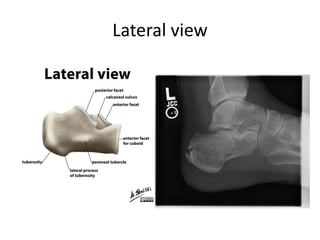
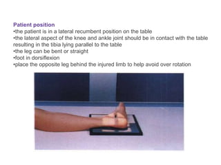
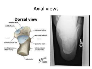
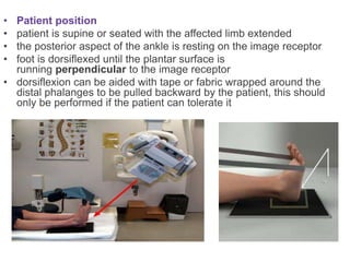
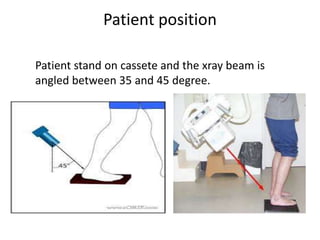
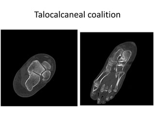


This document provides instructions for three radiographic views of the calcaneum: the lateral view, which assesses the calcaneum and surrounding joints from the side; the axial view, which images the talocalcaneal joint and plantar calcaneum from above; and the Harris view, a specialized projection used to detect talocalcaneal coalition. Patient positioning instructions are outlined for each view, including recumbent, supine, seated, or standing positions and manipulation of the foot and leg.


![Patient care [autosaved]](https://cdn.slidesharecdn.com/ss_thumbnails/patientcareautosaved-150405120334-conversion-gate01-thumbnail.jpg?width=640&height=640&fit=bounds)


![Polymer [ बहुलक ] Chemistry Notes PDF - Irfanullah Mehar - JJ Sir Chemistry.pdf](https://cdn.slidesharecdn.com/ss_thumbnails/polymerchemistrynotespdf-irfanullahmehar-jjsirchemistry-260210172118-3f9b37f7-thumbnail.jpg?width=640&height=640&fit=bounds)


