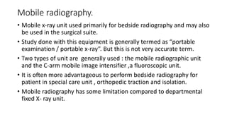
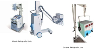
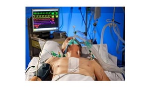
Bedside Radiography: Special
condition and Environments
Mobile radiography.
• Mobile x-ray unit used primarily for bedside radiography and may also
be used in the surgical suite.
• Study done with this equipment is generally termed as “portable
examination / portable x-ray”. But this is not very accurate term.
• Two types of unit are generally used : the mobile radiographic unit
and the C-arm mobile image intensifier ,a fluoroscopic unit.
• It is often more advantageous to perform bedside radiography for
patient in special care unit , orthopedic traction and isolation.
• Mobile radiography has some limitation compared to departmental
fixed X- ray unit.
Mobile Radiography Units.
Portable Radiography Unit.
Cont..
• Follow these guidelines:
• Check with the nurse in charge to inquire about patients condition.
• Confirm the order in the patient’s chart , if applicable .
• Always greet the patient (if applicable ) , check the name and D.O.B
and identification band.
• Inspect and prepare room before bringing x-ray machine .
Special Care Units
• Intensive care Unit (ICU)
• Coronary care Unit (CCU)
• Neonatal Intensive Care Unit (NICU)
• Post anesthesia Care Unit/ Recovery (PACU)
• Emergency Department (ED) /Trauma Unit
• High Dependency Unit (HDU).
Treatment Situations Involving specialty Equipment
Special Beds and Mattresses.
• Alternating pressure mattress / Rocking beds – needs to be turned
off during exposure to avoid motion artifacts .
• Air mattress – must be fully inflated to firm up and level the bed
before placing the IR under patient.
• Patient laying on an alcohol or water filled pad to raise or lower body
temperature , it is important to place IR on the top of the pad to avoid
artifacts.
• Must be careful while placing IR in between of patient and Mylar
warming blanket.
Orthopedic Traction.
• Don’t attempt to alter a patient’s traction.
• Sudden release of traction may result in serious harm.
• Don’t bump against bed.
• Take help of nursing staff.
• Encourage patient to co-operate and use Trapeze bar to raise patient's
torso.
Tracheostomies:
• Monitor patient closely.
• Never untie the tapes holding a tracheostomy tube in place.
Mechanical Ventilation:
• An endotracheal tube is passed through the mouth into the trachea.
• The tube is connected to ventilator.
• While performing radiograph don’t turn off ventilator to eliminate
respiratory motion.
• Pay attention to breathing rhythm .
• Sense the breathing rhythm correctly and keep short exposure.
• Position patient carefully without applying tension to tubing .
• If patient is in AMBU bag , instruct nursing staff to stop squeezing
while exposure is made.
Nasogastric and nasoenteric Tubes.
• Tubes passes through the nose and down into the stomach or small
intestine.
• Has several purpose as feeding , decompression , radiographic
examination.
• Levine and Salem-sump tubes are often sued.
• Don’t disturb the tube while taking radiograph .
• If it has to be disconnected from suction ,temporarily take permission
from the nurse.
• While giving oral contrast through tube make sure not to displace
tube from its location .
Correct placement of NG tube
Incorrect placement of NG tube
Closed chest Drainage.
• Used to remove air or fluid accumulated in the pleural space.
• While taking radiograph:
• Ensure that chest suction and drainage apparatus is not disturbed.
• Take care that the chest tube is not dislodged when positioning the IR
for the chest radiograph.
• Ensure that drainage unit remains below the level of the patient’s
chest .
Pacemaker :
• You should not elevate or abduct the patient’s left arm for 24 hours
after surgery to prevent dislodging the pacemaker and catheter.
Placement of central lines.
• Swan - Ganz catheter is correctly placed when its tip lies in the right
or left pulmonary artery. Catheter measure cardiac output and heart
pressure. Inserted through subclavian , jugular or femoral vein .
• Central venous catheter (CVC)- These is advanced into the venecava
through veins . It facilitate the administration of long term drugs,
dialysis ,blood transfusion , drawing blood and monitoring central
venous pressure.
• Make sure to position patient and tube correctly in order to show
correct placement of catheter in radiograph .
• Avoid patient rotation .
Swan - Ganz catheter
Central venous catheter
Bedside radiography and patient care

Bedside radiography and patient care

  • 1.
  • 2.
    Mobile radiography. • Mobilex-ray unit used primarily for bedside radiography and may also be used in the surgical suite. • Study done with this equipment is generally termed as “portable examination / portable x-ray”. But this is not very accurate term. • Two types of unit are generally used : the mobile radiographic unit and the C-arm mobile image intensifier ,a fluoroscopic unit. • It is often more advantageous to perform bedside radiography for patient in special care unit , orthopedic traction and isolation. • Mobile radiography has some limitation compared to departmental fixed X- ray unit.
  • 3.
  • 4.
    Cont.. • Follow theseguidelines: • Check with the nurse in charge to inquire about patients condition. • Confirm the order in the patient’s chart , if applicable . • Always greet the patient (if applicable ) , check the name and D.O.B and identification band. • Inspect and prepare room before bringing x-ray machine .
  • 5.
    Special Care Units •Intensive care Unit (ICU) • Coronary care Unit (CCU) • Neonatal Intensive Care Unit (NICU) • Post anesthesia Care Unit/ Recovery (PACU) • Emergency Department (ED) /Trauma Unit • High Dependency Unit (HDU).
  • 7.
    Treatment Situations Involvingspecialty Equipment Special Beds and Mattresses. • Alternating pressure mattress / Rocking beds – needs to be turned off during exposure to avoid motion artifacts . • Air mattress – must be fully inflated to firm up and level the bed before placing the IR under patient. • Patient laying on an alcohol or water filled pad to raise or lower body temperature , it is important to place IR on the top of the pad to avoid artifacts. • Must be careful while placing IR in between of patient and Mylar warming blanket.
  • 9.
    Orthopedic Traction. • Don’tattempt to alter a patient’s traction. • Sudden release of traction may result in serious harm. • Don’t bump against bed. • Take help of nursing staff. • Encourage patient to co-operate and use Trapeze bar to raise patient's torso.
  • 11.
    Tracheostomies: • Monitor patientclosely. • Never untie the tapes holding a tracheostomy tube in place.
  • 12.
    Mechanical Ventilation: • Anendotracheal tube is passed through the mouth into the trachea. • The tube is connected to ventilator. • While performing radiograph don’t turn off ventilator to eliminate respiratory motion. • Pay attention to breathing rhythm . • Sense the breathing rhythm correctly and keep short exposure. • Position patient carefully without applying tension to tubing . • If patient is in AMBU bag , instruct nursing staff to stop squeezing while exposure is made.
  • 14.
    Nasogastric and nasoentericTubes. • Tubes passes through the nose and down into the stomach or small intestine. • Has several purpose as feeding , decompression , radiographic examination. • Levine and Salem-sump tubes are often sued. • Don’t disturb the tube while taking radiograph . • If it has to be disconnected from suction ,temporarily take permission from the nurse. • While giving oral contrast through tube make sure not to displace tube from its location .
  • 15.
    Correct placement ofNG tube Incorrect placement of NG tube
  • 16.
    Closed chest Drainage. •Used to remove air or fluid accumulated in the pleural space. • While taking radiograph: • Ensure that chest suction and drainage apparatus is not disturbed. • Take care that the chest tube is not dislodged when positioning the IR for the chest radiograph. • Ensure that drainage unit remains below the level of the patient’s chest .
  • 17.
    Pacemaker : • Youshould not elevate or abduct the patient’s left arm for 24 hours after surgery to prevent dislodging the pacemaker and catheter.
  • 18.
    Placement of centrallines. • Swan - Ganz catheter is correctly placed when its tip lies in the right or left pulmonary artery. Catheter measure cardiac output and heart pressure. Inserted through subclavian , jugular or femoral vein . • Central venous catheter (CVC)- These is advanced into the venecava through veins . It facilitate the administration of long term drugs, dialysis ,blood transfusion , drawing blood and monitoring central venous pressure. • Make sure to position patient and tube correctly in order to show correct placement of catheter in radiograph . • Avoid patient rotation .
  • 19.
    Swan - Ganzcatheter
  • 20.