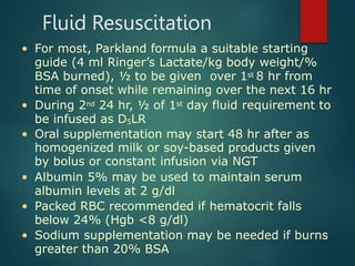
Burns are classified based on depth and extent of tissue damage. First degree burns involve only the epidermis, while second degree burns extend deeper into the dermis and may cause blistering. Third degree burns extend through the entire thickness of skin. Proper first aid and fluid resuscitation are important to prevent further tissue damage. Hospitalization is recommended for burns covering over 10% of total body surface area or involving sensitive areas like the hands, face or genitals. Management involves wound care, pain control, nutrition and physical therapy.
This document provides information on burns and sudden death. It begins by defining burns and classifying them based on depth and extent. It then discusses the pathophysiology of burns, including edema, cardiac, renal and immunologic effects. Burn management in the hospital is outlined, including fluid resuscitation, airway management, infection control, pain relief and nutrition. Complications of burns like shock, infections and scarring are also summarized. The document concludes by defining sudden death and noting it is important in forensic analysis of unexpected deaths within 24 hours of burn onset.
This document provides information on burns and sudden death. It defines burns and classifies them based on depth. It describes the pathophysiology of burns including effects on the skin, cardiovascular, renal and immune systems. Common causes of burns and initial first aid measures are outlined. Criteria for hospitalization, standard hospital management including fluid resuscitation and infection control are summarized. Complications of burns and causes of sudden death involving various body systems like cardiovascular, respiratory and gastrointestinal are also reviewed.
Burns are caused by thermal injury and result in skin and tissue damage. They are classified by depth and extent of body surface area affected. Common types include scalds, flames, chemicals, electricity, and radiation. Management involves assessing airway/breathing, fluid resuscitation to prevent shock, wound care, pain relief, and infection control. Resuscitation aims to stabilize the patient and replace fluid losses using formulas like Parkland or Brooke, followed by acute wound management and later rehabilitation.
This document provides an overview of burns, including definitions, classifications, pathophysiology, management, and complications. It defines burns as thermal injuries to the skin and tissues. Burns are classified based on depth and extent of damage. First, second, and third degree burns are described. Hospitalization is generally recommended for burns over 10% of total body surface area. The pathophysiology involves fluid shifts, cardiac, metabolic, immunologic, and renal effects. Burn management includes airway control, fluid resuscitation, wound care, infection prevention, pain relief, and nutrition. Complications can include shock, infection, renal failure, and scarring.
1) Burns are classified based on depth of tissue damage, with first degree affecting only the epidermis and third degree extending into deeper tissues.
2) Burn injuries cause fluid shifts, increased metabolism, and immune dysfunction that must be addressed through resuscitation and wound management.
3) Initial treatment involves fluid resuscitation, airway protection, infection control and pain management. Hospitalization is needed for severe or complicated burns.
1) A burn is a thermal injury caused by heat, chemicals, electricity or other sources that damages skin and tissue through local and systemic effects.
2) Thermal burns are caused by heat and can be first, second, or third degree depending on depth of tissue damage. Chemical and electrical burns damage tissues through other means.
3) Initial management of burns involves stopping the burning process, cooling the affected area, and providing fluid resuscitation for severe burns over 15% of total body surface area. Comprehensive examination determines burn size, depth and other injuries for treatment.
This document provides information on burns, including definitions, types, classification, pathophysiology, assessment, and management. It defines burns as thermal injuries to the skin and tissues. Burns are classified based on depth and extent of damage. First and second degree burns involve the epidermis and dermis, while third degree burns extend deeper. Burn severity is also classified according to percentage of total body surface area affected. Management involves fluid resuscitation, wound care, pain control, and nutrition support. Complications can impact various organ systems. The goal is to prevent infection, contractures, and other issues through proper acute care and rehabilitation.
This document provides a classification and overview of burn injuries. It discusses:
1. The classification of burns based on etiology including thermal, electrical, chemical, radiation, and inhalation injuries.
2. The degrees of burn injuries from first to fourth degree based on depth of tissue damage.
3. Key aspects of burn management including emergent resuscitation focusing on airway, circulation and fluid replacement to maintain organ function in the first 24-48 hours.
4. Wound care including open and closed methods and use of antimicrobial agents like silver sulfadiazine cream.
This document discusses the anesthetic management of burns. It covers the pathophysiology of burns including the zones of burn injury and complications like inhalation injury. It describes the severity classification of burns and guidelines for fluid resuscitation. Anesthetic concerns in burns include difficult airway management due to facial burns, vascular access issues, temperature control, and fluid management. Drugs may have unpredictable responses due to reduced plasma proteins. Regional anesthesia can be used but general anesthesia is more common, requiring careful monitoring and management of respiratory and hemodynamic parameters.
A complete review for all medical students and doctors working in burn unit in any hospital. #Emergency #BurnProtocol #protocol #Burns #Abhishek #MUSTKNOW #knowledge #Medical #Health
1. Burn injuries cause cell destruction of the skin layers and depletion of fluids and electrolytes from the body. Mortality rates are higher for children under 4 and adults over 65.
2. Preexisting medical conditions like heart, lung, and kidney disease negatively impact recovery from burn injuries and increase mortality.
3. Burn management involves assessing the size, depth, and cause of the burns and providing fluid resuscitation, wound care, pain management, and nutrition. The goal is to restore fluid and electrolyte balance, promote wound healing, and maximize patient function and independence.
1) Burns can result from direct contact with flames, hot liquids, gases, chemicals, electricity, or radiation. They cause tissue injuries by denaturing proteins.
2) Burn injuries affect the skin, which acts as a protective barrier and regulates temperature and fluid balance. Deeper burns extend beyond the epidermis into the dermis.
3) Proper evaluation and treatment of burn injuries requires assessing burn depth, size, inhalation injury, and associated complications affecting various organ systems. Early fluid resuscitation is critical.
The document discusses the structured approach to presenting burn cases including relevant anatomy, classification of burns, complications affecting various organ systems, and the three phases of burn management with a focus on the priorities in the resuscitative phase including airway management, breathing, circulation, fluid resuscitation, and endpoints of successful resuscitation. Specific formulas for fluid resuscitation and indications for endotracheal intubation are also outlined.
This document discusses the classification, pathophysiology, clinical signs, and management of burns. It describes burns as injuries caused by heat, electricity, or chemicals that can range from superficial damage of the epidermis to full thickness damage involving muscle and fascia. Burns are classified based on their depth and severity into four degrees. Management involves cooling the burn, applying antibacterial creams, managing shock, preventing infection, and promoting wound healing through dressings and nutrition. Smoke inhalation injuries also require airway management and bronchodilation treatments.
1. Burns can be classified based on depth and percentage of total body surface area affected. Deeper burns involving more surface area lead to worse outcomes.
2. Burns trigger a complex pathophysiological response involving fluid shifts, immune dysfunction, and metabolic alterations. Locally, burns cause tissue damage and fluid accumulation. Systemically, burns induce hypovolaemic shock, immune suppression increasing infection risk, and a hypermetabolic state.
3. Managing the pathophysiological effects of burns, especially fluid shifts and immune dysfunction, is important for treatment and recovery from burn injuries. Deeper and more extensive burns have more severe local and systemic pathophysiological consequences.
The document discusses different types of burn injuries including thermal, chemical, smoke inhalation, and electrical burns. It describes the pathophysiology and clinical manifestations in the emergent and acute phases after a burn. Key aspects are fluid and electrolyte shifts leading to shock in the emergent phase and wound healing through debridement, grafting, and rehabilitation in the acute phase. Complications include infection and contractures.
1. A burn is an injury to the skin or flesh caused by heat, electricity, chemicals, friction or radiation. The severity depends on the temperature and duration of exposure.
2. About 2.4 million people suffer burns annually in the US, with 700,000 cases requiring medical treatment. The main causes are thermal, electrical, chemical and radiation burns.
3. Burns are classified by depth and extent of the affected body surface area. Depth is classified as superficial, partial-thickness, or full-thickness. Extent is classified using methods like the Rule of Nines or Lund and Browder chart.
Burn and scald injuries can be caused by heat, electricity, chemicals, or radiation. Thermal burns are the most common and are classified as superficial, partial thickness, or full thickness depending on the depth of tissue damage. A severe burn over 25% of the total body surface area can cause systemic effects like shock due to fluid loss, decreased blood pressure, and increased heart rate. Complications include infection, respiratory failure, renal failure, and contractures. The severity of the burn is estimated using methods like the Rule of Nines or Lund and Browder chart which divide the body into sections and assign a percentage of total body surface area to each.
The document summarizes the pathophysiology of burns in three phases. The initial ebb phase occurs in the first 24 hours and involves hypotension, low cardiac output, and hypoventilation. The flow phase follows and involves increases in cardiac output and oxygen consumption. A hypermetabolic hyperdynamic response peaks at 10-14 days. Systemic effects include metabolic, cardiac, renal, blood, immunologic, lung, GI, and infectious responses. Burn-induced inflammatory mediators cause widespread vascular permeability and organ dysfunction. Successful resuscitation is needed to avoid multi-organ failure from hypovolemia and infection risk due to impaired immunity and skin barrier function.
A burn is a cutaneous injury caused by heat, electricity, chemicals, friction, or radiation. Burns are classified based on the depth of skin involvement, ranging from superficial 1st degree burns only involving the epidermis to full thickness 3rd degree burns extending into subcutaneous tissue. Management involves stopping the burning process, assessing airway and breathing needs, administering fluid resuscitation based on formulas like Parkland that account for total body surface area burned, and treating wounds appropriately based on depth. Proper initial burn management and resuscitation are critical to prevent complications and optimize healing outcomes.
1) Burns can result from heat, chemicals, electricity, or radiation and cause damage to the skin and underlying tissues. The very young, old, and careless are at high risk of severe burns.
2) Initial burn management involves assessing the airway, giving oxygen, establishing IV access, giving fluids resuscitation based on the Parkland formula, monitoring vitals and urine output, and giving pain medications.
3) Providing anesthesia for burn patients poses challenges due to potential airway issues, pulmonary insufficiency, fluid shifts, and altered drug metabolism. Careful attention to the airway, adequate vascular access and fluid resuscitation, temperature control, and effects of medications are important.
Burns are classified by depth and extent of injury. Superficial burns only affect the epidermis while deep burns damage the dermis. Full thickness burns destroy the entire dermis. Management involves fluid resuscitation, wound care, prevention of infection and complications. Local wound care includes cleaning, silver sulfadiazine cream and dressing changes. For deep burns, debridement and skin grafting may be needed. Monitoring of vitals, urine output and blood work is important. Escarotomy may be required for circumferential full thickness limb burns to prevent limb loss.
Burns are injuries caused by heat, chemicals, electricity or radiation. They can occur at any age and socioeconomic group. Thermal burns result from flame, hot liquids or objects, while chemical burns are caused by strong acids or alkalis. Electrical burns occur from electricity passing through the body. Radiation burns come from exposure to radiation sources. Burns are classified by depth and extent of body surface area affected. Management involves cooling the wound, establishing airways, fluid resuscitation, and calculating fluid needs using formulas based on total body surface area burned. The goal is adequate circulation and urine output to prevent shock.
Sepsis is a life-threatening condition that arises when the body's response to infection causes injury to its own tissues. Globally, sepsis kills about 8 million people annually. Early recognition and treatment are key to improving outcomes. The first hours after diagnosis are especially critical, as mortality increases by about 8% every hour that antibiotics are delayed. Prompt administration of broad-spectrum antibiotics and fluid resuscitation can significantly reduce mortality from sepsis.
The document discusses temperature management in surgical patients. It covers normal body temperature, factors that can alter temperature, thermoregulatory responses to heat and cold, and classifications of hypothermia and hyperthermia. It also summarizes complications of hypothermia, prevention and treatment of intraoperative hypothermia, malignant hyperthermia, and management of hyperthermia and fever.
This document outlines key concepts related to healthcare supply chain and inventory management. It discusses the typical players in the supply chain including manufacturers, distributors, group purchasing organizations, and e-distributors. It also describes how materials flow through the supply chain and some contemporary issues in medical inventory management like just-in-time systems and single vs multiple vendors. Key aspects of effective inventory management are identified such as inventory accounting systems, lead times, costs including holding, ordering and shortage costs, and the economic order quantity model.
Introduction to quantitative decision-making methods in healthcare managementMUKESH SUNDARARAJAN
This document provides an introduction to quantitative decision-making methods in healthcare management. It discusses the historical development of management techniques from scientific management to modern approaches like operations research, management information systems, and total quality management. The document also outlines the nature of the healthcare industry, the healthcare process, and the types of decisions made by healthcare managers at strategic, tactical, and operational levels. These decisions involve areas like capacity, personnel, inventory, scheduling, and quality assurance. The document concludes by noting some distinctive characteristics of healthcare services like the simultaneous production and consumption of care.
This document defines health and productivity management (HPM) as a systematic approach for companies to quantify, evaluate, and optimize their investment in employee health. HPM aims to reduce health-related costs like medical expenses and lost productivity from absenteeism and presenteeism. The history of HPM shows early studies linking health factors like iron deficiency to lost productivity. Common health risks like poor diet and smoking drive chronic diseases responsible for most health costs. Implementing strategies to improve lifestyle behaviors and reduce health risks through workplace programs can help control costs and improve employee health.
The document discusses the need for healthcare project management training and the benefits it provides. It notes that recent US legislation and industry trends have led to an increased number of healthcare projects. Good project management is required to implement projects successfully and achieve goals like improved quality and reduced costs. However, healthcare workers often lack project management skills since they are more familiar with operational versus project work. The document advocates for training clinical leaders in project management principles and provides suggestions for developing effective training programs.
This document discusses various decision making tools and techniques that can be used in healthcare facilities. It outlines the typical decision making process and some common causes of poor decisions. It then covers different types of decisions based on certainty, uncertainty and risk. Various decision tools are presented, including payoff tables, expected value, decision trees, and techniques for decisions with non-monetary attributes. Examples are provided to illustrate how to apply these tools and techniques to make optimal decisions under different conditions.
Inductive transducers work by changing the inductance of a coil due to changes in a measured quantity like displacement, force, or pressure. There are two main types - self-generating transducers that produce a voltage signal from relative motion in a magnetic field, and non-self generating transducers that require an external power source to energize coils whose inductance changes. Inductive transducers work by changing the self-inductance of a coil, producing eddy currents in a nearby conductor, or altering the mutual inductance between two coils due to the measured quantity. They are used for proximity sensing, touchpads, and detecting metal objects or movement.
The document discusses different types of ultrasound transducers. The essential element of each transducer is a piezoelectric crystal that generates and receives ultrasound waves. Transducers come in various shapes, sizes, and features depending on the body part being imaged. The main types of transducers discussed are linear, convex, phased array, pencil, endocavitary, transesophageal, and 4D transducers. Each type has a different piezoelectric crystal arrangement, aperture, frequency, and intended medical applications.
This document discusses three main types of photoelectric cells: photoemissive cells, photovoltaic cells, and photoconductive cells. Photoemissive cells contain a photosensitive cathode and anode inside an evacuated glass bulb, and emit electrons when light hits the cathode. Photovoltaic cells, also called true cells, generate their own voltage when light hits a semiconductor-metal junction without needing an external voltage. Photoconductive cells have a semiconductor like selenium between two electrodes, and their resistance decreases when light hits, allowing current to flow in an external circuit. All three types of cells convert light energy into electrical energy or signals based on the photoelectric effect.
Piezoelectric transducers work by exploiting the piezoelectric effect, where certain materials generate an electric voltage when subjected to mechanical stress. They can be used to measure dynamic changes by converting mechanical energy into electrical signals. Common piezoelectric materials used in transducers include barium titanate, lead zirconate titanate, Rochelle salt, and quartz. While offering high frequency response and transient response, piezoelectric transducers also have low output and high impedance. Applications include dynamic measurement, studying high-speed phenomena, vibration detection, and use in medical devices, printers, lighters, and phone screens.
A scintillation counter detects and measures ionizing radiation by using a scintillator material that produces light pulses when excited by incident radiation. It consists of a scintillator that generates photons when hit by radiation, a photomultiplier tube that converts the light to an electrical signal, and electronics to process the signal. Scintillation counters are used in applications like radiation monitoring, medical imaging, and nuclear security to detect radioactive contamination. Variants mounted on vehicles can provide rapid response in security situations involving dirty bombs or radioactive waste.
Spectrophotometry involves measuring the intensity of light at selected wavelengths to analyze substances. It relies on substances absorbing light at characteristic wavelengths. A spectrophotometer uses light sources, monochromators, and detectors to isolate wavelengths and measure light intensity. Key concepts include Beer's Law which states absorbance is directly proportional to concentration. Spectrophotometers are used in applications like measuring analyte concentration, detecting impurities, and studying chemical kinetics through observation of absorbance changes over time.
This document discusses different types of photoelectric transducers, including photoemissive, photoconductive, and photovoltaic devices. It focuses on photomultiplier tubes, describing their construction and working principle of electron multiplication through secondary emission at dynode stages. Photomultiplier tubes can amplify current by 105 to 109 times, achieving high luminous sensitivity down to 10-5 lumens. The document also covers photoconductive cells, whose resistance varies with light intensity, allowing their use in light-controlled circuits.
Photodiodes are semiconductor devices that convert light into an electrical current. When light strikes the photodiode's PN junction, electrons are excited creating free charge carriers. This results in a photocurrent that can be measured. Photodiodes operate in reverse bias and can be used as solar cells, photodetectors, or LEDs. Common materials used include silicon, germanium, indium gallium arsenide, and lead sulfide, depending on the desired wavelength detection range. Photodiodes find applications in devices like optical mice, smoke detectors, and infrared remote controls.
Inductive transducers work by changing the inductance of a coil due to changes in a measured quantity like displacement, force, or pressure. There are two main types - self-generating transducers that produce a voltage signal from relative motion in a magnetic field, and non-self generating transducers that require an external power source to energize coils whose inductance changes with motion. Inductive transducers work by changing the self-inductance of a coil, producing eddy currents in a nearby conductor, or altering the mutual inductance between two coils as a function of the measured quantity.
This document discusses training, promotion, and transfer in an organizational context. It defines training as a systematic process aimed at developing employee skills, abilities, and attitudes. The purpose of training includes improving performance, reducing waste, and developing manpower. Promotion is defined as a change to a better job with higher status and responsibilities, and is used to reward performance, boost morale, and retain skilled employees. Both seniority and merit are discussed as bases for promotion, as well as a combination of the two. Transfer is defined as a lateral shift between jobs, departments, or locations with no change in pay or status. Different types of transfers are outlined, including production, replacement, remedial, and versatility transfers.
The document describes the different departments found in hospitals and provides brief descriptions of their functions. It discusses 20 departments including casualty, cardiology, critical care, ENT, geriatrics, gastroenterology, general surgery, gynecology, hematology, maternal/neonatal/pediatrics, neurology, oncology, ophthalmology, orthopedics, urology, psychiatry, outpatient, inpatient, central sterilization unit, housekeeping, catering and food services, medical social work, physiotherapy, pharmacy, nutrition and dietetics, microbiology, diagnostic imaging, medical records, medical maintenance and engineering, information technology and communication, human resources, finance, and administration.
This document provides an overview of human resource management. It defines HRM and discusses its key functions such as acquisition, development, compensation, and industrial relations. It also outlines the nature and scope of HRM, highlighting that it is a pervasive, future-oriented, and people-focused function. The document further discusses human resource planning, recruitment, selection, and placement processes. It compares personnel management with HRM and lists the objectives and importance of effective HRM. Finally, it notes some emerging issues and the role of HR professionals in adapting to changing business environments.
1. The document discusses measurement systems and instrumentation. It covers topics like order of instruments, instrument classification, units of measurement, standards of measurement, dimensions of measurement, and errors in measurement.
2. Instruments can be classified as mechanical, electrical, or electronic. They can also be categorized as absolute, secondary, digital, or analogue instruments.
3. The seven base SI units are meter, kilogram, second, Kelvin, mole, candela, and ampere. Derived units are formed by combining base units.
4. Standards provide defined relationships to measurement units and are used to calibrate other instruments. Primary standards define measurement units while secondary and working standards are calibrated against primary standards.
This document provides first aid guidelines for insect, scorpion, snake, and animal bites. It describes symptoms of bites and stings from bees, wasps, ants, scorpions, centipedes, snakes, and animals. Treatment recommendations include washing the affected area, applying antiseptic or cold compress, keeping it elevated, and seeking medical help for snake or scorpion bites. Guidelines are given for proper tick removal and when to see a doctor after insect bites depending on rash or disease risk in the region.
This document provides guidance on first aid for animal bites, insect stings, and snake bites. It advises remaining calm, cleaning and dressing wounds, immobilizing affected areas as needed, and seeking medical help for serious injuries or allergic reactions. Precautions are described such as giving animals space, not provoking them, and calling for assistance if a situation cannot be handled alone.
How to Manage Reception Report in Odoo 17Celine George
A business may deal with both sales and purchases occasionally. They buy things from vendors and then sell them to their customers. Such dealings can be confusing at times. Because multiple clients may inquire about the same product at the same time, after purchasing those products, customers must be assigned to them. Odoo has a tool called Reception Report that can be used to complete this assignment. By enabling this, a reception report comes automatically after confirming a receipt, from which we can assign products to orders.
How to Download & Install Module From the Odoo App Store in Odoo 17Celine George
Custom modules offer the flexibility to extend Odoo's capabilities, address unique requirements, and optimize workflows to align seamlessly with your organization's processes. By leveraging custom modules, businesses can unlock greater efficiency, productivity, and innovation, empowering them to stay competitive in today's dynamic market landscape. In this tutorial, we'll guide you step by step on how to easily download and install modules from the Odoo App Store.
Andreas Schleicher presents PISA 2022 Volume III - Creative Thinking - 18 Jun...EduSkills OECD
Andreas Schleicher, Director of Education and Skills at the OECD presents at the launch of PISA 2022 Volume III - Creative Minds, Creative Schools on 18 June 2024.
A Free 200-Page eBook ~ Brain and Mind Exercise.pptxOH TEIK BIN
(A Free eBook comprising 3 Sets of Presentation of a selection of Puzzles, Brain Teasers and Thinking Problems to exercise both the mind and the Right and Left Brain. To help keep the mind and brain fit and healthy. Good for both the young and old alike.
Answers are given for all the puzzles and problems.)
With Metta,
Bro. Oh Teik Bin 🙏🤓🤔🥰
This document provides an overview of wound healing, its functions, stages, mechanisms, factors affecting it, and complications.
A wound is a break in the integrity of the skin or tissues, which may be associated with disruption of the structure and function.
Healing is the body’s response to injury in an attempt to restore normal structure and functions.
Healing can occur in two ways: Regeneration and Repair
There are 4 phases of wound healing: hemostasis, inflammation, proliferation, and remodeling. This document also describes the mechanism of wound healing. Factors that affect healing include infection, uncontrolled diabetes, poor nutrition, age, anemia, the presence of foreign bodies, etc.
Complications of wound healing like infection, hyperpigmentation of scar, contractures, and keloid formation.
CapTechTalks Webinar Slides June 2024 Donovan Wright.pptxCapitolTechU
Slides from a Capitol Technology University webinar held June 20, 2024. The webinar featured Dr. Donovan Wright, presenting on the Department of Defense Digital Transformation.
THE SACRIFICE HOW PRO-PALESTINE PROTESTS STUDENTS ARE SACRIFICING TO CHANGE T...indexPub
The recent surge in pro-Palestine student activism has prompted significant responses from universities, ranging from negotiations and divestment commitments to increased transparency about investments in companies supporting the war on Gaza. This activism has led to the cessation of student encampments but also highlighted the substantial sacrifices made by students, including academic disruptions and personal risks. The primary drivers of these protests are poor university administration, lack of transparency, and inadequate communication between officials and students. This study examines the profound emotional, psychological, and professional impacts on students engaged in pro-Palestine protests, focusing on Generation Z's (Gen-Z) activism dynamics. This paper explores the significant sacrifices made by these students and even the professors supporting the pro-Palestine movement, with a focus on recent global movements. Through an in-depth analysis of printed and electronic media, the study examines the impacts of these sacrifices on the academic and personal lives of those involved. The paper highlights examples from various universities, demonstrating student activism's long-term and short-term effects, including disciplinary actions, social backlash, and career implications. The researchers also explore the broader implications of student sacrifices. The findings reveal that these sacrifices are driven by a profound commitment to justice and human rights, and are influenced by the increasing availability of information, peer interactions, and personal convictions. The study also discusses the broader implications of this activism, comparing it to historical precedents and assessing its potential to influence policy and public opinion. The emotional and psychological toll on student activists is significant, but their sense of purpose and community support mitigates some of these challenges. However, the researchers call for acknowledging the broader Impact of these sacrifices on the future global movement of FreePalestine.
2. Definition
• Burns are a result of the effects of thermal
injury on the skin and other tissues
• Human skin can tolerate temperatures up to
42-440 C (107-1110 F) but above these, the
higher the temperature the more severe the
tissue destruction
• Below 450 C (1130 F), resulting changes are
reversible but >450 C, protein damage
exceeds the capacity of the cell to repair
3. Classification According to Depth
• First-degree Burns (mild): epidermis
Pain, erythema & slight swelling, no blisters
Tissue damage usually minimal, no scarring
Pain resolves in 48-72 hours
• Superficial Second-degree Burns: entire epidermis &
variable dermis
Vesicles and blisters characteristic
Extremely painful due to exposed nerve endings
Heal in 7-14 days if without infection
• Midlevel to Deep Second-degree Burns:
Few dermal appendages left
There are some fluid & metabolic effects
• Full-thickness or Third-Degree: entire epidermis and
dermis, no residual epidermis
Painless, extensive fluid & metabolic deficits
Heal only by wound contraction, if small, or if big,
by skin grafting or coverage by a skin flap
5. Classification According to Extent
• Mild: 10%
• Moderate:
10-30%
• Severe: > 30%
• Hospitalization
for > 10% of
body surface area
Infant Rule of Nines
(for quick assessment of
total body surface area
affected by burns)
Anatomic
structure
Surface
area
Head 18%
Anterior Torso 18%
Posterior Torso 18%
Each Leg 14%
Each Arm 9%
Perineum 1%
6. Kinds of Burns
• Scald Burn: most frequent in home injuries; hot
water, liquids and foods are most common causes;
above 65o C, cell death
• Flame Burn: due to gasoline, kerosene, liquified
petroleum gas (LPG) or burning houses
• Chemical Burn: common in industries and
laboratories but may also occur at home; acid is
more common than alkali
• Electrical Burn: worse than the other types; with
entrance and exit wounds; may stop the heart and
depress the respiratory center; may cause
thrombosis and cataracts
• Radiation Burn: from X-ray, radioactive radiation
and nuclear bomb explosions
9. Burn Photos
Electrical Burns
Entrance Wounds
Entrance wound of electrical
burns from an overheated tool
Severe swelling
peaks 24-72 hrs after
Electrical Burns
Exit Wounds
Electrical burns mummified
1st 2 fingers later removed
10. Physiological Response
• Typically, biphasic response
• The initial period of hypofunction manifests as: (a)
Hypotension, (b) Low cardiac output, (c) Metabolic acidosis,
(d) Ileus, (e) Hypoventilation, (f) Hyperglycemia, (g) Low
oxygen consumption and (h) Inability to thermoregulate
• This ebb phase occurs usually in the first 24 hours and
responds to fluid resuscitation
• The flow phase, resuscitation, follows and is characterized by
gradual increases in (a) Cardiac output, (b) Heart rate, (c)
Oxygen consumption and (d) Supranormal increases of
temperature
• This hypermetabolic hyperdynamic response peaks in 10-14
days after the injury after which condition slowly recedes to
normal as the burn wounds heal naturally or surgically closed
by applying skin grafting
11. Pathologic Features
• Zone of coagulation (necrosis): Superficial area of
coagulation necrosis and cell death on exposure to
temperatures >450 (primary injury)
• Zone of stasis (vascular thrombosis): Local capillary
circulation is sluggish, depending on the adequacy of the
resuscitation, can either remain viable or proceed to cell
death (secondary injury)
• Zone of hyperemia (increased capillary permeability)
12. Burn Pathophysiology: Edema
• Injured tissue Increased permeability of entire
vascular tree loss of water, electrolytes and
proteins from the vascular compartment severe
hemoconcentration
• Protein leakage resultant hypoproteinemia,
increased osmotic pressure in the interstitial space
• Decreased cell membrane potential cause inward
shift of Na+ and H2O cellular swelling
• In the injured skin, effect maximal 30 min after the
burn but capillary integrity not restored until 8-12
hours after, usually resolved by 3-5 days
• In non-injured tissues, only mild and transient
leaks even for burns >40% BSA
13. Burn Pathophysiology: Cardiac
• Cardiac output decreases due to:
1) Decreased preload induced by fluid shifts
2) Increased systemic vascular resistance caused
by both hypovolemia and systemic
catecholamine release
3) A myocardial depressant factor has been
described that impairs cardiac function
• Cardiac output normal within 12-18 hours, with
successful resuscitation
• After 24 hours, it may increase up to 2 ½
times the normal and remain elevated until
several months after the burn is closed
14. Burn Pathophysiology: Blood
• The red-cell mass decreases due to direct losses
• Immediate, 1-2 hours after, and delayed, 2-7 days
postburn, hemolysis occurs due to damaged cells
and increased fragility
• Anemia within 4-7 days is common and expected,
typically, will persist until wound healing occur;
depressed erythropoietin levels documented
• Early mild thrombocytopenia (sequestration)
followed by thrombocytosis (2-4x normal) and
elevated fibrinogen, factor V and factor VIII levels
commonly by end of the 1st week
• A “normal” platelet or fibrinogen level may be an
early sign of disseminated intravascular coagulation
• Persistent thrombocytopenia is associated with poor
prognosis -- suspect sepsis
15. Burn Pathophysiology: Metabolic
• Severe catabolism with breakdown of muscle
protein for gluconeogenesis as acute response
• Prostaglandins and cytokines implicated in
increased core temperature of 1-20 C and in
initiating acceleration of nitrogen catabolism
• Plasma levels of catecholamines, glucagon and
cortisol all increase, maximal in patients with
50-60% TBSAB, while insulin and thyroid hormone
levels decrease
• Hypermetabolic response may approach 200% of
BMR remaining elevated for months after burn
closed
• Early enteral feeding associated with lessening of
the hypermetabolic response
16. Burn Pathophysiology: Renal
• Renal blood flow and GFR decrease soon after
due to hypovolemia, decreased cardiac output,
and elevated systemic vascular oliguria and
antidiuresis develops during 1st 12-24 hours
• Followed by a usually modest diuresis as the
capillary leaks seal, plasma volume normalizes,
and cardiac output increases after successful
resuscitation and coinciding with onset of the
postburn hypermetabolic state, and
hyperdynamic circulation
17. Burn Pathophysiology: Immunologic
• Mechanical barrier to infection is impaired because
of skin destruction
• Immunoglobulin levels decreased as part of general
leak and leukocyte chemotaxis, phagocytosis, and
cytotoxic activity impaired
• The reticuloendothelial system's depressed
bacterial clearance is due to decreases in opsonic
function
• These changes, together with a non-perfused,
bacterially-colonized eschar overlying a wound full
of proteinaceous fluid, put the patient in a
significant risk for infection
18. First Aid Measures in Burns
Extinguish flames by rolling in the ground, cover
child with blanket, coat or carpet
After determining airway is patent, remove
smoldering clothes and constricting accessories
during edema phase in the 1st 24-72 hours after
Brush off remaining chemical if powdered or solid
then wash or irrigate abundantly with water
Cover burn wounds with clean, dry sheet and
apply cold (not iced) wet compresses to small
injuries; significant burns (>15-20% BSA)
decreases body temperature which
contraindicates use of cold compress dressings
If burn caused by hot tar, mineral oil to remove it
19. Outpatient Management
• For 1st and 2nd degree burns less
than 10% BSA
• Blisters should be left intact and
dressed with silver sulfadiazine
cream
• Dressings should be changed daily
washing with lukewarm water to
remove any cream left
20.
21. Recommendations for Hospitalization
1. Total burns >10% BSA or >2% full
thickness, halved for <2 or >40 yr
2. Hands, face, feet or genitalia involved
3. Evidence or suspicion of inhalation injury
4. Associated injuries present
5. Suspicion that burn inflicted
6. Burn is infected
7. Burn circumferential
8. History of prior medical illness
9. Patient is comatose
10.Patient or family unable to cope with
situation
22. Hospital Management
1. General assessment and
cardiopulmonary stabilization
2. Resuscitation
3. Establishment of IV lines and blood
studies
4. Wound care and infection control
5. Pain relief and psychological support
6. Nutritional support
7. Physical Therapy/Occupational
Therapy
23. Airway compromise?
Respiratory distress?
Circulatory compromise?
Intubation, 100% O2
IV access, fluids Multiple trauma?
Yes No
Evaluate & treat
injuries Burns >15% or
complicated burns?
Yes
No
Burn care, tetanus prophylaxis,
analgesia
IV access;
fluid replacement
Circumferential full
thickness burns?
Escharotomy
Yes
Yes
No
No
24. Initial Procedures
• Fluid infusion must be started immediately
• NGT insertion to prevent gastric dilatation,
vomiting and aspiration
• Urinary catheter to measure urine output
• Weight important and has to be taken daily
• Local treatment delayed till respiratory
distress and shock controlled
• Hematocrit and bacterial cultures necessary
25. Fluid Resuscitation
• For most, Parkland formula a suitable starting
guide (4 ml Ringer’s Lactate/kg body weight/%
BSA burned), ½ to be given over 1st 8 hr from
time of onset while remaining over the next 16 hr
• During 2nd 24 hr, ½ of 1st day fluid requirement to
be infused as D5LR
• Oral supplementation may start 48 hr after as
homogenized milk or soy-based products given
by bolus or constant infusion via NGT
• Albumin 5% may be used to maintain serum
albumin levels at 2 g/dl
• Packed RBC recommended if hematocrit falls
below 24% (Hgb <8 g/dl)
• Sodium supplementation may be needed if burns
greater than 20% BSA
26. Inhalation Injury
• Three syndromes:
1. Early CO poisoning, airway obstruction &
pulmonary edema major concerns
2. ARDS usually at 24-48 hrs or much later
3. Pneumonia and pulmonary emboli as late
complications (days to weeks)
• Assessment:
1. Observation (swelling or carbonaceous material
in nasal passages
2. Laboratory determination of
carboxyhemoglobin and ABGs
• Treatment:
1. Maintain patent airway by early ET intubation,
adequate ventilation and oxygenation
2. Aggressive pulmonary toilet and chest
physiotherapy
27. Infection Control
• Tetanus prophylaxis: 250-500 IU TIG or 3000
units equine ATS ANST IM; Toxoid also
• Antibiotic of choice is one that will include
Pseudomonas in its spectrum; most frequent
pathogens in burns are Staphylococcus aureus,
Pseudomonas aeruginosa and the Klebsiella-
Enterobacter species
• Topical therapy:
0.5% Silver nitrate dressing
Mafenide acetate or Sulfacetamide acetate
cream
Silver sulfadiazine cream
Povidone-iodine ointment
Gentamicin cream or ointment
28. Pain Relief and Adjustment
• Important to provide adequate
analgesia, anxiolytics and
psychological support to:
a) Reduce early metabolic stress
b) Decrease potential for
posttraumatic stress syndrome
c) Allow future stabilization and
rehabilitation
• Family support patient through
grieving process and help accept
long-term changes in appearance
29. Nutritional Support
• Shriners Burn Institute at Galveston,
Texas Guidelines for Caloric Intake
Infants
1000 kcal/m2 BSA burned +
2100 kcal/m2 total BSA
2-15 years
1300 kcal/m2 BSA burned +
1800 kcal/m2 total BSA
Adolescents
1500 kcal/m2 BSA burned +
1500 kcal/m2 total BSA
30. Complications of Burns
• Burn Shock
• Pulmonary complications due to
inhalation injury
• Acute Renal Failure
• Infections and Sepsis
• Curling’s ulcer in large burns over
30% usually after 9th day
• Extensive and disabling scarring
• Psychological trauma
• Cancer called Marjolin’s ulcer, may
take 21 years to develop