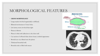
PATHOLOGY OF
BRONCHIECTASIS
By: Ms. Saili Gaude
Principal
Shivam College of Nursing
DEFINITION
◦ Abnormal and irreversible
dilatation of the bronchi and
bronchioles developing secondary
to inflammatory weakening of the
bronchial wall.
ETIOPATHOGENESIS
◦ 2 basic mechanisms
ENDOBRONCHIAL
OBSTRUCTION
INFECTION
1) ENDOBRONCHIAL OBSTRUCTION
◦ Obstruction of the bronchi by foreign body, tumors, enlarged ,lymph nodes
Resorption of air distal to obstruction
Atelectasis
Retention of secretions
2) INFECTION
Secondary to obstruction
Growth of micro organisms
Infection
ETIOLOGY
◦ Both mechanisms are widely seen in various clinical settings
1) HEREDITY
2) OBSTRUCTION
3) SECONDARY COMPLICATION
1) HEREDITY
◦ 1) Congenital bronchiectasis – caused by developmental defect of the bronchi
◦ 2) Cystic fibrosis – defect of the exocrine glands causing increased mucus secretion leading to
bronchiectasis
◦ 3) Hereditary Immune Deficiency diseases- high incidence of bronchiectasis
◦ 4) Immotile cilia syndrome- ultrastructural changes in the cilia leads to ineffective airway
clearance
◦ 5) Atopic bronchial asthma – leads to bronchiectasis in some cases
2) OBSTRUCTION
◦ 1) Foreign body obstruction
◦ 2) Endotracheal Tumors
◦ 3) Compression by enlarged lymphnodes
◦ 4) Post inflammatory scarring of bronchi
3) SECONDARY COMPLICATIONS
◦ Bronchiectasis may occur as a result of the complication of many respiratory disorders.
◦ Pneumonia
◦ Tuberculosis
◦ Bronchitis
PATHOGENESIS
Inflammation
Impaired
mucociliary
clearance
Bronchial
obstruction
Inflammation
and bronchial
damage
Bronchiectasis
MORPHOLOGICAL FEATURES
◦ GROSS MORPHOLOGY
◦ Lungs maybe involved segmentally or diffusely
◦ Bilateral involvement of lower lobes
◦ Left airway more involved than right
◦ Pleura is fibrotic
◦ Pleura is thick with adhesions to the chest wall
◦ Cut section of affected lobes shows honey combed appearance
◦ Bronchi are very dilated near the pleura
◦ Thickened bronchial wall.
◦ Bronchia tube is filled with mucus
◦ BRONCHOGRAPHIC APPEARANCE OF THE AIRWAYS
◦ 1) CYLINDRICAL – tube like bronchial dilation
◦ 2) FUSIFORM- spindle shaped bronchial dilation
◦ 3) SACCULAR- rounded sac like bronchial dilation
◦ 4) VARICOSE- irregular shaped bronchial dilation
◦ MICROSCOPIC FEATURES
◦ Bronchial epithelium – normal, ulcerated or metaplastic
◦ Bronchial wall- infiltrated with inflammatory cells
◦ Destruction of normal muscle and elastic tissues
◦ Replaced by fibrosis
◦ Lung parenchyma shows fibrosis
◦ Interstitial pneumonia
◦ Adherent pleura with fibrous tissue
CLINICAL FEATURES
◦ Persistent productive cough
◦ Thick tenacious sputum
◦ Clubbing of fingers
◦ Crackles
◦ Wheezing
◦ Airway obstruction
DIAGNOSTIC TESTS
◦ Clinical history
◦ Radiographic features
◦ X ray
◦ HRCT thorax
◦ Pulmonary function test- reveals obstructive pattern , reduced FEV1 and FVC
◦ Cough test – ability , strength and effectiveness of coughing checked
◦ Bronchial biopsy
◦ Sputum culture and sensitivity
◦ Alpha anti trypsin levels
TREATMENT
◦ Antimicrobial therapy
◦ Maintenance of hygiene
◦ Clearance of secretions by postural drainage
◦ Mucolytics
◦ Anti inflammatory therapy
◦ Bronchodilation – corticosteroids and bronchodilators
◦ Resection of affected lung

BRONCHIECTASIS PATHOLOGY FOR NURSES.pptx

  • 1.
    PATHOLOGY OF BRONCHIECTASIS By: Ms.Saili Gaude Principal Shivam College of Nursing
  • 2.
    DEFINITION ◦ Abnormal andirreversible dilatation of the bronchi and bronchioles developing secondary to inflammatory weakening of the bronchial wall.
  • 3.
    ETIOPATHOGENESIS ◦ 2 basicmechanisms ENDOBRONCHIAL OBSTRUCTION INFECTION
  • 4.
    1) ENDOBRONCHIAL OBSTRUCTION ◦Obstruction of the bronchi by foreign body, tumors, enlarged ,lymph nodes Resorption of air distal to obstruction Atelectasis Retention of secretions
  • 5.
    2) INFECTION Secondary toobstruction Growth of micro organisms Infection
  • 6.
    ETIOLOGY ◦ Both mechanismsare widely seen in various clinical settings 1) HEREDITY 2) OBSTRUCTION 3) SECONDARY COMPLICATION
  • 7.
    1) HEREDITY ◦ 1)Congenital bronchiectasis – caused by developmental defect of the bronchi ◦ 2) Cystic fibrosis – defect of the exocrine glands causing increased mucus secretion leading to bronchiectasis ◦ 3) Hereditary Immune Deficiency diseases- high incidence of bronchiectasis ◦ 4) Immotile cilia syndrome- ultrastructural changes in the cilia leads to ineffective airway clearance ◦ 5) Atopic bronchial asthma – leads to bronchiectasis in some cases
  • 8.
    2) OBSTRUCTION ◦ 1)Foreign body obstruction ◦ 2) Endotracheal Tumors ◦ 3) Compression by enlarged lymphnodes ◦ 4) Post inflammatory scarring of bronchi
  • 9.
    3) SECONDARY COMPLICATIONS ◦Bronchiectasis may occur as a result of the complication of many respiratory disorders. ◦ Pneumonia ◦ Tuberculosis ◦ Bronchitis
  • 10.
  • 11.
    MORPHOLOGICAL FEATURES ◦ GROSSMORPHOLOGY ◦ Lungs maybe involved segmentally or diffusely ◦ Bilateral involvement of lower lobes ◦ Left airway more involved than right ◦ Pleura is fibrotic ◦ Pleura is thick with adhesions to the chest wall ◦ Cut section of affected lobes shows honey combed appearance ◦ Bronchi are very dilated near the pleura ◦ Thickened bronchial wall. ◦ Bronchia tube is filled with mucus
  • 12.
    ◦ BRONCHOGRAPHIC APPEARANCEOF THE AIRWAYS ◦ 1) CYLINDRICAL – tube like bronchial dilation ◦ 2) FUSIFORM- spindle shaped bronchial dilation ◦ 3) SACCULAR- rounded sac like bronchial dilation ◦ 4) VARICOSE- irregular shaped bronchial dilation
  • 13.
    ◦ MICROSCOPIC FEATURES ◦Bronchial epithelium – normal, ulcerated or metaplastic ◦ Bronchial wall- infiltrated with inflammatory cells ◦ Destruction of normal muscle and elastic tissues ◦ Replaced by fibrosis ◦ Lung parenchyma shows fibrosis ◦ Interstitial pneumonia ◦ Adherent pleura with fibrous tissue
  • 15.
    CLINICAL FEATURES ◦ Persistentproductive cough ◦ Thick tenacious sputum ◦ Clubbing of fingers ◦ Crackles ◦ Wheezing ◦ Airway obstruction
  • 16.
    DIAGNOSTIC TESTS ◦ Clinicalhistory ◦ Radiographic features ◦ X ray ◦ HRCT thorax ◦ Pulmonary function test- reveals obstructive pattern , reduced FEV1 and FVC ◦ Cough test – ability , strength and effectiveness of coughing checked ◦ Bronchial biopsy ◦ Sputum culture and sensitivity ◦ Alpha anti trypsin levels
  • 17.
    TREATMENT ◦ Antimicrobial therapy ◦Maintenance of hygiene ◦ Clearance of secretions by postural drainage ◦ Mucolytics ◦ Anti inflammatory therapy ◦ Bronchodilation – corticosteroids and bronchodilators ◦ Resection of affected lung