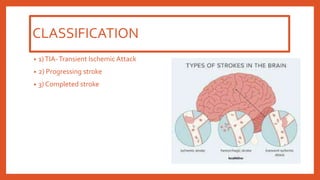
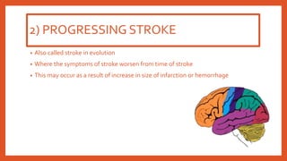
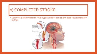
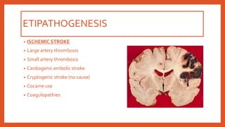
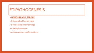
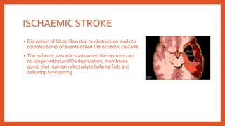
BY: MS. SAILI GAUDE
PRINCIPAL
SHIVAM COLLEGE OF NURSING, AMIRGADH
DEFINITIONS
• Episodes of focal brain dysfunction due to focal ischemia or infarction
• Common medical emergency
CLASSIFICATION
• 1)TIA-Transient Ischemic Attack
• 2) Progressing stroke
• 3) Completed stroke
1)TRANSIENT ISCHEMICATTACK
• Describes patients whose symptoms lasts for few seconds or minutes and resolves
within 24 hours.
• Symptoms are due to transient temporary impairment of blood flow to a specific
part of the brain
• It is an indication that a complete stroke may take place soon
2) PROGRESSING STROKE
• Also called stroke in evolution
• Where the symptoms of stroke worsen from time of stroke
• This may occur as a result of increase in size of infarction or hemorrhage
2) COMPLETED STROKE
• Describes stroke where the focal hypoxic defect persists but does not progress any
further
PATHOLOGIC CHANGES
• 1) Hypoxic encephalopathy : diffuse hypoxia is
present. Patient may recover without damage
• 2) Cerebral Infarction : Localised area of tissue
necrosis due to occlusion of blood supply.
• 3) Red infarct – multiple petechial hemorrhage
is seen
• 4) Pale infarct – associated with ischemic injury
ETIPATHOGENESIS
• ISCHEMIC STROKE
• Large artery thrombosis
• Small artery thrombosis
• Cardiogenic embolic stroke
• Cryptogenic stroke (no cause)
• Cocaine use
• Coagulopathies
ETIPATHOGENESIS
• HEMORRHAGIC STROKE
• Intracerebral hemorrhage
• Subarachnoid hemorrhage
• Cerebral aneurysm
• Arterio venous malformations
ISCHAEMIC STROKE
• Disruption of blood flow due to obstruction leads to
complex series of events called the ischemic cascade
• The ischemic cascade starts when the neurons can
no longer withstand O2 deprivation, membrane
pump that maintain electrolyte balance fails and
cells stop functioning
HEMMORHAGIC STROKE
• Pathologic changes depends on the type of cerebro vascular disease.
• Here normal brain metabolism is disrupted by the brain getting exposed to
blood due to hemorrhage
• It can occur due to :
• 1. Increased ICP
• 2. Secondary ischemia of the brain due to reduced perfusion pressure
• 3.Vasospasm of the blood vessels
GROSS MORPHOLOGY
• Single or multiple small cavity like infarction are seen
• Lake like spaces 1.5 cm wide present in the white mater and grey mater
• Can be clinically silent or cause severe neurologic deficit
MICROSCOPIC
• Cavities with loss of tissues are scattered with fat laden macrophages surrounded
by gliosis
• Hemorrhage infarct have extravasation and resorption of blood
• Increased intracerebral hematomas

STROKE AND ITS TYPES - PATHOLOGY LECTURE

  • 1.
    BY: MS. SAILIGAUDE PRINCIPAL SHIVAM COLLEGE OF NURSING, AMIRGADH
  • 2.
    DEFINITIONS • Episodes offocal brain dysfunction due to focal ischemia or infarction • Common medical emergency
  • 3.
    CLASSIFICATION • 1)TIA-Transient IschemicAttack • 2) Progressing stroke • 3) Completed stroke
  • 4.
    1)TRANSIENT ISCHEMICATTACK • Describespatients whose symptoms lasts for few seconds or minutes and resolves within 24 hours. • Symptoms are due to transient temporary impairment of blood flow to a specific part of the brain • It is an indication that a complete stroke may take place soon
  • 5.
    2) PROGRESSING STROKE •Also called stroke in evolution • Where the symptoms of stroke worsen from time of stroke • This may occur as a result of increase in size of infarction or hemorrhage
  • 6.
    2) COMPLETED STROKE •Describes stroke where the focal hypoxic defect persists but does not progress any further
  • 7.
    PATHOLOGIC CHANGES • 1)Hypoxic encephalopathy : diffuse hypoxia is present. Patient may recover without damage • 2) Cerebral Infarction : Localised area of tissue necrosis due to occlusion of blood supply. • 3) Red infarct – multiple petechial hemorrhage is seen • 4) Pale infarct – associated with ischemic injury
  • 8.
    ETIPATHOGENESIS • ISCHEMIC STROKE •Large artery thrombosis • Small artery thrombosis • Cardiogenic embolic stroke • Cryptogenic stroke (no cause) • Cocaine use • Coagulopathies
  • 9.
    ETIPATHOGENESIS • HEMORRHAGIC STROKE •Intracerebral hemorrhage • Subarachnoid hemorrhage • Cerebral aneurysm • Arterio venous malformations
  • 10.
    ISCHAEMIC STROKE • Disruptionof blood flow due to obstruction leads to complex series of events called the ischemic cascade • The ischemic cascade starts when the neurons can no longer withstand O2 deprivation, membrane pump that maintain electrolyte balance fails and cells stop functioning
  • 11.
    HEMMORHAGIC STROKE • Pathologicchanges depends on the type of cerebro vascular disease. • Here normal brain metabolism is disrupted by the brain getting exposed to blood due to hemorrhage • It can occur due to : • 1. Increased ICP • 2. Secondary ischemia of the brain due to reduced perfusion pressure • 3.Vasospasm of the blood vessels
  • 13.
    GROSS MORPHOLOGY • Singleor multiple small cavity like infarction are seen • Lake like spaces 1.5 cm wide present in the white mater and grey mater • Can be clinically silent or cause severe neurologic deficit
  • 14.
    MICROSCOPIC • Cavities withloss of tissues are scattered with fat laden macrophages surrounded by gliosis • Hemorrhage infarct have extravasation and resorption of blood • Increased intracerebral hematomas