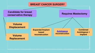
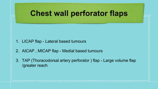
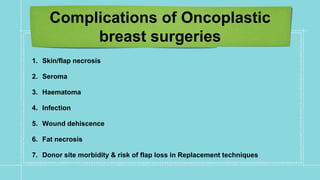
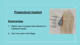
BREAST RECONSTRUCTION
SURGERIES
- Dr.J SAHRUDAI ,
- 2nd Yr Post graduate
Dept of Gen surgery ,
Kurnool Medical college , Kurnool
Why..??
Preservation of body image
Increase in survival rates
Breast cancer in young women
Awareness and demand for risk reducing mastectomies
BREAST CANCER SURGERY
Candidate for breast
conservative therapy
Requires Mastectomy
Volume
Displacement
Volume
Replacement
Expander/implant
based
reconstruction
Autologous
reconstruction
Combined
Autologous +
implant
Oncoplastic breast surgery
Indications of OPBS
1. Loss of >20% of breast volume
2. Poor breast:tumour ratio
3. Central/upper inner quadrant tumours
4. Prior breast surgery/scar/deformities
5. Large pendulous breast keen on reduction
Contraindications for OPBS
1. Large tumours - Insufficient residual breast tissue
2. Multicentric disease
3. Prior irradiation
4. Prior augmentation mammoplasty
PRIOR TO OPBS
1. Tumour location
2. Volume of tissue to be excised
3. Breast size & glandular density
4. Contralateral symmetrization by reduction
5. Risk factors - smoking, diabetes , obesity
Levels of oncoplasty
Level -I
<20% of Breast volume is
excised
No skin resection
Level -II
20-50% of breast volume is excised
Extensive Skin excision
Mammoplasty techniques for re
shaping
Oncoplastic techniques
Volume displacement
“ADJACENT TISSUE
REARRANGEMENT ”
by moving local glandular or
dermoglandular tissue into the
defect
Volume Replacement
Autologous tissue from extra
mammary site is brought in to replace
the lost volume
Volume Displacement Technique
Procedure steps
1. Skin Incision
2. Skin undermining
3. NAC undermining
4. Full thickness excision
5. Glandular reapproximation
6. Deepithelialization & NAC repositioning
Based on location of
tumour
Upper pole tumours
1. Inferior pedicel inverted T-mammoplasty
2. Benelli round block
3. Batwing mastopexy
Upper outer quadrant tumours
1. Lateral (Racquet) mammoplasty
2. Benelli round block
Lower outer quadrant tumours
1. Superior pedicle inverted-T
mammoplasty
2. J-mammoplasty
Lower pole tumours
1. Superior pedicle inverted-T
mammoplasty
2. Vertical scar mammoplasty
3. Inframammary fold pasty
Lower inner quadrant tumours
1. Superior pedicle inverted -T mammoplasty
2. V- mammoplasty
Upper inner quadrant tumours
1. Benelli round block
2. Batwing mastopexy
Central quadrant tumours
1. Inverted-T/Vertical scar mammoplasty with/without NAC
resection
2. Grisotti technique
Volume Replacement
techniques
1. >50% loss of volume or in patients with small/medium sized
breasts
2. Replaced with locally available tissue around mammary area
3. Based on supply by perforating vessels
Chest wall perforator flaps
1. LICAP flap - Lateral based tumours
2. AICAP , MICAP flap - Medial based tumours
3. TAP (Thoracodorsal artery perforator ) flap - Large volume flap
/greater reach
1. Larger Defects ;
2. Latissimus dorsi miniflap - M/C used
3. Omental flap , Upper abdominal advancement flaps , immediate
fat grafting
4. Free flap techniques - Transverse upper gracilis flap (TUG)
Complications of Oncoplastic
breast surgeries
1. Skin/flap necrosis
2. Seroma
3. Haematoma
4. Infection
5. Wound dehiscence
6. Fat necrosis
7. Donor site morbidity & risk of flap loss in Replacement techniques
Breast reconstruction options
AUTOGENOUS ALLOPLASTIC
Abdomen based flaps
TRAM flap-single,double
pedicle,freeflap
DIEP flap
LD Myocutaneous flap
Gluteal flap-Sup,Inf
Lateral thigh flap
1. Silicone gel implant
2. Silicone implant
with saline fill
3. Smooth wall
4. Textured wall
5. Round
6. Anatomic mound
COMBINATION PROCEDURE
1. LD flap with
implant
2. TRAM flap
with implant
Timing of reconstruction
Immediate Reconstruction
Advantage
1. Psychological benefit
2. Better aesthetics d/t
preservation of inframammary
fold
3. Skin flaps are more pliable
Disadvantage
1. Chemo & Radio interfere
with post op healing
2. Delay in CT/RT …. Adverse
Onco logical outcomes
Delayed Reconstruction
Advantage
1. Better oncological
outcomes
Disadvantage
1. Psychology issues
2. Loss of native skin envelope
3. New Inframammary fold needed
4. Exploration/Dissection of irradiated tissue
Delayed-Immediate
1. Immediate placement of tissue expanders
2. Delayed reconstruction in case of radiation / immediate
reconstruction (implants or autologous) in case of no radiation.
ALLOPLASTIC / IMPLANT
RECONSTRUCTION
INDICATIONS
1. Bilateral reconstruction
2. Augmentation in addition to reconstruction
3. Pt not suited for long surgery
4. Lack of adequate abdominal tissue
5. Avoiding additional scars on back/abdomen
6. small breast mound with minimal ptosis
Relative Contraindications
Young age
Very large & ptotic breast
Need for adjuvant radiation theraphy
Silicon allergy & implant fear
Previous failed implants
Types of Implants
Textured vs Smooth
Reduces implant movement &
maintains orientation
Reduces Rate & degree of
capsular contracture
Anatomical vs Round
Decrease upper pole fullness
(Normal breasts have
supraareolar flattening)
Increase projection than rounds
implants of same volume
Location of placement
Subpectoral implant
Advantage
1. Less risk of infection
2. Used in poorly perfumed mastectomy skin flaps
Disadvantage
1. Breast pain
2. Muscle spasm
Prepectoral implant
Disadvantage
1. Higher risk of surgical site infections in
irradiated breast.
2. Can’t be used in thin flaps.
Complications of implant based
flaps
1. Haematoma , seroma
2. Wound dehiscence
3. Flap necrosis
4. Implant rupture - Linguini sign on MRI
5. Capsular contracture
High risk of
complications in
1. Preop radiation
2. Post op radiation
3. Obesity
4. Large breast size
5. Smoking
Abdomen based flaps
ADVANTAGES
1. Similar consistency
2. Suitable for breast of all sizes
3. Improvement of abdominal
contour in some patients
4. Suitable even for ptotic
breasts
5. Acceptable donor site scar
DISADVANTAGES
1. High metabolic rate
2. Time consuming procedure
3. Long recovery period
4. Abdominal weakness
5. Not suitable for - smokers ,
previous abdominal surgeries,
liposuction.
Abdomen based flaps
1. Myocutaneous flaps - Pedicle , Free
2. Muscle sparing flaps
3. Perforator based flaps
Pedicled TRAM flap
1. Based on superior
epigastric vessels
2. Tunneled into
ipsilateral / contra
lateral mastectomy
defect
3. Disadvantage-
Epigastric muscle
bulge raised by the
pedicle
Free TRAM flap
1. Based on inferior epigastric
vessels
2. Microvascular anastomosis
to Internal mammary
vessels / thoracodorsal
vessels
Ms-TRAM flap
1. Only muscle fibers around the
pedicle are included in the flap
2. Based on inferior epigastric
vessels
Perforator based flaps
Superficial inferior
epigastric
Artery ( SIEA ) flap
Deep inferior epigastric
Artery (DIEP) flap
Gluteal based flaps
1. Less adipose tissue in
abdomen
2. Previous abdominal surgery
3. Disadvantage - contour
deformity , muscle weakness
, sciatica
Disadvantages
1. Difficulty in flap harvest
2. Intraop positing changes
3. Short pedicle length
4. Size discrepancy btw gluteal vein
& Internal mammary vein
Thigh based flaps
1. Myocutaneous or True perforator based flaps
2. Profunda femoris artery perforator based flaps
3. Transverse upper gracilis TUG
4. Vertical upper gracilis VUG
5. Diagonal upper gracilis DUG
Latissimus Dorsi flap Reconstruction
Indications
1. Small breast
2. Minor breast ptosis
3. Unavailable abdominal donor
site
4. Salvage of previous breast
reconstruction
Contraindications
1. Previous lateral thoracotomy
2. Bilateral reconstruction
3. Significant breast ptosis
4. Planned post op radiation
theraphy
5. Very large breast who don’t
desire reduction
1. Based on Thoracodorsal
vessels
2. Can be Pedicled or Free flap
(if accidentally damaged
during axillary dissection)
3. Extended Latissimus flap -
Harvesting of supramuscular
and subscapular fat
4. To increase volume of LD
flap.
Preop imaging
CT angiogram
MR angiogram
Provide accurate location and intramuscular course of
perforators.
1mm slices instead of standard 3mm slices
Monitoring
1. Assess - Colour , Turgor , temperature , capillary refill
2. Cool,pale flap with minimal or No bleeding from wound edges &
capillary refill <3secs - Arterial insufficiency
3. Hyperaemic /purple , edematous,warm flap ,Rapid dark oozing
from edges with <2secs capillary refill - Venous congestion
Fluorescent angiography- Intraop to assess vascular supply ,
ICG injected & probe used to assess perfusion of cutaneous
territory of flap
Near infrared spectroscopy- Probe applied to skin flap ,
measures oxgenation of superficial tissue (2cm depth)
Implantable Doppler devices - Probe being attached to at a
level or just distal to anastomosis & is connected to an external
device
Nipple areolar reconstruction
Commonly performed 2-3 months after final breast mound
reconstruction is completed
1. Nipple sharing - Graft from opposite nipple
2. Composite graft - Ear & hallux
3. Prosthetic material - Alloderm & polyurethane
4. Local flap - most popular - Use of local breast
skin/fat to build a papule
Nipple
reconstruct
ion
Areolar Reconstruction
1. Tattooing by using pigments to simulate areola
2. Skin grafting by doughnout shaped full thickeness skin graft applied
around nipple to gives colour & texture contrast
3. Insitu skin grafting by lifting the skin surrounding the nipple & then
sewing back down
Radiological surveillance
1. Bilateral Annual mammography is recommended after OPBS with
delay of 12 months.
2. No Indication for ipsilateral imaging following mastectomy either
implant based or autologous
3. Because recurrences can occur in mastectomy flaps.
“Thank you….”

Breast reconstruction surgeries . PPT

  • 1.
    BREAST RECONSTRUCTION SURGERIES - Dr.JSAHRUDAI , - 2nd Yr Post graduate Dept of Gen surgery , Kurnool Medical college , Kurnool
  • 2.
    Why..?? Preservation of bodyimage Increase in survival rates Breast cancer in young women Awareness and demand for risk reducing mastectomies
  • 3.
    BREAST CANCER SURGERY Candidatefor breast conservative therapy Requires Mastectomy Volume Displacement Volume Replacement Expander/implant based reconstruction Autologous reconstruction Combined Autologous + implant
  • 4.
    Oncoplastic breast surgery Indicationsof OPBS 1. Loss of >20% of breast volume 2. Poor breast:tumour ratio 3. Central/upper inner quadrant tumours 4. Prior breast surgery/scar/deformities 5. Large pendulous breast keen on reduction
  • 5.
    Contraindications for OPBS 1.Large tumours - Insufficient residual breast tissue 2. Multicentric disease 3. Prior irradiation 4. Prior augmentation mammoplasty
  • 6.
    PRIOR TO OPBS 1.Tumour location 2. Volume of tissue to be excised 3. Breast size & glandular density 4. Contralateral symmetrization by reduction 5. Risk factors - smoking, diabetes , obesity
  • 7.
    Levels of oncoplasty Level-I <20% of Breast volume is excised No skin resection Level -II 20-50% of breast volume is excised Extensive Skin excision Mammoplasty techniques for re shaping
  • 8.
    Oncoplastic techniques Volume displacement “ADJACENTTISSUE REARRANGEMENT ” by moving local glandular or dermoglandular tissue into the defect Volume Replacement Autologous tissue from extra mammary site is brought in to replace the lost volume
  • 9.
    Volume Displacement Technique Proceduresteps 1. Skin Incision 2. Skin undermining 3. NAC undermining 4. Full thickness excision 5. Glandular reapproximation 6. Deepithelialization & NAC repositioning Based on location of tumour
  • 10.
    Upper pole tumours 1.Inferior pedicel inverted T-mammoplasty 2. Benelli round block 3. Batwing mastopexy
  • 11.
    Upper outer quadranttumours 1. Lateral (Racquet) mammoplasty 2. Benelli round block
  • 12.
    Lower outer quadranttumours 1. Superior pedicle inverted-T mammoplasty 2. J-mammoplasty
  • 13.
    Lower pole tumours 1.Superior pedicle inverted-T mammoplasty 2. Vertical scar mammoplasty 3. Inframammary fold pasty
  • 14.
    Lower inner quadranttumours 1. Superior pedicle inverted -T mammoplasty 2. V- mammoplasty
  • 15.
    Upper inner quadranttumours 1. Benelli round block 2. Batwing mastopexy
  • 16.
    Central quadrant tumours 1.Inverted-T/Vertical scar mammoplasty with/without NAC resection 2. Grisotti technique
  • 17.
    Volume Replacement techniques 1. >50%loss of volume or in patients with small/medium sized breasts 2. Replaced with locally available tissue around mammary area 3. Based on supply by perforating vessels
  • 18.
    Chest wall perforatorflaps 1. LICAP flap - Lateral based tumours 2. AICAP , MICAP flap - Medial based tumours 3. TAP (Thoracodorsal artery perforator ) flap - Large volume flap /greater reach
  • 19.
    1. Larger Defects; 2. Latissimus dorsi miniflap - M/C used 3. Omental flap , Upper abdominal advancement flaps , immediate fat grafting 4. Free flap techniques - Transverse upper gracilis flap (TUG)
  • 20.
    Complications of Oncoplastic breastsurgeries 1. Skin/flap necrosis 2. Seroma 3. Haematoma 4. Infection 5. Wound dehiscence 6. Fat necrosis 7. Donor site morbidity & risk of flap loss in Replacement techniques
  • 21.
    Breast reconstruction options AUTOGENOUSALLOPLASTIC Abdomen based flaps TRAM flap-single,double pedicle,freeflap DIEP flap LD Myocutaneous flap Gluteal flap-Sup,Inf Lateral thigh flap 1. Silicone gel implant 2. Silicone implant with saline fill 3. Smooth wall 4. Textured wall 5. Round 6. Anatomic mound COMBINATION PROCEDURE 1. LD flap with implant 2. TRAM flap with implant
  • 22.
    Timing of reconstruction ImmediateReconstruction Advantage 1. Psychological benefit 2. Better aesthetics d/t preservation of inframammary fold 3. Skin flaps are more pliable Disadvantage 1. Chemo & Radio interfere with post op healing 2. Delay in CT/RT …. Adverse Onco logical outcomes
  • 23.
    Delayed Reconstruction Advantage 1. Betteroncological outcomes Disadvantage 1. Psychology issues 2. Loss of native skin envelope 3. New Inframammary fold needed 4. Exploration/Dissection of irradiated tissue
  • 24.
    Delayed-Immediate 1. Immediate placementof tissue expanders 2. Delayed reconstruction in case of radiation / immediate reconstruction (implants or autologous) in case of no radiation.
  • 25.
    ALLOPLASTIC / IMPLANT RECONSTRUCTION INDICATIONS 1.Bilateral reconstruction 2. Augmentation in addition to reconstruction 3. Pt not suited for long surgery 4. Lack of adequate abdominal tissue 5. Avoiding additional scars on back/abdomen 6. small breast mound with minimal ptosis
  • 26.
    Relative Contraindications Young age Verylarge & ptotic breast Need for adjuvant radiation theraphy Silicon allergy & implant fear Previous failed implants
  • 27.
    Types of Implants Texturedvs Smooth Reduces implant movement & maintains orientation Reduces Rate & degree of capsular contracture Anatomical vs Round Decrease upper pole fullness (Normal breasts have supraareolar flattening) Increase projection than rounds implants of same volume
  • 28.
    Location of placement Subpectoralimplant Advantage 1. Less risk of infection 2. Used in poorly perfumed mastectomy skin flaps Disadvantage 1. Breast pain 2. Muscle spasm
  • 29.
    Prepectoral implant Disadvantage 1. Higherrisk of surgical site infections in irradiated breast. 2. Can’t be used in thin flaps.
  • 30.
    Complications of implantbased flaps 1. Haematoma , seroma 2. Wound dehiscence 3. Flap necrosis 4. Implant rupture - Linguini sign on MRI 5. Capsular contracture High risk of complications in 1. Preop radiation 2. Post op radiation 3. Obesity 4. Large breast size 5. Smoking
  • 31.
    Abdomen based flaps ADVANTAGES 1.Similar consistency 2. Suitable for breast of all sizes 3. Improvement of abdominal contour in some patients 4. Suitable even for ptotic breasts 5. Acceptable donor site scar DISADVANTAGES 1. High metabolic rate 2. Time consuming procedure 3. Long recovery period 4. Abdominal weakness 5. Not suitable for - smokers , previous abdominal surgeries, liposuction.
  • 32.
    Abdomen based flaps 1.Myocutaneous flaps - Pedicle , Free 2. Muscle sparing flaps 3. Perforator based flaps
  • 33.
    Pedicled TRAM flap 1.Based on superior epigastric vessels 2. Tunneled into ipsilateral / contra lateral mastectomy defect 3. Disadvantage- Epigastric muscle bulge raised by the pedicle
  • 34.
    Free TRAM flap 1.Based on inferior epigastric vessels 2. Microvascular anastomosis to Internal mammary vessels / thoracodorsal vessels
  • 35.
    Ms-TRAM flap 1. Onlymuscle fibers around the pedicle are included in the flap 2. Based on inferior epigastric vessels
  • 36.
    Perforator based flaps Superficialinferior epigastric Artery ( SIEA ) flap Deep inferior epigastric Artery (DIEP) flap
  • 37.
    Gluteal based flaps 1.Less adipose tissue in abdomen 2. Previous abdominal surgery 3. Disadvantage - contour deformity , muscle weakness , sciatica
  • 38.
    Disadvantages 1. Difficulty inflap harvest 2. Intraop positing changes 3. Short pedicle length 4. Size discrepancy btw gluteal vein & Internal mammary vein
  • 39.
    Thigh based flaps 1.Myocutaneous or True perforator based flaps 2. Profunda femoris artery perforator based flaps 3. Transverse upper gracilis TUG 4. Vertical upper gracilis VUG 5. Diagonal upper gracilis DUG
  • 40.
    Latissimus Dorsi flapReconstruction Indications 1. Small breast 2. Minor breast ptosis 3. Unavailable abdominal donor site 4. Salvage of previous breast reconstruction Contraindications 1. Previous lateral thoracotomy 2. Bilateral reconstruction 3. Significant breast ptosis 4. Planned post op radiation theraphy 5. Very large breast who don’t desire reduction
  • 41.
    1. Based onThoracodorsal vessels 2. Can be Pedicled or Free flap (if accidentally damaged during axillary dissection) 3. Extended Latissimus flap - Harvesting of supramuscular and subscapular fat 4. To increase volume of LD flap.
  • 42.
    Preop imaging CT angiogram MRangiogram Provide accurate location and intramuscular course of perforators. 1mm slices instead of standard 3mm slices
  • 43.
    Monitoring 1. Assess -Colour , Turgor , temperature , capillary refill 2. Cool,pale flap with minimal or No bleeding from wound edges & capillary refill <3secs - Arterial insufficiency 3. Hyperaemic /purple , edematous,warm flap ,Rapid dark oozing from edges with <2secs capillary refill - Venous congestion
  • 44.
    Fluorescent angiography- Intraopto assess vascular supply , ICG injected & probe used to assess perfusion of cutaneous territory of flap Near infrared spectroscopy- Probe applied to skin flap , measures oxgenation of superficial tissue (2cm depth) Implantable Doppler devices - Probe being attached to at a level or just distal to anastomosis & is connected to an external device
  • 45.
    Nipple areolar reconstruction Commonlyperformed 2-3 months after final breast mound reconstruction is completed 1. Nipple sharing - Graft from opposite nipple 2. Composite graft - Ear & hallux 3. Prosthetic material - Alloderm & polyurethane 4. Local flap - most popular - Use of local breast skin/fat to build a papule Nipple reconstruct ion
  • 46.
    Areolar Reconstruction 1. Tattooingby using pigments to simulate areola 2. Skin grafting by doughnout shaped full thickeness skin graft applied around nipple to gives colour & texture contrast 3. Insitu skin grafting by lifting the skin surrounding the nipple & then sewing back down
  • 47.
    Radiological surveillance 1. BilateralAnnual mammography is recommended after OPBS with delay of 12 months. 2. No Indication for ipsilateral imaging following mastectomy either implant based or autologous 3. Because recurrences can occur in mastectomy flaps.
  • 48.