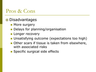
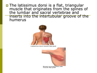
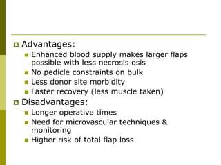
Breast Reconstruction
Introduction
 Objective is to restore symmetry by
recreating:
 Volume
 Shape
 Position
 Must address:
 Volume deficit
 Skin deficiency
History
 The first attempt at true breast
reconstruction occurred in 1895, when
Vincent Czerny transplanted a large
lipoma from his patient's flank to the
mastectomy site
 Latissimus dorsi myocutaneous flap ------
Tansini in 1906
History
 In 1963, the silicone breast implant was
introduced for breast augmentation and
was quickly adopted for breast
reconstruction --------------Cronin and
Gerow
History
 1970’s -------- Rediscovery of the lat dorsi flap
Discovery of tissue expanders
 1980’s -------Vertical rectus abdominis flap
Pedicled TRAM flap
Gluteal
 1990’s DIEP –-------Deep inferior epigastric
perforator free flap
Pros & Cons
 Advantages
 Don’t need to wear an external prosthesis
 Better self esteem
 ‘Feel whole again’
 Less grief
 Fewer sexual problems
Pros & Cons
 Disadvantages
 More surgery
 Delays for planning/organisation
 Longer recovery
 Unsatisfying outcome (expectations too high)
 Other scars if tissue is taken from elsewhere,
with associated risks
 Specific surgical side effects
Patient Selection
 Young healthy patients with early-stage disease -
------ Best candidates for reconstruction
 Older patients with advanced disease -----------
Poorer candidates
“All women at least need to be
presented with the options
before being excluded”
Obesity
Moderate: <25% above ideal body
weight
1
Severe: >25% over ideal body
weight
5
Small Vessel Disease
Light to moderate smoking (1+
pack/day for 2-10 yr)
1
Chronic heavy smoking (10-20 pack-
years)
2
Chronic heavy smoking (20-30 pack-
years)
5
Autoimmune disease (e.g.,
scleroderma, Raynaud's)
8
Diabetes mellitus: non–insulin
dependent
5
Diabetes mellitus: insulin dependent 10
Risk factor severity score ---- Devised by CARL HARTRAMPF
Psychosocial Problems
Unstable emotional state (life crisis) 2
Personality disorder 3
Substance abuse 5
Abdominal Scars
If “planned out” of flap design 0.5
Disruption of vascular perforators:
transection of superior epigastric
vessels (e.g., chevron incision,
abdominoplasty)
10
Patient's Attitude
Patient unwilling or unable to invest
the time required for healing or
objects to an abdominal scar
10
Surgeon's Inexperience
<10 TRAM flaps 1
Major System Disease Process
Chronic lung disease 10
Severe cardiovascular disease 10
Risk factor severity score ---- Devised by CARL HARTRAMPF
 Each risk is given a numerical weight
 The total is added to derive a numerical score
 Poor Candidate ----- Any patient with a score
greater than 5 or with three or more risk factors
 Marginal Candidates ------ With two risk factors
 Of the risk factors listed, advanced age, obesity,
smoking, concomitant disease, and the patient's
psychological/emotional state are the most
important to consider
Timing (Delayed Vs Immediate)
IMMEDIATE MASTECTOMY REQUIRED
CONSIDER PROGNOSTIC FACTORS
FOR LOCAL RECURRENCE
Adverse Factors
•Large primary small breast
•Involvement of pectoralis
•Large area peau d orange
•High grade tumor
•Radiotherapy planned post
operatively
•Known axillary involvement
Favourable factors
•Small primary larger breast
•Low grade tumor
•Clinically clear axilla
•No radiotherapy likely
Immediate reconstruction
Delayed reconstruction
Factors Affecting Choice of
Reconstruction Procedures
Patient Factors
 Age
 Medical conditions
 Previous abdominal or
thoracic surgery
 Coronary artery disease
 Chronic obstructive
pulmonary disease
 Medications
 Chronic corticosteroid use
 Obesity
 Body morphology
 Occupation
 Social activities
 Financial resources
 Support systems
 Expectations/desires
 Disease Factors
 Stage of disease
 Type of tumor
 Need for adjuvant therapy
 Miscellaneous Factors
 Experience of the surgeon
 Availability of equipment
(e.g., microscope)
 Religious beliefs regarding
blood transfusion
 Blood banking facilities
Surgical Planning
 Best cosmetic results are achieved with immediate
autologous reconstruction
 Requires preoperative planning
 Skin sparing mastectomy offers the best outcomes
 No increased risk of recurrence
BUT
 May be compromised by unexpected pathological findings:
 Positive margins
 Nodal involvement
 Need for re-excision excision
 Need for post chemo/radiotherapy
 NOTE: Autologous reconstructions can usually withstand
postoperative radiation but significant atrophy occurs in up
to 1/3. Implants on the other hand can harden and/or
extrude (60% long term failure with XRT at any time)
Surgical Planning
 Best candidates for immediate reconstruction are
those with:
 T2 or less (<5cm)
 N1 or less (mobile ipsilateral axillary nodes) (stage II)
 Given the potential for trouble, many favour a
delayed approach:
 Skin sparing mastectomy
 Saline expander implants
 Complete all treatment
 Allows the patient more time to take it all in
Surgical Options
 Autogenous
 Abdominal-based flaps
 TRAM
 Single pedicle
 Double pedicle
 Free flap
 Deep inferior epigastric
perforator flap
 Upper abdominal
horizontal flap
 Vertical abdominal flap
 Latissimus dorsi
musculocutaneous flap
 Gluteal flap
 Superiorly based
 Inferiorly based
 Rubens flap
 Alloplastic
 Silicone gel implant
 Silicone implant with
saline fill
 Smooth wall
 Textured wall
 Round shaped
 Anatomic shaped
 Silicone injection
 Combination
procedures
 Latissimus dorsi flap
with implant
 TRAM flap with implant
Surgical Options
 Autogenous tissue will usually provide
better symmetry than an implant
 Only 35% of TRAM flap reconstructions
required a symmetry procedure versus
55% of implant reconstructions in one
study -----
Giacalone PL, Bricout N, Dantas MJ, et
al 2002
Surgical Options
 The choice of procedure for a given
patient is affected by her:
 Age
 Health
 Contralateral breast size and shape
 Personal preference
 Expertise of the reconstructive surgeon
Surgical Options
 The reconstructive plan must
accommodate
 The size and shape of the opposite breast
 The position on the chest wall
 The location of the inframammary crease
 The height, size, and color of the nipple-
areolar complex
 The amount of breast ptosis
Breast Reconstruction With Implants
 Initially popularized for breast
augmentation --------- use of the silicone
implant was extended to breast
reconstruction after mastectomy
procedures
 In the original description, the implant
was inserted into the subcutaneous breast
wound
 The use of saline-filled implants precludes
subcutaneous placement because these
implants visibly ripple the skin
 Placement of the implant in a submuscular
plane beneath the pectoralis major,
superior portion of the rectus
abdominis, and serratus anterior
muscles provides better protection
against implant extrusion, as well as
decreased risk for capsular contracture
and implant displacement
 Avoid sub–rectus abdominis placement because
the tight fascial amalgamation can flatten the
implant inferiorly and displace it supariorly
 Some surgeons place expenders or implants in
the subpectoral plane, without elevation of the
serratus muscle, by adding a sheet of acellular
dermis that is sutured in place between the
inframammary fold and the edge of the pectoralis
major
 In approximating the pectoralis major to the
serratus anterior muscles, preservation of the
clavipectoral fascia, when possible, facilitates
prevention of the suture pulling through muscle
and ripping muscle fibers
Tissue expanders
 In women with a large
contralateral breast --
------------Permanent
or temporary tissue
expanders are used
 These expanders are
silicone envelopes
with an integrated or
remote port for
episodic injection of
saline in the
outpatient setting
Tissue expanders
 Most surgeons overinflate past the desired size
 Overinflation affords a larger skin envelope that
gives some ptosis in the end result
 At a secondary procedure, the expander is
exchanged for a permanent implant
 Waiting at least 6 weeks from the last expansion
to implant exchange is believed to limit the rapid
shrinkage of expanded skin
Permanent Becker Silicone/Saline
Implant Expander With A Remote Port
 An alternative technique involves the use
of a permanent Becker silicone/saline
implant expander with a remote port
 This implant expander is filled to the
desired breast size over the course of
several weeks postoperatively
 Later, the port can be taken out as an
outpatient procedure
Breast Reconstruction With Implants
 Indications
 Bilateral reconstruction
 Patient requesting augmentation in addition to
reconstruction
 Patient not suited for long surgery
 Lack of adequate abdominal tissue
 Patient unwilling to have additional scars on
either her back or abdomen
 Small breast mound with minimal ptosis
 Patient preference
Breast Reconstruction With Implants
 Relative Contraindications
 Young age (may need an implant replaced
multiple times)
 Patient unwilling to adhere to follow-up
 Very large breast
 Very ptotic breast
Breast Reconstruction With Implants
 Contraindications:
 Silicon allergy
 Implant fear
 Previous failed implants
 Need for adjuvant radiation therapy
 Suture line tension
 Thin flaps of marginal viability
Breast Reconstruction With Implants
 Advantages:
 No donor site morbidity
 Decreased operating time
 Decreased recovery time
 No additional scar
 Disadvantages:
 Implant rupture/leak/extrusion
 Infection
 Flap necrosis
 Capsular contracture
 Long reconstruction period (expanders)
 Reoperations for implant-related problems related
problems
Breast Reconstruction With Implants
 Complications:
 Exposure
 Extrusion
 Infection
 Asymmetry
 Capsular contracture
 Malposition of the implant
 Rupture
 Pain
Latissimus Dorsi Reconstruction
 First described by Professor Iginio Tansini
for chest wall coverage in 1906, the
latissimus dorsi myocutaneous flap was
not commonly used until the 1970s
 The latissimus dorsi is a flat, triangular
muscle that originates from the spines of
the lumbar and sacral vertebrae and
inserts into the intertubular groove of the
humerus
 Its blood supply comes
from the thoracodorsal
artery and from multiple
segmental perforators off
the lumbar intercostal
arteries
 These arteries provide
musculocutaneous
perforating vessels that
penetrate the
subcutaneous tissue to
supply a territory of skin
directly overlying the
muscle
Latissimus Dorsi Reconstruction
 Indications
 Small breast
 Minor breast ptosis
 Abdominal donor site unavailable (e.g., scars,
lack of tissue)
 Salvage of previous breast reconstruction
Latissimus Dorsi Reconstruction
 Relative Contraindications
 Planned postoperative radiation therapy
 Bilateral reconstruction
 Significant breast ptosis
 Contraindications
 Previous lateral thoracotomy
 Very large breast in a patient who does not
desire reduction
Latissimus Dorsi Reconstruction
 Advantages
 Reliable
 Faster recovery cf TRAM
 Less morbidity cf TRAM
 Disadvantages
 New scar
 May still need implant
Transverse Rectus Abdominis
Myocutaneous Flap
 TRAM flap is the most commonly
performed autogenous reconstructive
procedure
 First suggested in 1979 by Robbins, the
TRAM flap was popularized in 1982 by
Hartrampf and coworkers
Transverse Rectus Abdominis
Myocutaneous Flap
 Skin paddle design is placed over the lower part of the
abdomen to take advantage of:
 Large amount of adipose tissue available
 Favorable scar location
 Long pedicle for ease in transposition
 The flap is divided
into four regions
on the basis of
entrance of the
perforating vessels
 The most reliable
zones are either
directly over the
muscle (zone I) or
directly adjacent to
this zone (zones II,
III)
Transverse Rectus Abdominis
Myocutaneous Flap
Transverse Rectus Abdominis
Myocutaneous Flap
 Zone IV is the contralateral
tissue farthest away from
the musculocutaneous
perforators and, in most
cases, must be discarded,
especially in obese patients
and smokers
 Perfusion studies have
further delineated that
zone III, which has an
axial blood supply, has
more reliable perfusion
than zone II, which is
supplied across the midline
and therefore random
Transverse Rectus Abdominis
Myocutaneous Flap
 Indications:
 Need for large volume of tissue
 Previous injury to superior epigastric artery
 Contraindications:
 Division of deep inferior epigastric artery from previous
surgery (eg hernia or paramedian incisions) paramedian
incisions)
 Similar as for pedicled, however free preferred if at
higher risk of flap failure eg smoking or obese. Perfusion
from inferior epigastric is better.
 Advantages:
 Enhanced blood supply makes larger flaps
possible with less necrosis osis
 No pedicle constraints on bulk
 Less donor site morbidity
 Faster recovery (less muscle taken)
 Disadvantages:
 Longer operative times
 Need for microvascular techniques &
monitoring
 Higher risk of total flap loss
 Complications:
 Total flap failure < 3%, most < 1%, usually
secondary to venous thrombosis
 Fat necrosis < 5%
 Hernia ~ 5%
 Other free options:
 DIEP –
 Deep inferior epigastric perforator flap (skin & fat only)
 1.5 hours longer than free TRAM
 Decreased rate of hernia or weakness
 Gluteus maximus flap –
 Based on superior & inferior gluteal arteries
 TFL (Tensor Fascia Lata) flap –
 Based on transverse branch of based on transverse branch
of lateral circumflex femoral artery
 DCIA (Deep Circumflex Iliac Artery/Rubens) DCIA (Deep
Circumflex Iliac Artery/Rubens)
Reconstruction of a Partial
Mastectomy
 Although all of the previously discussed
options for total breast reconstruction can
be used for reconstructing a partial
mastectomy wound, common options for
partial mastectomy wounds include a
latissimus dorsi muscle flap or local
rearrangement of breast tissue
Reconstruction of a Partial
Mastectomy
 The disadvantage of immediate
reconstruction after partial mastectomy is
the risk for a positive margin reported
days later on routine pathologic
evaluation, which could require sacrifice of
the entire breast and flap
 Additional risks associated with immediate
partial breast reconstruction include fat
necrosis and nipple necrosis
 The rate of fat necrosis could be worsened
by radiotherapy.
Nipple-Areola Reconstruction
 If patients desire reconstruction of the nipple-
areola complex, it is performed as a second stage
 If the patient is to receive adjunctive
chemotherapy or radiation therapy, most
surgeons prefer to wait until after completion of
such therapy
 Changes in breast mound shape and position on
the chest wall are expected after surgery and in
response to radiation. Therefore, proper position
of the nipple may not be able to be determined
until 2 to 3 months after the initial surgery
Nipple-Areola Reconstruction
 Numerous different techniques have been
described, but all have similar limitations
 Within 12 months, most undergo at least
a 50% reduction in projection. Therefore,
at the initial surgery the nipple is made
larger than desired
 The pigmented areola was originally
reconstructed with split-thickness skin
grafts from the hyperpigmented upper
medial aspect of the thigh, labia majora,
or retroauricular regions
Nipple-Areola Reconstruction
 This has been replaced with medical
tattooing ------------ Pigment is matched
to the native nipple areola from the other
side
 Tattooing is performed 3 to 6 weeks after
creation of the nipple
Thank you!!!!

breast reconstruction techniques detailed

  • 1.
  • 2.
    Introduction  Objective isto restore symmetry by recreating:  Volume  Shape  Position  Must address:  Volume deficit  Skin deficiency
  • 3.
    History  The firstattempt at true breast reconstruction occurred in 1895, when Vincent Czerny transplanted a large lipoma from his patient's flank to the mastectomy site  Latissimus dorsi myocutaneous flap ------ Tansini in 1906
  • 4.
    History  In 1963,the silicone breast implant was introduced for breast augmentation and was quickly adopted for breast reconstruction --------------Cronin and Gerow
  • 5.
    History  1970’s --------Rediscovery of the lat dorsi flap Discovery of tissue expanders  1980’s -------Vertical rectus abdominis flap Pedicled TRAM flap Gluteal  1990’s DIEP –-------Deep inferior epigastric perforator free flap
  • 6.
    Pros & Cons Advantages  Don’t need to wear an external prosthesis  Better self esteem  ‘Feel whole again’  Less grief  Fewer sexual problems
  • 7.
    Pros & Cons Disadvantages  More surgery  Delays for planning/organisation  Longer recovery  Unsatisfying outcome (expectations too high)  Other scars if tissue is taken from elsewhere, with associated risks  Specific surgical side effects
  • 8.
    Patient Selection  Younghealthy patients with early-stage disease - ------ Best candidates for reconstruction  Older patients with advanced disease ----------- Poorer candidates “All women at least need to be presented with the options before being excluded”
  • 9.
    Obesity Moderate: <25% aboveideal body weight 1 Severe: >25% over ideal body weight 5 Small Vessel Disease Light to moderate smoking (1+ pack/day for 2-10 yr) 1 Chronic heavy smoking (10-20 pack- years) 2 Chronic heavy smoking (20-30 pack- years) 5 Autoimmune disease (e.g., scleroderma, Raynaud's) 8 Diabetes mellitus: non–insulin dependent 5 Diabetes mellitus: insulin dependent 10 Risk factor severity score ---- Devised by CARL HARTRAMPF
  • 10.
    Risk factor severityscore ---- Devised by CARL HARTRAMPF Psychosocial Problems Unstable emotional state (life crisis) 2 Personality disorder 3 Substance abuse 5 Abdominal Scars If “planned out” of flap design 0.5 Disruption of vascular perforators: transection of superior epigastric vessels (e.g., chevron incision, abdominoplasty) 10 Patient's Attitude Patient unwilling or unable to invest the time required for healing or objects to an abdominal scar 10
  • 11.
    Surgeon's Inexperience <10 TRAMflaps 1 Major System Disease Process Chronic lung disease 10 Severe cardiovascular disease 10 Risk factor severity score ---- Devised by CARL HARTRAMPF
  • 12.
     Each riskis given a numerical weight  The total is added to derive a numerical score  Poor Candidate ----- Any patient with a score greater than 5 or with three or more risk factors  Marginal Candidates ------ With two risk factors  Of the risk factors listed, advanced age, obesity, smoking, concomitant disease, and the patient's psychological/emotional state are the most important to consider
  • 13.
    Timing (Delayed VsImmediate) IMMEDIATE MASTECTOMY REQUIRED CONSIDER PROGNOSTIC FACTORS FOR LOCAL RECURRENCE Adverse Factors •Large primary small breast •Involvement of pectoralis •Large area peau d orange •High grade tumor •Radiotherapy planned post operatively •Known axillary involvement Favourable factors •Small primary larger breast •Low grade tumor •Clinically clear axilla •No radiotherapy likely Immediate reconstruction Delayed reconstruction
  • 14.
    Factors Affecting Choiceof Reconstruction Procedures Patient Factors  Age  Medical conditions  Previous abdominal or thoracic surgery  Coronary artery disease  Chronic obstructive pulmonary disease  Medications  Chronic corticosteroid use  Obesity  Body morphology  Occupation  Social activities  Financial resources  Support systems  Expectations/desires  Disease Factors  Stage of disease  Type of tumor  Need for adjuvant therapy  Miscellaneous Factors  Experience of the surgeon  Availability of equipment (e.g., microscope)  Religious beliefs regarding blood transfusion  Blood banking facilities
  • 15.
    Surgical Planning  Bestcosmetic results are achieved with immediate autologous reconstruction  Requires preoperative planning  Skin sparing mastectomy offers the best outcomes  No increased risk of recurrence BUT  May be compromised by unexpected pathological findings:  Positive margins  Nodal involvement  Need for re-excision excision  Need for post chemo/radiotherapy  NOTE: Autologous reconstructions can usually withstand postoperative radiation but significant atrophy occurs in up to 1/3. Implants on the other hand can harden and/or extrude (60% long term failure with XRT at any time)
  • 16.
    Surgical Planning  Bestcandidates for immediate reconstruction are those with:  T2 or less (<5cm)  N1 or less (mobile ipsilateral axillary nodes) (stage II)  Given the potential for trouble, many favour a delayed approach:  Skin sparing mastectomy  Saline expander implants  Complete all treatment  Allows the patient more time to take it all in
  • 17.
    Surgical Options  Autogenous Abdominal-based flaps  TRAM  Single pedicle  Double pedicle  Free flap  Deep inferior epigastric perforator flap  Upper abdominal horizontal flap  Vertical abdominal flap  Latissimus dorsi musculocutaneous flap  Gluteal flap  Superiorly based  Inferiorly based  Rubens flap  Alloplastic  Silicone gel implant  Silicone implant with saline fill  Smooth wall  Textured wall  Round shaped  Anatomic shaped  Silicone injection  Combination procedures  Latissimus dorsi flap with implant  TRAM flap with implant
  • 18.
    Surgical Options  Autogenoustissue will usually provide better symmetry than an implant  Only 35% of TRAM flap reconstructions required a symmetry procedure versus 55% of implant reconstructions in one study ----- Giacalone PL, Bricout N, Dantas MJ, et al 2002
  • 19.
    Surgical Options  Thechoice of procedure for a given patient is affected by her:  Age  Health  Contralateral breast size and shape  Personal preference  Expertise of the reconstructive surgeon
  • 20.
    Surgical Options  Thereconstructive plan must accommodate  The size and shape of the opposite breast  The position on the chest wall  The location of the inframammary crease  The height, size, and color of the nipple- areolar complex  The amount of breast ptosis
  • 21.
  • 22.
     Initially popularizedfor breast augmentation --------- use of the silicone implant was extended to breast reconstruction after mastectomy procedures
  • 23.
     In theoriginal description, the implant was inserted into the subcutaneous breast wound  The use of saline-filled implants precludes subcutaneous placement because these implants visibly ripple the skin
  • 24.
     Placement ofthe implant in a submuscular plane beneath the pectoralis major, superior portion of the rectus abdominis, and serratus anterior muscles provides better protection against implant extrusion, as well as decreased risk for capsular contracture and implant displacement
  • 25.
     Avoid sub–rectusabdominis placement because the tight fascial amalgamation can flatten the implant inferiorly and displace it supariorly  Some surgeons place expenders or implants in the subpectoral plane, without elevation of the serratus muscle, by adding a sheet of acellular dermis that is sutured in place between the inframammary fold and the edge of the pectoralis major  In approximating the pectoralis major to the serratus anterior muscles, preservation of the clavipectoral fascia, when possible, facilitates prevention of the suture pulling through muscle and ripping muscle fibers
  • 26.
    Tissue expanders  Inwomen with a large contralateral breast -- ------------Permanent or temporary tissue expanders are used  These expanders are silicone envelopes with an integrated or remote port for episodic injection of saline in the outpatient setting
  • 27.
    Tissue expanders  Mostsurgeons overinflate past the desired size  Overinflation affords a larger skin envelope that gives some ptosis in the end result  At a secondary procedure, the expander is exchanged for a permanent implant  Waiting at least 6 weeks from the last expansion to implant exchange is believed to limit the rapid shrinkage of expanded skin
  • 28.
    Permanent Becker Silicone/Saline ImplantExpander With A Remote Port  An alternative technique involves the use of a permanent Becker silicone/saline implant expander with a remote port  This implant expander is filled to the desired breast size over the course of several weeks postoperatively  Later, the port can be taken out as an outpatient procedure
  • 29.
    Breast Reconstruction WithImplants  Indications  Bilateral reconstruction  Patient requesting augmentation in addition to reconstruction  Patient not suited for long surgery  Lack of adequate abdominal tissue  Patient unwilling to have additional scars on either her back or abdomen  Small breast mound with minimal ptosis  Patient preference
  • 30.
    Breast Reconstruction WithImplants  Relative Contraindications  Young age (may need an implant replaced multiple times)  Patient unwilling to adhere to follow-up  Very large breast  Very ptotic breast
  • 31.
    Breast Reconstruction WithImplants  Contraindications:  Silicon allergy  Implant fear  Previous failed implants  Need for adjuvant radiation therapy  Suture line tension  Thin flaps of marginal viability
  • 32.
    Breast Reconstruction WithImplants  Advantages:  No donor site morbidity  Decreased operating time  Decreased recovery time  No additional scar  Disadvantages:  Implant rupture/leak/extrusion  Infection  Flap necrosis  Capsular contracture  Long reconstruction period (expanders)  Reoperations for implant-related problems related problems
  • 33.
    Breast Reconstruction WithImplants  Complications:  Exposure  Extrusion  Infection  Asymmetry  Capsular contracture  Malposition of the implant  Rupture  Pain
  • 34.
    Latissimus Dorsi Reconstruction First described by Professor Iginio Tansini for chest wall coverage in 1906, the latissimus dorsi myocutaneous flap was not commonly used until the 1970s
  • 35.
     The latissimusdorsi is a flat, triangular muscle that originates from the spines of the lumbar and sacral vertebrae and inserts into the intertubular groove of the humerus
  • 36.
     Its bloodsupply comes from the thoracodorsal artery and from multiple segmental perforators off the lumbar intercostal arteries  These arteries provide musculocutaneous perforating vessels that penetrate the subcutaneous tissue to supply a territory of skin directly overlying the muscle
  • 37.
    Latissimus Dorsi Reconstruction Indications  Small breast  Minor breast ptosis  Abdominal donor site unavailable (e.g., scars, lack of tissue)  Salvage of previous breast reconstruction
  • 38.
    Latissimus Dorsi Reconstruction Relative Contraindications  Planned postoperative radiation therapy  Bilateral reconstruction  Significant breast ptosis  Contraindications  Previous lateral thoracotomy  Very large breast in a patient who does not desire reduction
  • 39.
    Latissimus Dorsi Reconstruction Advantages  Reliable  Faster recovery cf TRAM  Less morbidity cf TRAM  Disadvantages  New scar  May still need implant
  • 40.
    Transverse Rectus Abdominis MyocutaneousFlap  TRAM flap is the most commonly performed autogenous reconstructive procedure  First suggested in 1979 by Robbins, the TRAM flap was popularized in 1982 by Hartrampf and coworkers
  • 41.
    Transverse Rectus Abdominis MyocutaneousFlap  Skin paddle design is placed over the lower part of the abdomen to take advantage of:  Large amount of adipose tissue available  Favorable scar location  Long pedicle for ease in transposition
  • 42.
     The flapis divided into four regions on the basis of entrance of the perforating vessels  The most reliable zones are either directly over the muscle (zone I) or directly adjacent to this zone (zones II, III) Transverse Rectus Abdominis Myocutaneous Flap
  • 43.
    Transverse Rectus Abdominis MyocutaneousFlap  Zone IV is the contralateral tissue farthest away from the musculocutaneous perforators and, in most cases, must be discarded, especially in obese patients and smokers  Perfusion studies have further delineated that zone III, which has an axial blood supply, has more reliable perfusion than zone II, which is supplied across the midline and therefore random
  • 44.
    Transverse Rectus Abdominis MyocutaneousFlap  Indications:  Need for large volume of tissue  Previous injury to superior epigastric artery  Contraindications:  Division of deep inferior epigastric artery from previous surgery (eg hernia or paramedian incisions) paramedian incisions)  Similar as for pedicled, however free preferred if at higher risk of flap failure eg smoking or obese. Perfusion from inferior epigastric is better.
  • 45.
     Advantages:  Enhancedblood supply makes larger flaps possible with less necrosis osis  No pedicle constraints on bulk  Less donor site morbidity  Faster recovery (less muscle taken)  Disadvantages:  Longer operative times  Need for microvascular techniques & monitoring  Higher risk of total flap loss
  • 46.
     Complications:  Totalflap failure < 3%, most < 1%, usually secondary to venous thrombosis  Fat necrosis < 5%  Hernia ~ 5%
  • 47.
     Other freeoptions:  DIEP –  Deep inferior epigastric perforator flap (skin & fat only)  1.5 hours longer than free TRAM  Decreased rate of hernia or weakness  Gluteus maximus flap –  Based on superior & inferior gluteal arteries  TFL (Tensor Fascia Lata) flap –  Based on transverse branch of based on transverse branch of lateral circumflex femoral artery  DCIA (Deep Circumflex Iliac Artery/Rubens) DCIA (Deep Circumflex Iliac Artery/Rubens)
  • 48.
    Reconstruction of aPartial Mastectomy  Although all of the previously discussed options for total breast reconstruction can be used for reconstructing a partial mastectomy wound, common options for partial mastectomy wounds include a latissimus dorsi muscle flap or local rearrangement of breast tissue
  • 49.
    Reconstruction of aPartial Mastectomy  The disadvantage of immediate reconstruction after partial mastectomy is the risk for a positive margin reported days later on routine pathologic evaluation, which could require sacrifice of the entire breast and flap  Additional risks associated with immediate partial breast reconstruction include fat necrosis and nipple necrosis  The rate of fat necrosis could be worsened by radiotherapy.
  • 50.
    Nipple-Areola Reconstruction  Ifpatients desire reconstruction of the nipple- areola complex, it is performed as a second stage  If the patient is to receive adjunctive chemotherapy or radiation therapy, most surgeons prefer to wait until after completion of such therapy  Changes in breast mound shape and position on the chest wall are expected after surgery and in response to radiation. Therefore, proper position of the nipple may not be able to be determined until 2 to 3 months after the initial surgery
  • 51.
    Nipple-Areola Reconstruction  Numerousdifferent techniques have been described, but all have similar limitations  Within 12 months, most undergo at least a 50% reduction in projection. Therefore, at the initial surgery the nipple is made larger than desired  The pigmented areola was originally reconstructed with split-thickness skin grafts from the hyperpigmented upper medial aspect of the thigh, labia majora, or retroauricular regions
  • 52.
    Nipple-Areola Reconstruction  Thishas been replaced with medical tattooing ------------ Pigment is matched to the native nipple areola from the other side  Tattooing is performed 3 to 6 weeks after creation of the nipple
  • 53.