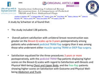
3. Anatomy of the breast
Boundaries
2nd and 6th ribs
Sternal edge and midaxillary line
Tail of Spence
Primarily adipose tissue, glandular tissue, and
suspensory ligaments
Mammary gland is a modified sweat gland – 15-
20 gland lobules drain into 15-20 lactiferous ducts
which open onto the nipple
4. Innervation
Via intercostal nerves 2-6
Long thoracic nerve – “winged
scapula”
Lymphatic drainage
Axillary nodes primarily
Also parasternal, clavicular,
and inguinal nodes
5.
6. Patient evaluation
Patient variables:
-Body habitus (enough tissue vs. too much)
-Past surgical history (? Previous Abdominoplasty)
-Social history (smoking ?)
-Patients wishes and education
-Internet
-Opinion on implants
-“Save my muscle”
9. Pros
Implants
-Shorter operations
-Body Habitus not a factor
-Greater variability in
adjusting size
Autologous
-Less long term complications
-Breast will adjust with weight changes
-Natural appearance “Feels own breast”
10. Cons
•Implants
-Does not work with radiation
-Short term implant complications
-Long term implant Complications
-Capsular contracture
Autologous
-Longer surgery
-Body Habitus (BMI<35)
-preferred Non smokers
-Longer recovery
12. Radiation Therapy
- Repetitive radiation injury disrupts this highly
organized sequence of events (wound healing), resulting in
repetitive inflammatory responses and ongoing cellular
regeneration
-Radiation affects the skin and subcutaneous tissue (color or
turgor and blood vessels ).
-These changes affect the type of reconstruction and the results.
-Due to these changes it is important to include this variable when
discussing your reconstruction options.
13. Effects of radiation therapy on wound healing
Early side effects include erythema, dry
desquamation, hyperpigmentation and hair loss.
Late effects include skin atrophy, dryness,
telangiectasia, dyschromia, dyspigmentation, fibrosis,
and ulcers.
14. Why choose implants?
1. First choice for many women.
2. Rapid recovery.
3. Ability to modify breast volume
4. No donor site morbidity.
5. Decreased hospitalization.
6. Decreased pain.
7. Earlier return to daily activities.
15. Implant reconstruction can be
performed in:
One-stage reconstruction
Women with smaller breasts (A or B cup)
Women undergoing nipple or skin-sparing or
subcutaneous mastectomy.
16.
17.
18. Two-stage reconstruction
Most common method
1.When mastectomy skin requires tissue expansion
for the desired volume or symmetry.
2.Tissue expander is exchanged for a permanent
implant
29. Autogenous Tissue
Advantages:
Corrects skin deficiency
Normal subcutaneous tissue
No foreign material
Better for tolerance than implant for adjuvant radiotherapy
Disadvantages
Longer operative time
Higher morbidity
Donor site morbidity e.g donor site.
30. Flaps
Definition:
A flap is a vascularized block of tissue that is
mobilized from its donor site and transferred to
another location, adjacent or remote, for
reconstructive purposes.
34. Causes of flap failure
• Poor Blood Supply (arterial or venous)
• Too much tension.
• Local sepsis or a septicaemic patient.
• Tight dressing around the pedicle.
36. Indications :
women with stage I and stage II breast cancer.
Incidence of women seeking immediate reconstruction has
increased.
Advantage :
1. Aesthetic outcome usually is improved because the natural
breast contour is preserved.
2. Same risk of recurrence for women with and those
without reconstruction.
3. Reconstruction does not impede the ability to detect a
recurrence.
Immediate reconstruction
37. Delayed reconstruction
Patients with advanced breast cancer who are at significant risk
for local recurrence and need adjuvant therapy.
A. Inflammatory cancer.
B. Ulcerated tumors.
C. Metastatic disease
D. Women who are to receive postoperative radiation
38. Reconstruction in the setting
of radiation therapy
Drawbacks:
1. Perivascular and soft tissue fibrosis.
2. Delayed wound healing
3. Cutaneous pigmentation, and contracture.
Recommendation: delay reconstruction for 6 to 12 months
The combination of radiation and implant reconstruction is associated
with a higher incidence of failure; therefore autologous reconstruction
is often preferred.
Advantage:
Autologous reconstruction tolerates the effects of radiation relatively
well, although some shrinkage and fibrosis can occur.
39. Ideal recipient vessels for
microvascular breast
reconstruction?
Vascular systems
used
Internal
mammary
artery and vein
Modified
radical
mastectomy.
Thoracodorsal
artery and vein
Skin-sparing
mastectomy
Delayed
reconstruction
40. Flaps used in Breast reconstruction
Pedicled
Free
Perforators
42. Anatomy of LD
Origin
Posterior Iliac Crest
Spinous Processes Of The Lower 6 Thoracic Vertebrae,
Lumbar And Sacral Vertebrae
Thoracolumbar Fascia Arising From The Dorsal Iliac Crest.
Insertion
lesser tubercle and intertubercular groove of the humerus
between the teres major and pectoralis major muscles.
Function
An adductor and medial rotator of the arm. It also serves to pull
the shoulder inferiorly and posteriorly.
43. Blood Supply
2 separate vascular systems:
The dominant blood supply
Thoracodorsal artery ( terminal branch of subscapular artery)
The secondary blood supply
Segmental perforating branches off of the intercostal and
lumbar arteries. (disrupted in the process of harvesting the
latissimus, the viability of this portion the flap can be tenuous)
The extramuscular pedicle length varies between 6-16 cm and is
about 9 cm on average.
The intramuscular thoracodorsal artery reliably divides into
vertical and transverse branches, which allows the flap to be
divided into 2 separate muscle and skin paddles.
Anatomy of LD
45. (LD) Flap
Def.: Musculocutaneous Pedicled flap tunneled to mound a breast.
A very reliable flap.
Not a “first choice” for most plastic surgeons
BVS: Thor
Indications used for women who:
1.Have an elevated BMI
2.Had a previous abdominal operations that violated critical zones.
46. Advantages of the LD flap
in BR
Large volume of tissue is available for
reconstruction yet may still need implant.
Long vascular pedicle offers excellent range for
pedicled flaps.
Minimal donor site morbidity occurs( Scar and
seroma)
47. Disadvantages of LD Flaps
in BR
1. Requires implant use if large breast.
2. The donor scar on the back is large and seromas are not uncommon.
3. The skin of the LD flap is thicker and darker than the front of the chest, thus creating
a patch effect.
4. The flap may not be available if the thoracodorsal vessels were damaged
during axillary dissection, although a flap can still be raised on the serratus anterior
branch under some circumstances.
48. Latissimus Dorsi
Incision is made
in the upper
back.
The latissimus
muscle is
tunneled
through the
axilla and placed
into the
mastectomy
defect.
50. Latissismus Dorsi
Flap with Implant
The flap itself is only about one
inch thick usually requiring an
expander/ implant to be placed in
order to match the opposite
breast. The muscle/ tissue with an
expander are shaped into a breast
mound.
54. fleur-de-lis
pattern extended LD flap
Aim :
To reduce the need for an
implant, an extended LD
flap can be raised by
taking more adjacent soft
tissue, e.g. in a fleur-de-lis
pattern, but the scar is
longer and the distal
tissue is less reliable)
56. What is a TRAM flap?
Carl Hartrampf,MD
Definition
The TRAM (Transverse Rectus Abdominis Musculocutaneous) which
can be:
1. Pedicled Single or double
2. Free myocutaeous flap.
3. Muscle sparing (perforator)
58. Flap Blood Supply
The blood supply of the flap can be based:
-The superior epigastric (pedicle TRAM)
-The inferior epigastric (free TRAM) artery and
vein.
59. Indications FOR TRAM
1. Radical mastectomy defect with large tissue requirement
2. History of radiation to the chest wall
3. Large opposite breast (difficult to match with an implant)
4. Small opposite breast (difficult to match with an implant)
5. Previous failure of implant reconstruction
6. Excess lower abdominal tissue and patient desires abdominoplasty
Drawback:
Cannot use if the rectus muscle is divided superiorly (Kocher incision)
60. Indications
of
bipedicle TRAM flap
• Patient undergoing BILATERAL mastectomy
• Patient who requires a LARGE AMOUNT OF
TISSUE FOR A UNILATERAL RECONSTRUCTION
and is not a candidate for a free TRAM flap.
61. Relevant Anatomy
If Single Pedicle
• Only tissues directly over or immediately
adjacent to the muscle have adequate vascularity.
• If more tissues are needed, consider other
procedures (midabdominal TRAM, delay
procedure, double pedicle TRAM, super-charged
TRAM, free TRAM flap, deep inferior epigastric
perforator [DIEP] flap).
62. Contraindications
• Cardiac disease (ie, myocardial infarction, angina, congestive heart
failure)
• Pulmonary disease (ie, emphysema, chronic obstructive pulmonary
disease)
• History of PE or DVT
• Collagen-vascular disease, lupus, scleroderma, polyarteritis (small
vessel disease)
• Unstable psychiatric disease
• Obesity (>25% ideal body weight)
• Older patient (physiologic age older than 70 y)
• Cigarette smoking; unwilling to quit
• Previous abdominal surgery that has interrupted blood supply to the
TRAM flap e.g Kocher incision
63. Complications
1. Fat necrosis and/or partial flap loss (5-15% of patients)
2. Complete loss of TRAM tissue (< 1% of patients)
3. Seroma (fluid collection, usually in abdominal donor site)
4. Hematoma (bleeding at either chest or abdomen)
5. Infection
6. Hernia (1-5% of patients)
7. Abdominal bulge without hernia (5-15% of patients)
8. Deep venous thrombosis and/or pulmonary embolus (< 1%
of patients)
9. Death (< 1% of patients)
64. Supercharging
• Supercharging is the process of performing a microvascular
anastomosis of the inferior epigastric artery and vein of a pedicle
TRAM to the thoracodorsal artery and vein.
• This is sometimes necessary following a pedicle TRAM flap that is
showing evidence of compromised vascularity, such as venous
congestion or arterial insufficiency.
• The additional arterial and venous flow usually will compensate for
the inadequate inflow or outflow of the superior epigastric vessel
within the flap.
65. Abdomen (TRAM flap)
Pedicled on the
CONTRALATERAL superior
epigastric artery.
It can be ‘supercharged’ by
anastomosing the deep
inferior epigastric artery to
the thoracodorsal artery or
‘super-drained’ to the veins
to improve drainage
66. A study by Schwitzer et al found that:
• The study included 138 patients.
• Overall patient satisfaction with unilateral breast reconstruction was
greater on the Breast-Q scales up to 3 years postoperatively among
patients who underwent pedicled TRAM flap surgery than it was among
those who underwent either muscle-sparing TRAM or DIEP flap surgery.
• Satisfaction equalized for the three procedures 3 years or more
postoperatively, with the pedicled TRAM flap patients displaying higher
scores on the Breast-Q scales with regard to Satisfaction with Breasts and
Physical Well-being Chest and Upper Body, and the free-flap patients
showing higher scores on Satisfaction with Outcome and Physical Well-
being Abdomen and Trunk.
Jonathan A. Schwitzer, MD,
* H. Catherine Miller, BS,
* Andrea L. Pusic, MD,
* Evan Matros, MD,
* Babak J. Mehrara, MD,
* Colleen M.
McCarthy, MD,
* Peter A. Lennox, MD,
† Nancy Van Laeken, MD,
† and Joseph J. Disa, MD *
67. Surgically Treated Hernia following Abdominally Based Autologous Breast
Reconstruction: Prevalence, Outcomes, and Expenditures.
Plast Reconstr Surg. 2016; 137(3):749-57 (ISSN: 1529-4242)
Shubinets V; Fox JP; Sarik JR; Kovach SJ; Fischer JP
A study by Shubinets et al indicated that:
*Surgical repair of abdominal hernia within a 4-year postoperative
period is more common among patients who undergo pedicled TRAM
flap breast reconstruction than among those who undergo free TRAM
or DIEP flap reconstruction (7.0% vs 5.7% and 1.8%, respectively).
*The study also suggested that the development of a surgical-site
infection within 30 days of discharge is a risk factor for subsequent
surgical repair of abdominal hernia.
* The study involved 8246 women.
68. Immediate Unilateral Breast Reconstruction using Abdominally Based Flaps:
Analysis of 3,310 Cases.
J Reconstr Microsurg. 2019; 35(1):74-82 (ISSN: 1098-8947)
Kwok AC; Simpson AM; Ye X; Tatro E; Agarwal JP
• Kwok et al reported that those who were
treated with a pedicled TRAM flap procedure
had a lower rate of return to the operating
room for vascular anastomosis
revision (0.0%) than did patients who
underwent free TRAM flap (1.72% rate of
return), DIEP flap (2.66% rate of return), and
SIEA flap (5.64% rate of return) surgery.
69. TRAM delay procedure
Aim
to promote vascular reorganization within the flap
such that the principal blood supply is derived from
the superior epigastric artery and vein.
When?
2 to 3 weeks prior to the scheduled TRAM flap.
How?
Ligating the inferior epigastric artery and vein as
well as incising a portion of the cutaneous surface
of the proposed flap outline BUT keep it in place.
Benefit
To minimize the occurrence of fat or partial flap
necrosis.
80. Latissimus dorsi flap versus pedicled transverse rectus
abdominis myocutaneous
breast reconstruction: outcomes.
J Surg Res. 2015; 199(1):274-9 (ISSN: 1095-8673)
Teisch LF; Gerth DJ; Tashiro J; Golpanian S; Thaller SR
A study by Teisch et al comparing the use of
latissimus dorsi flaps with pedicled TRAM flaps for
breast reconstruction indicated that latissimus dorsi
flaps are more likely to result in postoperative
surgical site complications, while pTRAM flaps are
associated with a greater risk for pulmonary
complications and longer hospital stays.
A total of more than 29,000 latissimus dorsi and
pTRAM cases.
83. Advantages of the internal
mammary Artery as a
recipient vessel
1. No risk of injury to the
intercostobrachial nerve.
2. No associated risk
lymphedema
3. Higher perfusion pressure.
85. Microvascular complications
• Studies have demonstrated that the most important factor in
preventing thrombosis is meticulous surgical technique.
• The administration of pharmacologic agents such as heparin or
dextran is secondary in importance.
• Attention to the vascular pedicle during inset will minimize the
occurrence of a kink or twist that can compromise circulation.
• An end-to-end anastomosis is performed in the majority of cases;
however, an end-to-side anastomosis may be necessary in the event
of caliber mismatch.
86. TRAM—the free flap
• It is a musculocutaneous
free flap based on the
stouter (deep) inferior
epigastric artery.
• Advantage: the incidence of
fat necrosis is much
reduced compared to the
pedicled flap.
87. TRAM—the free flap
• It can be anastomosed to the
contralateral internal
thoracic/mammary vessels (usually
providing more transverse fullness)
or the ipsilateral thoracodorsal
artery (more vertical fullness).
• Disadvantage: It cannot be used in those
who have had previous abdominoplasty or
liposuction.
• But the presence of a gynaecological
Pfannenstiel scar (usually low and muscle
splitting) does not preclude the use of the
flap
88. DIEP (deep inferior epigastric
perforator) flaps
DIEP is a muscle sparing free TRAM.
• Advantage: leaving the muscle
mass and fascia behind, with less
donor site morbidity, specifically in
terms of the strength of trunk
flexion on the hip and the incidence
of hernias, although the difference
may become less pronounced with
time and specific training.
The incidence of fat necrosis is slightly
higher than for the TRAM flap
90. DIEP
• Illustration of a medial
perforator DIEP flap, in which
perfusion is more centralized
and has a bigger vascular
territory.
• These are useful for large
breast reconstructions.
• Medial perforator DIEP flaps
follow Hartrampf zones of
perfusion.
• Zone II is on the contralateral
hemi-abdomen
92. DIEP
• Illustration of a lateral
perforator DIEP flap, in
which perfusion is more
lateralized. These are
useful for small to
moderate sized and
bilateral breast
reconstructions.
• Lateral perforator DIEP
flaps follow Holm’s zones
of perfusion.
• Zone II is on the ipsilateral
hemi-abdomen.
93. TRAM flaps vs the DIEP flap
DIEPFree TRAMTRAM
1% to 5%,1% to 10%,1% and 20%,Abdominal
bulge/hernia
5% to 15%5% to 10%5% to 10%,Fat necrosis
2% to 10%2% to 5%No dataVenous
congestion
1% to 5%1% to 5%.0% to 2%Total flap loss
94. DIEP Flap Breast Reconstruction Using 3-dimensional
Surface Imaging and a Printed Mold
Koichi Tomita, MD, PhD, Kenji Yano, MD, PhD, Yuki Hata, MD, Akimitsu Nishibayashi,
MD, and Ko Hosokawa, MD, PhD
A study of 11 unilateral DIEP flap breast reconstructions, Tomita et al described the
successful use of three-dimensional (3-D) surface imaging for surgical planning and a
3-D printed mold (made from the contralateral breast) for intraoperative breast
shaping.
A 56-year-old woman (patient 6) underwent total mastectomy of the left breast. One year later, she underwent 2-stage delayed
reconstruction with a DIEP flap. The preoperative (A) and 4-month postoperative (B) views are shown.
96. The superficial inferior epigastric artery (SIEA) flap allows for harvest of the
lower abdominal fatty tissue based on the superficial inferior epigastric system.
Occasionally, the flow from the superficial system may be more robust than that
of the deep inferior epigastric system.
If the perforators of the deep inferior epigastrics are judged to be of insufficient
size or location because of either previous surgery or atypical anatomy, the
superficial epigastric system may serve as a logical alternate flow source.
The SIEA flap is not preferred over the DIEP flap because the superficial
artery is usually of much smaller caliber than the deep inferior epigastric artery,
and the superficial artery is usually tortuous in its proximal origin point from the
common femoral. The advantage of no muscular dissection may make the SIEA
flap a preferred choice.
Disadvantages:
the vascular issues associated with the pigtailed small artery and the higher
incidence of seroma due to dissection through the groin lymphatics make the
Superficial inferior epigastric artery flap (SIEA)
97. Color-flow Doppler and CT angiography have been used
for imaging of blood flow patterns in various perforator
flaps.
They may be of use when the integrity or pattern of
blood flow within the flap is in question.
However, these modalities are not typically employed in
routine clinical practice.
Imaging Studies
98. A study by Davis et al.
indicated that CT angiography carried out prior to microsurgical breast
reconstruction can be used to identify whether a DIEP flap will be at greater risk
for postoperative venous congestion. The investigators reported that flaps found
on CT angiography to have atypical venous connections between the deep and
superficial systems, ie, those in which the connections were narrow, tortuous, or
incomplete, had a five-fold risk of such congestion.
102. Superior gluteal artery
Perforator
Def.
A gluteal myocutaneous free flap based
on the superior gluteal artery.
Indications
Patients who have inadequate
abdominal soft tissue volume
Patients who have undergone prior
abdominal surgeries that have
compromised the abdominal
perforating vessels, the GAP flap is
selected.
103. Advantage:
The donor site is in an aesthetically
acceptable location with minimal
resultant contour changes in the
buttock.
The GAP flap allows for harvest of
substantial amounts of fatty tissue,
even in patients who are very thin.
The avoidance of gluteal muscle
sacrifice minimizes long-term morbidity
and shortens recovery.
SGAP
105. Site:
lower positioning of the flap on the buttock.
In this lower position, the feeding
vasculature emanates from the inferior
gluteal artery, which passes below the
piriformis muscle (in contradistinction to the
superior gluteal artery, which passes over it).
Inferior gluteal artery Perforator
Def.
A gluteal myocutaneous free flap based on
the inferior gluteal artery.
106. Advantage:
Have longer pedicles and leave a less-
obvious scar (though you have to sit on it).
Patients with thin upper buttocks and
adequate lower buttocks may benefit
from movement of the donor site to a
lower position with adequate
discussion of anticipated effects.
Disadvantage:
The sciatic nerve is exposed during the
dissection (Both pass below piriformis )
The donor site may suffer contour
effects that mimic the male buttock
shape.
IGAP
108. Combination
Stacked/layered DIEP flap
• For women who are too thin for breast volume restoration with a
routine single DIEP flap reconstruction, 2 flaps may be combined
into a single breast reconstruction with the stacked DIEP flap
technique. “Body lift" perforator flap: Stacked abdomen/hip flap
“Body lift" perforator flap: Stacked abdomen/hip flap
• For women with inadequate volume in the abdominal and gluteal
donor regions, the stacked abdomen/hip flap may be appropriate.
This procedure allows for the DIEP flap to be layered with the GAP
flap. This stacking method incorporates 4 flaps to provide sufficient
volume in bilateral breast reconstruction.
109. Lumbar perforator flap
In patients with adequate soft tissue in the lumbar region, this area
may be used as a donor site for free fat transfer. The perfusion of this
flap depends on the vasculature from the lumbar perforators that
penetrate the fascia superior to the gluteus medius and posterior
superior iliac spine.
A study by Opsomer et al reviewing the outcomes of 100 lumbar
perforator free flap breast reconstructions (72 patients) reported the
procedure to be an effective alternative for patients who are ineligible
for DIEP flap surgery. The report stated that the lumbar flap is a useful
tool in BRCA-positive patients, who tend to be younger and to have
less excess tissue available at conventional donor sites, although the
revision rate (22%, with nine flaps lost) was higher for the lumbar
perforator free flaps than it was for DIEP flaps.
110. Transverse Upper Gracilis
(TUG)
• Anatomy
• The gracilis is a long, thin, straplike muscle lying on the medial
aspect of the thigh.
• The gracilis arises from the outer surface of the inferior ramus of
the pubis and adjoining ischium and inserts into the medial surface
of the tibia below the condyle, contributing to the tendinous pes
anserinus.
• The muscle is innervated by a single motor nerve, the anterior
branch of the obturator nerve, which measures up to 12 cm in
length. This nerve often divides into superior and inferior segments
before entering the muscle, making possible the dissection of
functionally discrete units within the muscle.
• The gracilis functions as a thigh adductor and hip flexor and is a
superficially located muscle of the thigh adductor muscle group,
situated just posteromedial to the adductor longus.
113. Summary
• Patient’s evaluation.
• Need for radiotherapy.
• Immediate vs delayed reconstruction.
• Common Flaps : LD, TRAM and DIEP.
• Choice of Flap according to each case.
• Satisfaction is best with delayed flap reconstruction.
• Each type of Reconstruction has its Pros and Cos.
• There is no “the best option” yet there is “best for her
case”