Downloaded 813 times
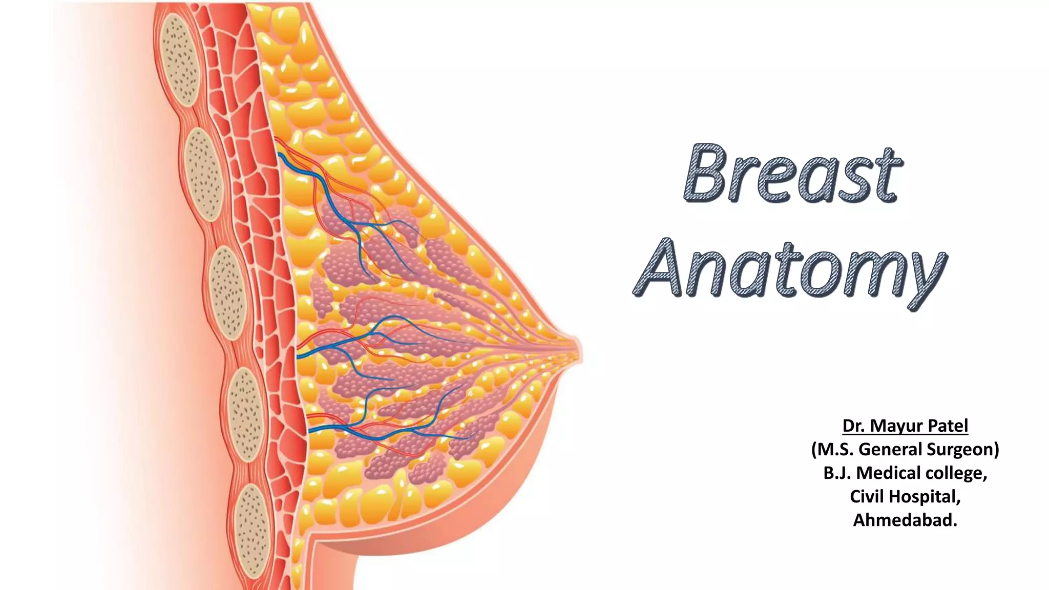
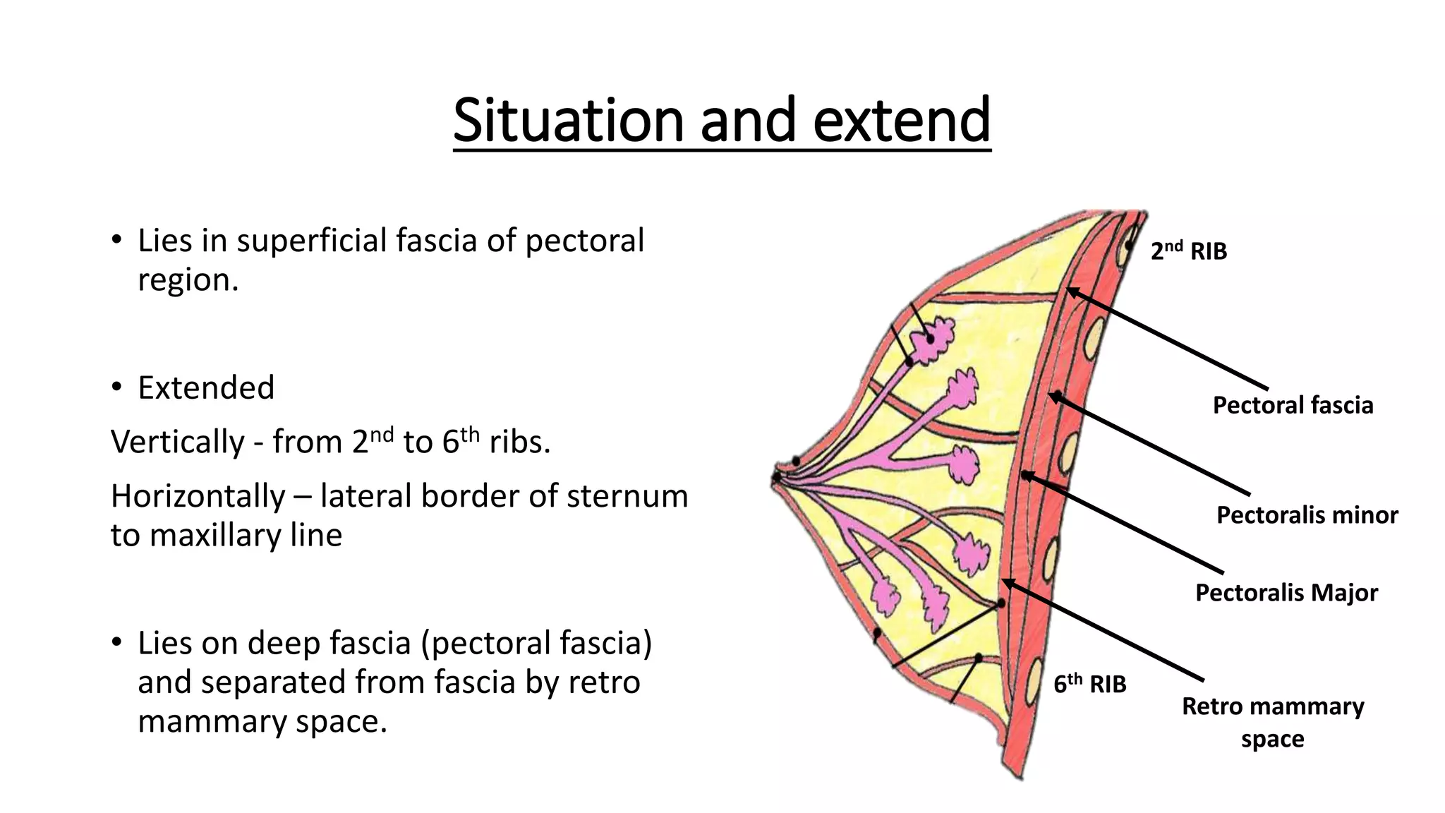
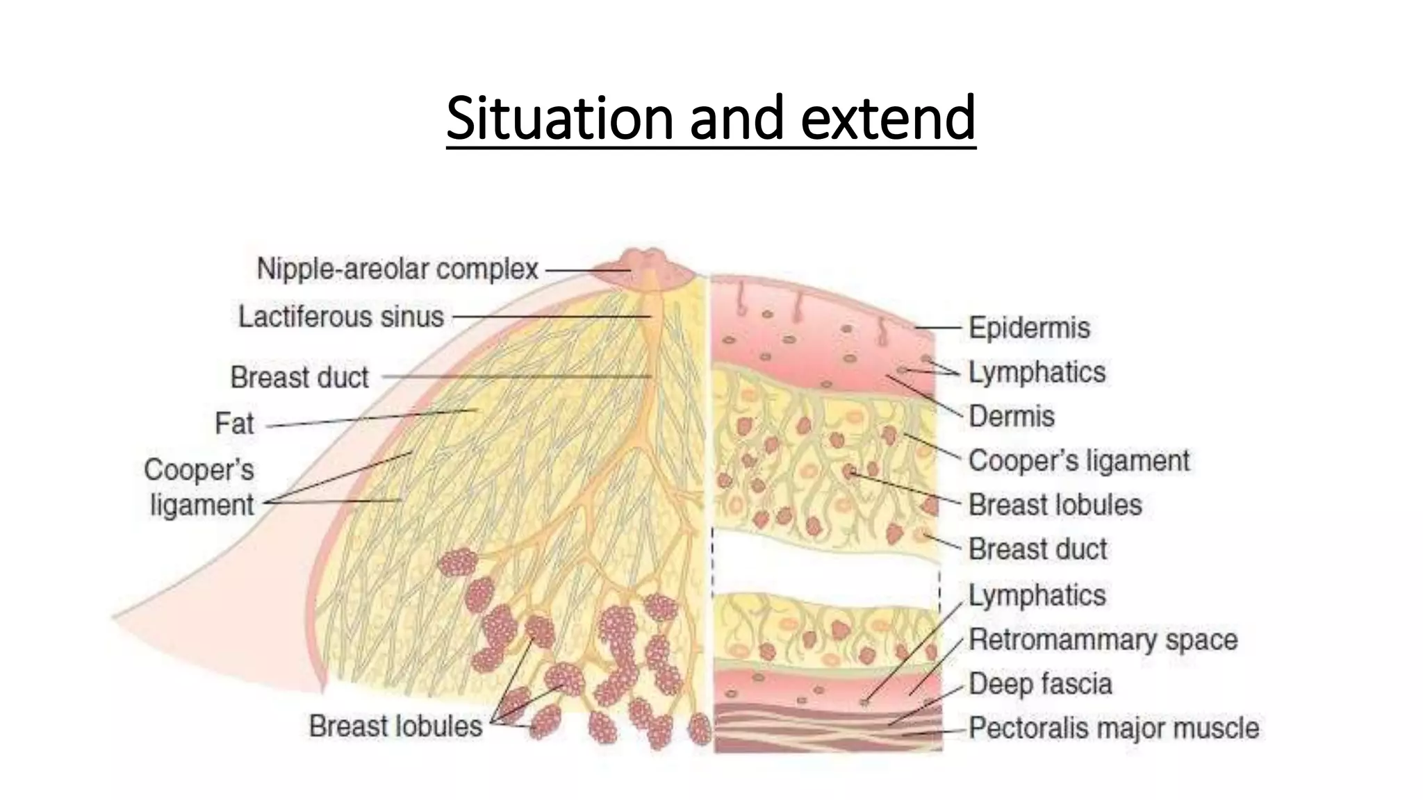
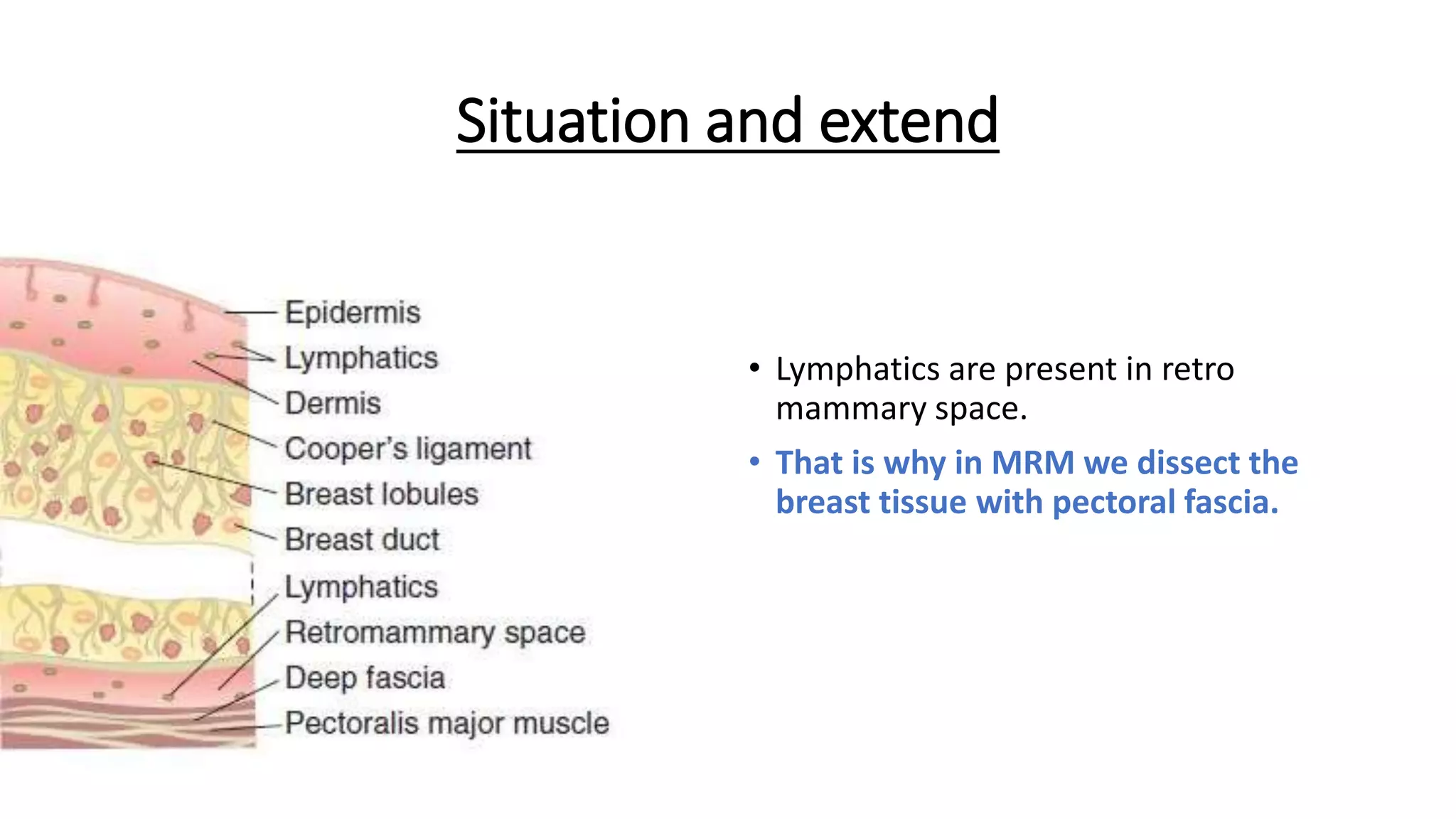

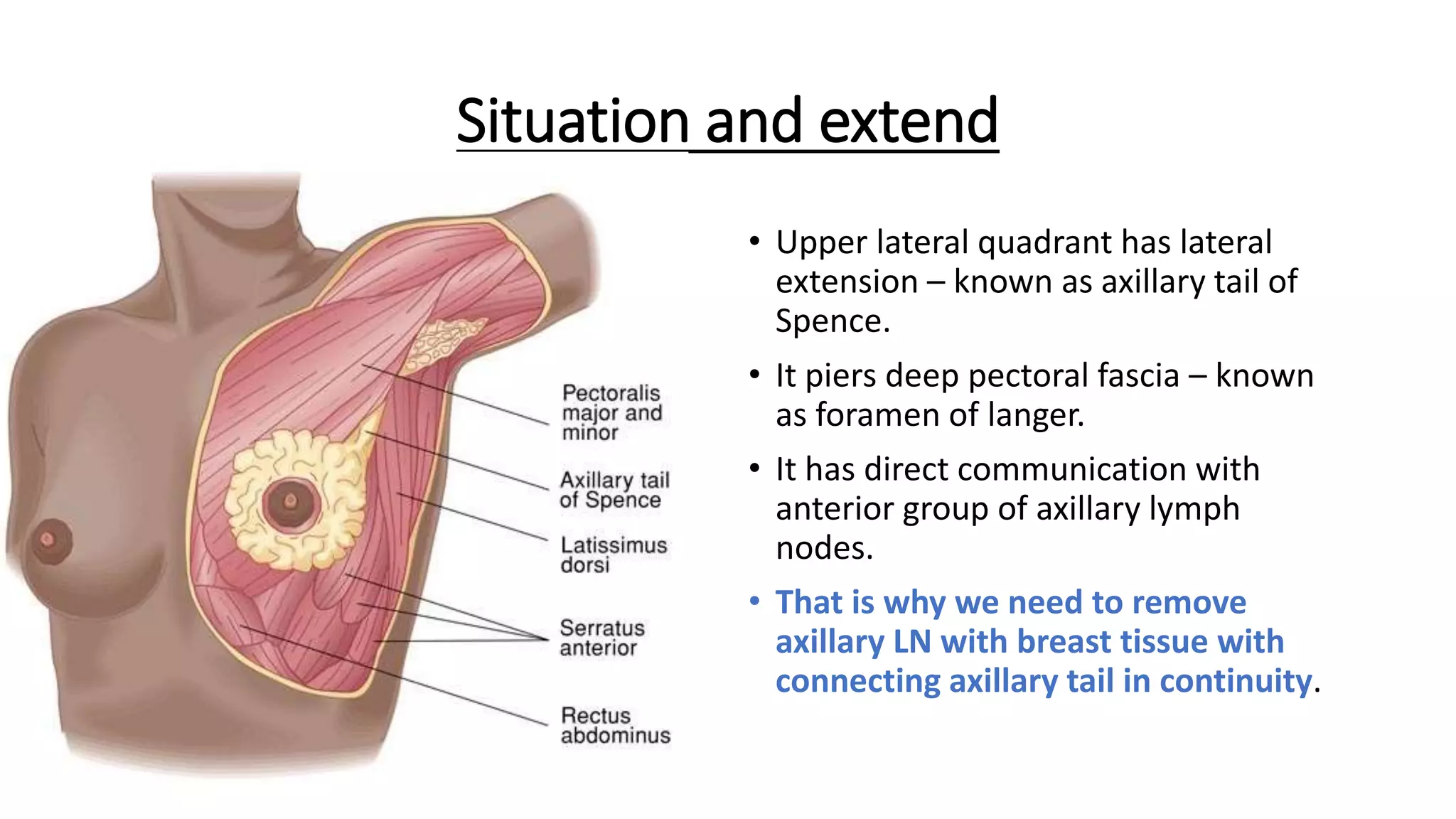


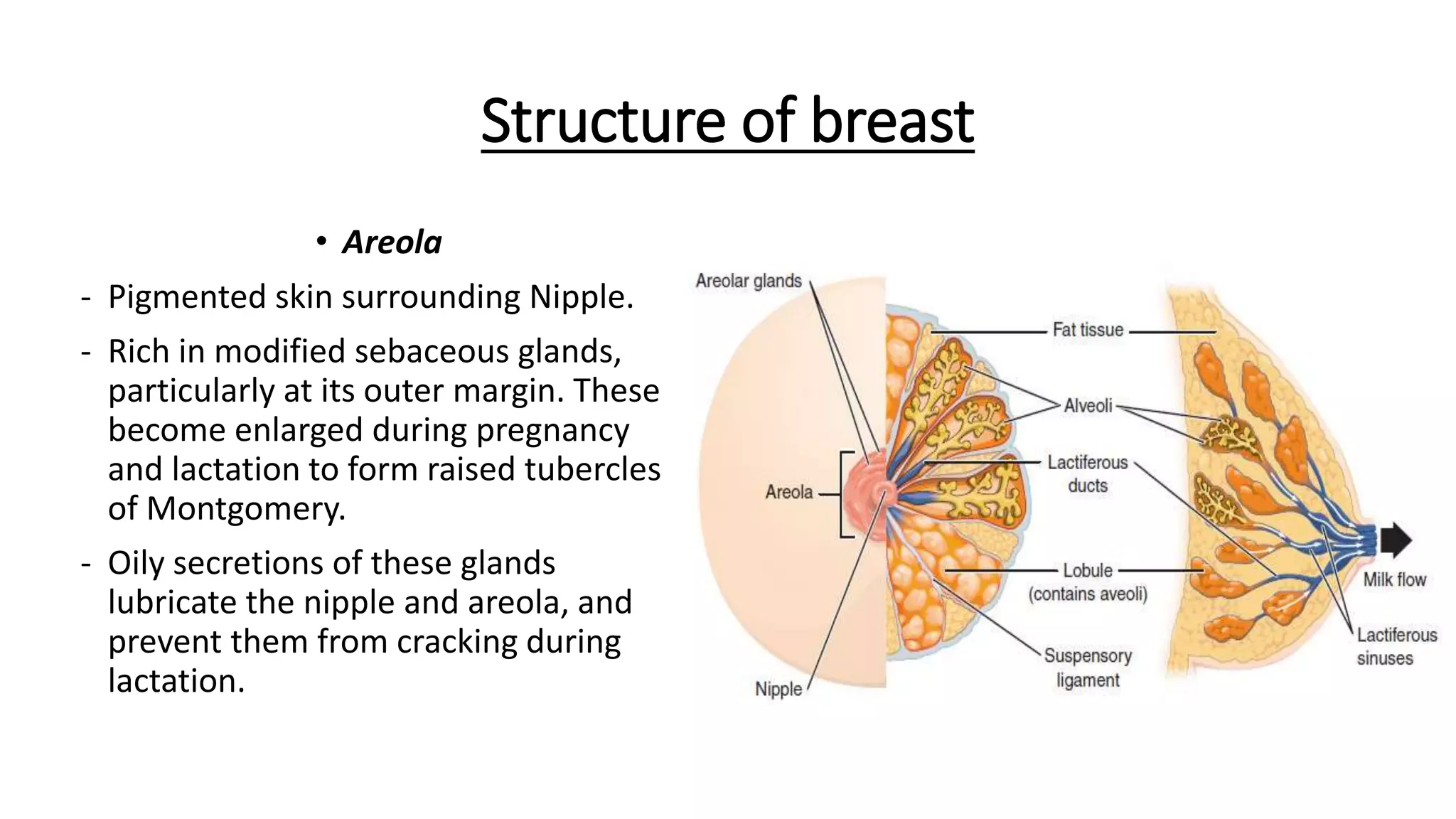


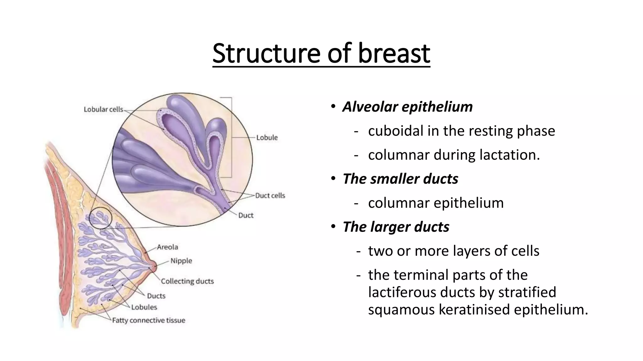


The document provides an in-depth overview of the anatomy and physiology of the breast, including its structure, blood supply, and lymphatic drainage. It details the locations and functions of various components such as the nipple, areola, and lobes of the gland, as well as surgical considerations and lymphatic pathways. Additionally, it discusses the vascular supply from multiple arteries and the intricate lymphatic drainage system connected to axillary and internal mammary lymph nodes.


