
Recommended
More Related Content
What's hot
What's hot (20)
Similar to Anatomy of the mammary gland MBBS
Similar to Anatomy of the mammary gland MBBS (20)
Recently uploaded
Recently uploaded (20)
Anatomy of the mammary gland MBBS
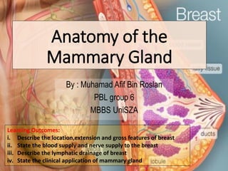
- 1. Anatomy of the Mammary Gland By : Muhamad Afif Bin Roslan PBL group 6 MBBS UniSZA Learning Outcomes: i. Describe the location,extension and gross features of breast ii. State the blood supply and nerve supply to the breast iii. Describe the lymphatic drainage of breast iv. State the clinical application of mammary gland 1
- 2. I. A:Location and extension of the breast • Lies within superficial fascia (in subcutaneous tissue) of anterior thoracic wall ➢ Upper 2/3: • Overlies pectoral fascia ➢ Lower 1/3: • Overlies fascia covering serratus anterior muscle • The circular base extends: ➢Transversely: • -From lateral border of sternum to midaxillary line ➢Vertically: • 2nd through 6th ribs Pectoral fascia Mammary gland Mid axillary line Lateral border of sternum 2
- 3. I .B: External features of the breast Areola •Circular pigmented area of skin •Presence of areolar glands •No fat tissue, no hair follicle • Areolar (Montgomery) glands Sebaceous glands forming rounded projections from surface of areola Nipple •Cylindrical prominence in the centre of areola •No fat, hair or sweat gland •Contain circular smooth muscle fibers •Tips are fissured with openings of lactiferous ducts •Position varies: ➢ Nulliparous women: 4th Intercostal space ➢ Multiparous women: varies The axillary process /tail (of Spence) Extends: ➢Along inferolateral edge of pectoralis major towards axillary fossa ➢Extension of breast tissue into axilla 3
- 4. I. C: Internal features of breast a. Suspensory Ligaments (of Cooper) •Fibrous connective tissue stroma •Well developed in the superior part of the gland Function: ➢Attaches mammary gland to dermis of overlying skin ➢Help to support the lobules of the gland b. Retromammary Space •Potential space or loose connective tissue plane between the breast and pectoral fascia •Contains small amount of fat & lymphatic tissue ✓ Allows the breast some degree of movement on the pectoral fascia Pectoral fascia Gland lobules 4
- 5. I. C: Internal features of breast ➢Glandular Tissue •Tissue that secretes milks •Arranged in lobes (15 to 20 lobes) •Embedded in connective tissue and fat •Each lobe is composed of several lobules •Each lobule is composed of several alveoli ➢Lactiferous duct •Excretory duct that drains each lobes •Each lobe has 1 lactiferous duct •Opens through a separate constricted opening on the surface of the nipple ➢ Lactiferous sinus (Ampulla) •Dilatation of lactiferous duct deep to areolar before it opens on the surface of the nipple ❖Stores droplet of milk ❖Allow expression of milk by compression lobules Lactiferous sinusLactiferous duct Lobe 5
- 6. II.A: Blood supply to breast 6
- 7. • Anterior & lateral cutaneous branches of 4th - 6th Intercostal nerves • These branches convey 2 fibers: II.B: Nerve supply to breast Fibers Area/part supplied Sensory fibers ▪ Skin of the breast Symphatetic fibers ▪ Blood vessels ▪ Smooth muscles in the overlying skin & nipple Intercostal nerves T4 7
- 8. III. Lymphatic drainage of breast ➢ Very important !!! Spread of breast cancer •Can be divided into: 1)Superficial lymphatic drainage: -Drains the skin of the breast EXCEPT areola & nipple 2)Deep lymphatic drainage: -Drains the glandular tissue of the breast INCLUDING areolar & nipple -Passes to the subareolar lymphatic plexus into axillary lymph nodes Superficial lymphatic drainage lymph nodes: 1)Axillary lymph nodes 2)Infraclavicular lymph nodes 3)Inferior deep cervical lymph nodes 4)Parasternal nodes 1 2 3 4 8
- 9. Quadrants of breast: ➢ Medial part: 1.Upper inner quadrant 2.Lower inner quadrant ➢ Lateral part : 3.Upper outer quadrant 4.Lower outer quadrant III. Lymphatic drainage of breast Deep lymphatic drainage: Quadrant Lymph nodes Lateral part (Upper & Lower outer quadrants) Axillary lymph nodes (>75%) •Mainly to anterior/pectoral nodes •Other axillary lymph nodes: - Interpectoral nodes - Deltopectoral nodes - Supraclavicular nodes - Inferior deep cervical nodes Medial part (Upper & Lower inner quadrants) • Parasternal nodes (Internal mammary nodes) • May drain to opposite breast Lower quadrants (Medial & lateral) Inferior phrenic lymph nodes (abdominal lymph nodes) Upper outer quadrant Upper inner quadrant Lower outer quadrant Lower inner quadrant MedialLateral 9
- 10. IV. Clinical importance of breast 1. Breast cancer: –Usually arise from ductal epithelium –Most common site is upper outer quadrant –Early symptom is presence of painless hard lump in breast –Spread of cancer cells : lymphatic / local / blood 2. Peau D’Orange: ➢A symptom of inflammatory breast cancer. ➢Cancer cells block the lymphatic vessels- fluid accumulation in the breast causing edema. ➢Skin appearance have ridges or appear pitted, like the skin of an orange. Paeu D’Orange appearance Breast carcioma 10
- 11. THANK YOU References : • Clinical Anatomy (Keith L. Moore) • Gray’s Anatomy for students (Richard L. Drake) • Dr Nurfarhana Che Lah, Anatomy of mammary gland,Lecture Notes Fakulti Perubatan UniSZA 2019 • Robbin’s Basic pathology of diseases Any QUESTIONS???? 11