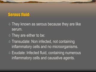
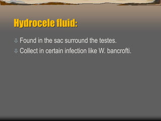
Body fluid
Dr. Mahadi H Abdallah
PhD Microbiology
Serous fluid:
 They known as serous because they are like
serum.
 They are either to be:
 Transudate: Non infected, not containing
inflammatory cells and no microorganisms.
 Exudate: Infected fluid, containing numerous
inflammatory cells and causative agents.
The serous fluid include:
 Pleural fluid.
 Peritoneal (Ascetic) fluid.
 Pericardial fluid.
 Synovial (joint) fluid.
 Hydrocele fluid.
The fluids are normally sterile so any pathogen
isolated is significant if the process done under
aseptic condition.
Pleural fluid:
 It is fluid fill the pleural cavity between the lung and the
inner chest wall, and it is non-purulent pleural effusion.
 Empyema: refer to the purulent infected pleural effusion
containing inflammtory cells (PMN cells) and
microorganisms.
 This caused by: Pneumonia, TB, other pulmonary
infection, malignancy, lymphoma, SLE, amoebic liver
abscess and rheumatoid disease.
Pleural fluid:
 Lab-diagnosis:
 Causative agent: all causative of pneumonia.
 Collection of specimen: by aspiration using
vacuum container (freeof O2) about 5-10ml
 Concentration: if clear use centrifuge to
concentrate.
 Direct examination:
Do 20% KOH for fungi.
Pleural fluid:
Z N stain.
Gram’s stain.
Cell count.
Culture: On routine media for aerobic and anaerobic
organisms.
Peritoneal fluid:
 It is fluid found in the peritoneal cavity.
 Peritoneum: is membrane covered an internal
organ.
 It is property:
Found in small amount.
WBCs 300 cells/ml.
Protein & specific cavity is low.
The normal consistency.
Peritoneal fluid:
 Mode of infection:
Perforation of bowel.
External inoculation (surgery-wound-trauma).
By blood stream.
As a complication of pelvic inflammatory disease.
Through infection within the abdominal viscera.
Peritoneal fluid:
 Ascites: is an increased amount of peritoneal effusion,
usually caused by enterococci, Clostridia and
Mycobacterium.
 Aeitology:
 In children: Pneumococci, S. pyogens, Enterobacteria
and Staphylococci.
 In adult: E. coli, B. fragilis, Clostridia and Streptococci
and Enterococci.
 In sexual active female: N. gonorrhoea, C. trachomatis.
Peritoneal fluid:
 Lab-diagnosis:
 Collection: By aspiration or during surgery
(collect about 10ml) transported in anaerobic
container.
 Culture: In blood culture bottle for both aerobic
and anaerobic organisms then subculture on
suitable routine media.
Pericardial fluid:
 It is the fluid found in the pericardial space
between pericardium (it is tissue surround &
protect the heart and major blood vessel), and
epicardium (it is the membrane sac covered the
heart muscle) and it is about 15-20 ml.
 A etiology:
Bacteria: Mycoplasma, Mycobacteria, Chlamydia, S.
aureus, Pneumococci, viridans strept, Enterococci
and Enterobacteria.
Pericardial fluid:
– Fungi: Candida, Aspergillus and C. neoformans.
– Viral: Enterovirus, Echovirus, Adenovirus and
Influenza virus.
– Also by malignancy, SLE, trauma, uraemia &
rheumatic fever.
Synovial (join) fluid:
 It is fluid that fill the joins.
 Synovitis: is the inflammation of the synovial
membrane that lining the joints.
 Ostemytitis: is the infection of bone.
 Arthritis: is the infection of joints and its two
types:
Infective arthritis: due to infection.
Reactive arthritis: due to Ag-Ab reaction.
Synovial (join) fluid:
 Mode of infection:
Haematogenous spread of bacteria or fungi.
Extend from the infection of bone.
Injection of drugs (corticosteroid).
 Aetiology:
S. aureus, N. gonorrhoea, H. influenzae, E.coli, B.
fragelis, Actinomycetes, Clostridia, Mycoplasma,
Pneumococci, Strept (viridans, group A&B)
Synovial (join) fluid:
 Lab-diagnosis:
Collection of specimen: by aspiration with sterile
syringe.
Culture: part inoculated in to blood culture bottle and
other for smear.
Direct examination: Gram stain, ZN stain 20% KOH
for fungi
Hydrocele fluid:
 Found in the sac surround the testes.
 Collect in certain infection like W. bancrofti.
Bone marrow:
 Can used to diagnose Brucellosis, TB,
Histoplasmosis, Blastomycosis and Lishmania.
The end

Body fluid.Dr.Mahadi

  • 1.
    Body fluid Dr. MahadiH Abdallah PhD Microbiology
  • 2.
    Serous fluid:  Theyknown as serous because they are like serum.  They are either to be:  Transudate: Non infected, not containing inflammatory cells and no microorganisms.  Exudate: Infected fluid, containing numerous inflammatory cells and causative agents.
  • 3.
    The serous fluidinclude:  Pleural fluid.  Peritoneal (Ascetic) fluid.  Pericardial fluid.  Synovial (joint) fluid.  Hydrocele fluid. The fluids are normally sterile so any pathogen isolated is significant if the process done under aseptic condition.
  • 4.
    Pleural fluid:  Itis fluid fill the pleural cavity between the lung and the inner chest wall, and it is non-purulent pleural effusion.  Empyema: refer to the purulent infected pleural effusion containing inflammtory cells (PMN cells) and microorganisms.  This caused by: Pneumonia, TB, other pulmonary infection, malignancy, lymphoma, SLE, amoebic liver abscess and rheumatoid disease.
  • 5.
    Pleural fluid:  Lab-diagnosis: Causative agent: all causative of pneumonia.  Collection of specimen: by aspiration using vacuum container (freeof O2) about 5-10ml  Concentration: if clear use centrifuge to concentrate.  Direct examination: Do 20% KOH for fungi.
  • 6.
    Pleural fluid: Z Nstain. Gram’s stain. Cell count. Culture: On routine media for aerobic and anaerobic organisms.
  • 7.
    Peritoneal fluid:  Itis fluid found in the peritoneal cavity.  Peritoneum: is membrane covered an internal organ.  It is property: Found in small amount. WBCs 300 cells/ml. Protein & specific cavity is low. The normal consistency.
  • 8.
    Peritoneal fluid:  Modeof infection: Perforation of bowel. External inoculation (surgery-wound-trauma). By blood stream. As a complication of pelvic inflammatory disease. Through infection within the abdominal viscera.
  • 9.
    Peritoneal fluid:  Ascites:is an increased amount of peritoneal effusion, usually caused by enterococci, Clostridia and Mycobacterium.  Aeitology:  In children: Pneumococci, S. pyogens, Enterobacteria and Staphylococci.  In adult: E. coli, B. fragilis, Clostridia and Streptococci and Enterococci.  In sexual active female: N. gonorrhoea, C. trachomatis.
  • 10.
    Peritoneal fluid:  Lab-diagnosis: Collection: By aspiration or during surgery (collect about 10ml) transported in anaerobic container.  Culture: In blood culture bottle for both aerobic and anaerobic organisms then subculture on suitable routine media.
  • 11.
    Pericardial fluid:  Itis the fluid found in the pericardial space between pericardium (it is tissue surround & protect the heart and major blood vessel), and epicardium (it is the membrane sac covered the heart muscle) and it is about 15-20 ml.  A etiology: Bacteria: Mycoplasma, Mycobacteria, Chlamydia, S. aureus, Pneumococci, viridans strept, Enterococci and Enterobacteria.
  • 12.
    Pericardial fluid: – Fungi:Candida, Aspergillus and C. neoformans. – Viral: Enterovirus, Echovirus, Adenovirus and Influenza virus. – Also by malignancy, SLE, trauma, uraemia & rheumatic fever.
  • 13.
    Synovial (join) fluid: It is fluid that fill the joins.  Synovitis: is the inflammation of the synovial membrane that lining the joints.  Ostemytitis: is the infection of bone.  Arthritis: is the infection of joints and its two types: Infective arthritis: due to infection. Reactive arthritis: due to Ag-Ab reaction.
  • 14.
    Synovial (join) fluid: Mode of infection: Haematogenous spread of bacteria or fungi. Extend from the infection of bone. Injection of drugs (corticosteroid).  Aetiology: S. aureus, N. gonorrhoea, H. influenzae, E.coli, B. fragelis, Actinomycetes, Clostridia, Mycoplasma, Pneumococci, Strept (viridans, group A&B)
  • 15.
    Synovial (join) fluid: Lab-diagnosis: Collection of specimen: by aspiration with sterile syringe. Culture: part inoculated in to blood culture bottle and other for smear. Direct examination: Gram stain, ZN stain 20% KOH for fungi
  • 16.
    Hydrocele fluid:  Foundin the sac surround the testes.  Collect in certain infection like W. bancrofti.
  • 17.
    Bone marrow:  Canused to diagnose Brucellosis, TB, Histoplasmosis, Blastomycosis and Lishmania.
  • 18.