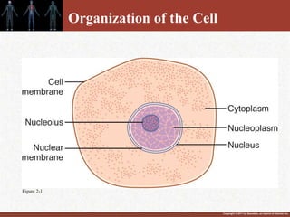
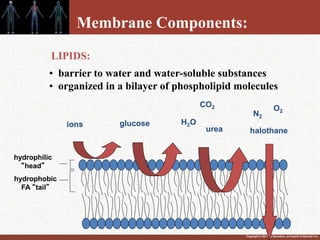
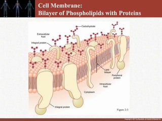
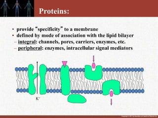
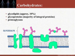
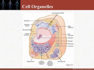
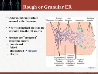
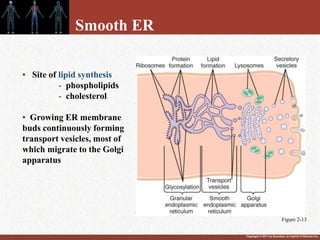
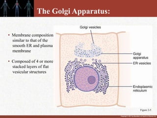
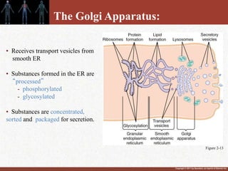
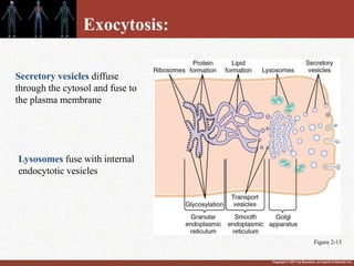
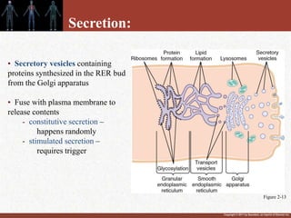
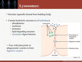
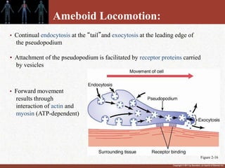
Copyright © 2011 by Saunders, an imprint of Elsevier Inc.
The Cell and Its Functions
UNIT 1
Chapter 2:
Slides by John E. Hall, PhD
Copyright © 2011 by Saunders, an imprint of Elsevier Inc.
Organization of the Cell
Figure 2-1
Copyright © 2011 by Saunders, an imprint of Elsevier Inc.
Cell Composition
Water
Ions
Proteins
Lipids
Carbohydrates
...70-85% of cell mass
...10-20%
...2-95%
...1-6%
Copyright © 2011 by Saunders, an imprint of Elsevier Inc.
Membrane Components:
• barrier to water and water-soluble substances
• organized in a bilayer of phospholipid molecules
hydrophilic
“head”
hydrophobic
FA “tail”
ions H2O
urea
CO2
LIPIDS:
O2
N2
halothane
glucose
Copyright © 2011 by Saunders, an imprint of Elsevier Inc.
Cell Membrane:
Bilayer of Phospholipids with Proteins
Figure 2-3
Copyright © 2011 by Saunders, an imprint of Elsevier Inc.
• provide “specificity” to a membrane
• defined by mode of association with the lipid bilayer
– integral: channels, pores, carriers, enzymes, etc.
– peripheral: enzymes, intracellular signal mediators
K+
Proteins:
Copyright © 2011 by Saunders, an imprint of Elsevier Inc.
• glycolipids (approx. 10%)
• glycoproteins (majority of integral proteins)
• proteoglycans
GLYCOCALYX
Carbohydrates:
Copyright © 2011 by Saunders, an imprint of Elsevier Inc.
• negative charge of the carbo chains repels other
negative charges
• involved in cell-cell attachments/interactions
• play a role in immune reactions
GLYCOCALYX
(-) (-)
(-)
(-)
(-)
(-)
(-)
Carbohydrates (Cont.):
Copyright © 2011 by Saunders, an imprint of Elsevier Inc.
• Present in membranes in varying amounts
• Generally decreases membrane FLUIDITY and
PERMEABILITY (except in plasma membrane)
• Increases membrane FLEXIBILITY and STABILITY
(-) (-)
(-)
(-)
(-)
(-)
(-)
Cholesterol
:
Copyright © 2011 by Saunders, an imprint of Elsevier Inc.
Cell Organelles
Figure 2-2
Copyright © 2011 by Saunders, an imprint of Elsevier Inc.
The Endoplasmic Reticulum:
• Network of tubular and flat vesicular structures
• Membrane is similar to (and contiguous with) the plasma membrane
• Space inside the tubules is called the endoplasmic matrix
Figure 2-4
Copyright © 2011 by Saunders, an imprint of Elsevier Inc.
• Outer membrane surface
covered with ribosomes
• Newly synthesized proteins are
extruded into the ER matrix
• Proteins are “processed”
inside the matrix
- crosslinked
- folded
- glycosylated (N-linked)
- cleaved
Rough or Granular ER
Figure 2-13
Copyright © 2011 by Saunders, an imprint of Elsevier Inc.
• Site of lipid synthesis
- phospholipids
- cholesterol
• Growing ER membrane
buds continuously forming
transport vesicles, most of
which migrate to the Golgi
apparatus
Smooth ER
Figure 2-13
Copyright © 2011 by Saunders, an imprint of Elsevier Inc.
The Golgi Apparatus:
• Membrane composition
similar to that of the
smooth ER and plasma
membrane
• Composed of 4 or more
stacked layers of flat
vesicular structures
Figure 2-5
Copyright © 2011 by Saunders, an imprint of Elsevier Inc.
• Receives transport vesicles from
smooth ER
• Substances formed in the ER are
“processed”
- phosphorylated
- glycosylated
• Substances are concentrated,
sorted and packaged for secretion.
Figure 2-13
The Golgi Apparatus:
Copyright © 2011 by Saunders, an imprint of Elsevier Inc.
Secretory vesicles diffuse
through the cytosol and fuse to
the plasma membrane
Exocytosis:
Lysosomes fuse with internal
endocytotic vesicles
Figure 2-13
Copyright © 2011 by Saunders, an imprint of Elsevier Inc.
Secretion:
• Secretory vesicles containing
proteins synthesized in the RER bud
from the Golgi apparatus
• Fuse with plasma membrane to
release contents
- constitutive secretion –
happens randomly
- stimulated secretion –
requires trigger
Figure 2-13
Copyright © 2011 by Saunders, an imprint of Elsevier Inc.
Lysosomes:
• Contain hydrolytic enzymes (acid hydrolases)
- phosphatases
- nucleases
- proteases
- lipid-degrading enzymes
- lysozymes digest bacteria
• Vesicular organelle formed from budding Golgi
• Fuse with pinocytotic or
phagocytotic vesicles to form
digestive vesicles
Figure 2-12
Copyright © 2011 by Saunders, an imprint of Elsevier Inc.
Lysosomal Storage Diseases
Absence of one or more hydrolases
• not synthesized
• inactive
• not properly sorted and packaged
Result: Lysosomes become engorged with undigested substrate
Examples:
• Acid lipase A deficiency
• I-cell disease (non-specific)
• Tay-Sachs disease (HEX A)
Copyright © 2011 by Saunders, an imprint of Elsevier Inc.
Peroxisomes:
• Similar physically to lysosomes
• Two major differences:
• formed by self-replication
• they contain oxidases
Function: oxidize substances (e.g. alcohol) that
may be otherwise poisonous
Copyright © 2011 by Saunders, an imprint of Elsevier Inc.
Secretory Granules
Figure 2-6
Copyright © 2011 by Saunders, an imprint of Elsevier Inc.
Mitochondria:
Primary function: extraction of energy from nutrients
Figure 2-7
Copyright © 2011 by Saunders, an imprint of Elsevier Inc.
The Nucleus: “Control Center” of the Cell
The double nuclear membrane and matrix are
contiguous with the endoplasmic reticulum
Figure 2-9
Copyright © 2011 by Saunders, an imprint of Elsevier Inc.
• 100 nm in diameter
• functional diameter
is ~9 nm
• (selectively)
permeable to
molecules of up to
44,000 MW
The nuclear membrane is permeated by
thousands of nuclear pores
Figure 2-9
Copyright © 2011 by Saunders, an imprint of Elsevier Inc.
Chromatin (condensed DNA) is found in the nucleoplasm
Nucleolus
• one or more per nucleus
• contains RNA and proteins
• not membrane delimited
• functions to form the granular “subunits” of ribosomes
Figure 2-1
Copyright © 2011 by Saunders, an imprint of Elsevier Inc.
• A-B. Molecules attach to
cell-surface receptors
concentrated in
clathrin-coated pits
• C-D. Receptor binding
induces invagination
• Endocytosis is ATP-
dependent and involves
recruitment of actin and
myosin
Receptor-mediated endocytosis:
Figure 2-11
Copyright © 2011 by Saunders, an imprint of Elsevier Inc.
Digestion of Substances in
Pinocytotic or Phagocytic Vesicles
Figure 2-12
Copyright © 2011 by Saunders, an imprint of Elsevier Inc.
Step 1.
• Carbohydrates are converted
into glucose
• Proteins are converted into
amino acids
• Fats are converted into
fatty acids
Step 2.
• Glucose, AA, and FA are
processed into AcetylCoA
Step 3.
• AcetylCoA reacts with O2 to
produce ATP
A maximum of 38 molecules of ATP
are formed per molecule of glucose
degraded.
ATP production
(More in Chapter 68)
Figure 2-14
Copyright © 2011 by Saunders, an imprint of Elsevier Inc.
The Use of ATP for Cellular Function
1. Membrane transport
2. Synthesis of
chemical
compounds
3. Mechanical work
• Under “standard” conditions G° is only -7.3 kcal/mole
• ATP concentration is ~10x that of ADP, the G is -12 kcal/mole
Figure 2-15
Copyright © 2011 by Saunders, an imprint of Elsevier Inc.
The Cytoskeleton
Intermediate Filaments:
Microtubules:
Thin Filaments:
Thick Filaments:
• Comprised of cell-specific fibrillar monomers
(e.g. vimentin, neurofilament proteins, keratins, nuclear
lamins)
• Heterodimers of a and b tubulin
• Make up spindle fibers, core of axoneme structure
• F-Actin
• Make up “stress fibers” in non-muscle cells
• Myosin (types I and II)
• Together with actin support cellular locomotion and
subcellular transport
Copyright © 2011 by Saunders, an imprint of Elsevier Inc.
Cilia and Ciliary Movements:
• Each cilium is an outgrowth of
the basal body and is covered by
an outcropping of the plasma
membrane.
• Occurs only on the inside surfaces
of the human airway and fallopian
tubes
• Each cilium is comprised of
11 microtubules
• 9 double tubules
• 2 single tubules axoneme
• Ciliary movement is
ATP-dependent
(also requires Ca2+ and Mg2+) Figure 2-17
Copyright © 2011 by Saunders, an imprint of Elsevier Inc.
Ameboid Locomotion:
• Continual endocytosis at the “tail”and exocytosis at the leading edge of
the pseudopodium
• Attachment of the pseudopodium is facilitated by receptor proteins carried
by vesicles
• Forward movement
results through
interaction of actin and
myosin (ATP-dependent)
Figure 2-16
Copyright © 2011 by Saunders, an imprint of Elsevier Inc.
Cell movement is influenced by
chemical substances…
high concentration
(positive)
Low concentration
(negative)
Chemotaxis

Bobby chapter 002

  • 1.
    Copyright © 2011by Saunders, an imprint of Elsevier Inc. The Cell and Its Functions UNIT 1 Chapter 2: Slides by John E. Hall, PhD
  • 2.
    Copyright © 2011by Saunders, an imprint of Elsevier Inc. Organization of the Cell Figure 2-1
  • 3.
    Copyright © 2011by Saunders, an imprint of Elsevier Inc. Cell Composition Water Ions Proteins Lipids Carbohydrates ...70-85% of cell mass ...10-20% ...2-95% ...1-6%
  • 4.
    Copyright © 2011by Saunders, an imprint of Elsevier Inc. Membrane Components: • barrier to water and water-soluble substances • organized in a bilayer of phospholipid molecules hydrophilic “head” hydrophobic FA “tail” ions H2O urea CO2 LIPIDS: O2 N2 halothane glucose
  • 5.
    Copyright © 2011by Saunders, an imprint of Elsevier Inc. Cell Membrane: Bilayer of Phospholipids with Proteins Figure 2-3
  • 6.
    Copyright © 2011by Saunders, an imprint of Elsevier Inc. • provide “specificity” to a membrane • defined by mode of association with the lipid bilayer – integral: channels, pores, carriers, enzymes, etc. – peripheral: enzymes, intracellular signal mediators K+ Proteins:
  • 7.
    Copyright © 2011by Saunders, an imprint of Elsevier Inc. • glycolipids (approx. 10%) • glycoproteins (majority of integral proteins) • proteoglycans GLYCOCALYX Carbohydrates:
  • 8.
    Copyright © 2011by Saunders, an imprint of Elsevier Inc. • negative charge of the carbo chains repels other negative charges • involved in cell-cell attachments/interactions • play a role in immune reactions GLYCOCALYX (-) (-) (-) (-) (-) (-) (-) Carbohydrates (Cont.):
  • 9.
    Copyright © 2011by Saunders, an imprint of Elsevier Inc. • Present in membranes in varying amounts • Generally decreases membrane FLUIDITY and PERMEABILITY (except in plasma membrane) • Increases membrane FLEXIBILITY and STABILITY (-) (-) (-) (-) (-) (-) (-) Cholesterol :
  • 10.
    Copyright © 2011by Saunders, an imprint of Elsevier Inc. Cell Organelles Figure 2-2
  • 11.
    Copyright © 2011by Saunders, an imprint of Elsevier Inc. The Endoplasmic Reticulum: • Network of tubular and flat vesicular structures • Membrane is similar to (and contiguous with) the plasma membrane • Space inside the tubules is called the endoplasmic matrix Figure 2-4
  • 12.
    Copyright © 2011by Saunders, an imprint of Elsevier Inc. • Outer membrane surface covered with ribosomes • Newly synthesized proteins are extruded into the ER matrix • Proteins are “processed” inside the matrix - crosslinked - folded - glycosylated (N-linked) - cleaved Rough or Granular ER Figure 2-13
  • 13.
    Copyright © 2011by Saunders, an imprint of Elsevier Inc. • Site of lipid synthesis - phospholipids - cholesterol • Growing ER membrane buds continuously forming transport vesicles, most of which migrate to the Golgi apparatus Smooth ER Figure 2-13
  • 14.
    Copyright © 2011by Saunders, an imprint of Elsevier Inc. The Golgi Apparatus: • Membrane composition similar to that of the smooth ER and plasma membrane • Composed of 4 or more stacked layers of flat vesicular structures Figure 2-5
  • 15.
    Copyright © 2011by Saunders, an imprint of Elsevier Inc. • Receives transport vesicles from smooth ER • Substances formed in the ER are “processed” - phosphorylated - glycosylated • Substances are concentrated, sorted and packaged for secretion. Figure 2-13 The Golgi Apparatus:
  • 16.
    Copyright © 2011by Saunders, an imprint of Elsevier Inc. Secretory vesicles diffuse through the cytosol and fuse to the plasma membrane Exocytosis: Lysosomes fuse with internal endocytotic vesicles Figure 2-13
  • 17.
    Copyright © 2011by Saunders, an imprint of Elsevier Inc. Secretion: • Secretory vesicles containing proteins synthesized in the RER bud from the Golgi apparatus • Fuse with plasma membrane to release contents - constitutive secretion – happens randomly - stimulated secretion – requires trigger Figure 2-13
  • 18.
    Copyright © 2011by Saunders, an imprint of Elsevier Inc. Lysosomes: • Contain hydrolytic enzymes (acid hydrolases) - phosphatases - nucleases - proteases - lipid-degrading enzymes - lysozymes digest bacteria • Vesicular organelle formed from budding Golgi • Fuse with pinocytotic or phagocytotic vesicles to form digestive vesicles Figure 2-12
  • 19.
    Copyright © 2011by Saunders, an imprint of Elsevier Inc. Lysosomal Storage Diseases Absence of one or more hydrolases • not synthesized • inactive • not properly sorted and packaged Result: Lysosomes become engorged with undigested substrate Examples: • Acid lipase A deficiency • I-cell disease (non-specific) • Tay-Sachs disease (HEX A)
  • 20.
    Copyright © 2011by Saunders, an imprint of Elsevier Inc. Peroxisomes: • Similar physically to lysosomes • Two major differences: • formed by self-replication • they contain oxidases Function: oxidize substances (e.g. alcohol) that may be otherwise poisonous
  • 21.
    Copyright © 2011by Saunders, an imprint of Elsevier Inc. Secretory Granules Figure 2-6
  • 22.
    Copyright © 2011by Saunders, an imprint of Elsevier Inc. Mitochondria: Primary function: extraction of energy from nutrients Figure 2-7
  • 23.
    Copyright © 2011by Saunders, an imprint of Elsevier Inc. The Nucleus: “Control Center” of the Cell The double nuclear membrane and matrix are contiguous with the endoplasmic reticulum Figure 2-9
  • 24.
    Copyright © 2011by Saunders, an imprint of Elsevier Inc. • 100 nm in diameter • functional diameter is ~9 nm • (selectively) permeable to molecules of up to 44,000 MW The nuclear membrane is permeated by thousands of nuclear pores Figure 2-9
  • 25.
    Copyright © 2011by Saunders, an imprint of Elsevier Inc. Chromatin (condensed DNA) is found in the nucleoplasm Nucleolus • one or more per nucleus • contains RNA and proteins • not membrane delimited • functions to form the granular “subunits” of ribosomes Figure 2-1
  • 26.
    Copyright © 2011by Saunders, an imprint of Elsevier Inc. • A-B. Molecules attach to cell-surface receptors concentrated in clathrin-coated pits • C-D. Receptor binding induces invagination • Endocytosis is ATP- dependent and involves recruitment of actin and myosin Receptor-mediated endocytosis: Figure 2-11
  • 27.
    Copyright © 2011by Saunders, an imprint of Elsevier Inc. Digestion of Substances in Pinocytotic or Phagocytic Vesicles Figure 2-12
  • 28.
    Copyright © 2011by Saunders, an imprint of Elsevier Inc. Step 1. • Carbohydrates are converted into glucose • Proteins are converted into amino acids • Fats are converted into fatty acids Step 2. • Glucose, AA, and FA are processed into AcetylCoA Step 3. • AcetylCoA reacts with O2 to produce ATP A maximum of 38 molecules of ATP are formed per molecule of glucose degraded. ATP production (More in Chapter 68) Figure 2-14
  • 29.
    Copyright © 2011by Saunders, an imprint of Elsevier Inc. The Use of ATP for Cellular Function 1. Membrane transport 2. Synthesis of chemical compounds 3. Mechanical work • Under “standard” conditions G° is only -7.3 kcal/mole • ATP concentration is ~10x that of ADP, the G is -12 kcal/mole Figure 2-15
  • 30.
    Copyright © 2011by Saunders, an imprint of Elsevier Inc. The Cytoskeleton Intermediate Filaments: Microtubules: Thin Filaments: Thick Filaments: • Comprised of cell-specific fibrillar monomers (e.g. vimentin, neurofilament proteins, keratins, nuclear lamins) • Heterodimers of a and b tubulin • Make up spindle fibers, core of axoneme structure • F-Actin • Make up “stress fibers” in non-muscle cells • Myosin (types I and II) • Together with actin support cellular locomotion and subcellular transport
  • 31.
    Copyright © 2011by Saunders, an imprint of Elsevier Inc. Cilia and Ciliary Movements: • Each cilium is an outgrowth of the basal body and is covered by an outcropping of the plasma membrane. • Occurs only on the inside surfaces of the human airway and fallopian tubes • Each cilium is comprised of 11 microtubules • 9 double tubules • 2 single tubules axoneme • Ciliary movement is ATP-dependent (also requires Ca2+ and Mg2+) Figure 2-17
  • 32.
    Copyright © 2011by Saunders, an imprint of Elsevier Inc. Ameboid Locomotion: • Continual endocytosis at the “tail”and exocytosis at the leading edge of the pseudopodium • Attachment of the pseudopodium is facilitated by receptor proteins carried by vesicles • Forward movement results through interaction of actin and myosin (ATP-dependent) Figure 2-16
  • 33.
    Copyright © 2011by Saunders, an imprint of Elsevier Inc. Cell movement is influenced by chemical substances… high concentration (positive) Low concentration (negative) Chemotaxis